
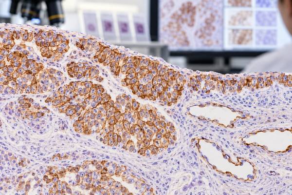
D2-40 immunohistochemistry (IHC) is an antibody stain that detects podoplanin, a cell-surface glycoprotein expressed by lymphatic endothelial cells, mesothelial cells, and several tumors. Pathologists use D2-40 most often as part of a panel when evaluating mesothelioma or lymphatic differentiation in a vascular lesion. In epithelioid mesothelioma, D2-40 commonly produces strong membranous staining, but it cannot distinguish malignant mesothelioma from benign reactive mesothelial cells because both can be positive. It is also not exclusive to mesothelioma: some squamous carcinomas, ovarian serous tumors, seminomas, and vascular neoplasms may express podoplanin. The stain becomes clinically useful when its pattern is combined with morphology and complementary markers such as calretinin, WT1, cytokeratin 5/6, claudin-4, BAP1, MTAP, ERG, CD31, or molecular testing. A positive D2-40 result therefore supports a differentiation pattern; it does not independently prove malignancy or a specific primary tumor.
- Membranous D2-40 positivity can support mesothelial differentiation, but reactive mesothelial cells are also positive.
- Mesothelioma diagnosis uses a panel of mesothelial and carcinoma markers; D2-40 should not be used alone.
- BAP1 or MTAP loss and CDKN2A deletion address malignant versus benign mesothelial proliferation more directly than D2-40.
- D2-40 also highlights lymphatic endothelium and can support lymphatic differentiation in selected vascular tumors.
- There is no universal D2-40 percentage cutoff for mesothelioma; localization, intensity, extent, morphology, and the comparison panel matter.
Table of Contents
- What D2-40 IHC detects
- D2-40 in mesothelioma
- Malignant versus reactive mesothelial cells
- D2-40 as a lymphatic endothelial marker
- Other D2-40-positive tumors
- How pathologists use marker panels
- Limitations, report meaning, and next steps
- Practical D2-40 scenarios and diagnostic decisions
What D2-40 IHC detects
D2-40 is a monoclonal antibody that recognizes podoplanin, a mucin-type transmembrane glycoprotein. Podoplanin is normally expressed in lymphatic endothelial cells and in mesothelial cells lining the pleura, peritoneum, and pericardium. It is also present in other specialized cell types and several neoplasms.
Because podoplanin sits at the cell membrane, a convincing positive D2-40 reaction is usually membranous. In mesothelial lesions, the staining may outline cell borders strongly. In lymphatic vessels, it highlights the thin endothelial lining and helps separate lymphatics from many blood vessels.
The test is tissue based. It has no serum reference range and does not measure the amount of podoplanin in the blood. Pathologists usually report positive versus negative staining and may qualify the extent or intensity.
D2-40 is a lineage/differentiation marker rather than a direct malignancy test. That distinction is particularly important in pleural biopsies, because both benign and malignant mesothelial cells can express podoplanin. The question “Are these cells mesothelial?” is different from “Are these mesothelial cells malignant?” and different tests help answer each question.
D2-40 in mesothelioma
Mesothelioma is a malignant tumor of mesothelial surfaces, most commonly the pleura. Its histologic subtypes include epithelioid, sarcomatoid, and biphasic forms. Epithelioid mesothelioma can resemble metastatic adenocarcinoma or squamous carcinoma, so immunohistochemistry is central to diagnosis.
D2-40 is one of the established positive markers of mesothelial differentiation. Other commonly used positive mesothelial markers include calretinin, WT1, and cytokeratin 5/6. The calretinin IHC test is particularly useful because a characteristic nuclear and cytoplasmic pattern supports mesothelial lineage.
Current guidelines recommend panels rather than a single marker. In an epithelioid tumor involving pleura, a pathologist may use at least two mesothelial markers and at least two carcinoma markers selected for the differential diagnosis. Claudin-4 is a highly useful broad carcinoma marker because most mesotheliomas are negative while many metastatic carcinomas are strongly positive. Organ-specific markers such as TTF-1, Napsin A, GATA3, PAX8, or others may be added when a particular metastatic primary is suspected.
D2-40 tends to be most useful in epithelioid mesothelioma. Sarcomatoid mesothelioma has a more challenging and often less complete mesothelial immunophenotype. In that setting, broad cytokeratin positivity and a carefully chosen exclusion panel may carry more weight, while D2-40 can still contribute in some cases.
Malignant versus reactive mesothelial cells
One of the most important facts about D2-40 is that reactive mesothelial cells are also positive. Therefore, D2-40 cannot establish that a mesothelial proliferation is malignant. This is a major difference from how patients sometimes interpret the word “tumor marker.”
When invasion into fat or other tissue is clearly visible on a biopsy, morphology can establish malignancy. Small biopsies and pleural-fluid cell blocks are harder because invasion may not be represented. In those cases, newer ancillary tests can provide evidence of malignancy.
Loss of nuclear BAP1 expression by IHC is highly specific for malignant mesothelial proliferation in the right setting, although not all mesotheliomas lose BAP1. MTAP loss can serve as a surrogate for CDKN2A deletion in many cases. FISH demonstrating homozygous deletion of CDKN2A is another highly specific finding. These tests are used to distinguish malignant mesothelioma from reactive mesothelial proliferation; D2-40 is used primarily to establish the mesothelial phenotype.
This stepwise logic is helpful when reading a report. “D2-40 positive” answers the lineage question. “BAP1 loss,” “MTAP loss,” or “CDKN2A homozygous deletion” addresses malignancy. A complete diagnosis may require both layers, along with morphology and radiologic distribution.
D2-40 as a lymphatic endothelial marker
D2-40 also labels normal lymphatic endothelium and is widely used to identify lymphatic channels in tissue. Blood-vessel endothelium is generally better marked by CD31 and ERG, whereas podoplanin is more associated with lymphatic differentiation.
In tumor specimens, D2-40 can help identify lymphatic invasion by carcinoma, although interpretation must be cautious because retraction artifact around tumor nests can mimic empty vascular spaces. Demonstrating a true D2-40-positive endothelial lining supports a lymphatic channel, but morphology still determines whether tumor is genuinely inside it.
Some vascular tumors show lymphatic differentiation and can be D2-40 positive. Lymphangiomas and lymphatic malformations typically label, while Kaposi sarcoma frequently expresses podoplanin. A subset of angiosarcomas can also show D2-40, reflecting mixed or lymphatic endothelial differentiation.
D2-40 should not replace broader endothelial markers in a suspected malignant vascular tumor. CD34 IHC, CD31, and ERG provide complementary information, and molecular studies may be decisive for specific vascular entities.
Other D2-40-positive tumors
Podoplanin expression is not confined to mesothelium and lymphatic endothelium. Several tumors can be positive, which limits specificity when D2-40 is used outside a focused panel.
Squamous cell carcinomas can express D2-40, sometimes strongly at the membrane. This is especially important in thoracic pathology because pleural mesothelioma can enter the differential with squamous carcinoma. Markers such as claudin-4, p40, p63, MOC31/BerEP4, and mesothelial markers help resolve the distinction.
Ovarian serous carcinomas may show D2-40 positivity, creating another potential overlap with mesothelioma in peritoneal disease. PAX8 and epithelial markers support Müllerian carcinoma, while WT1 can be positive in both and therefore cannot solve the problem alone. The WT1 IHC test must be interpreted with this overlap in mind.
Seminoma and some other germ-cell tumors can express D2-40. These tumors usually have a distinctive morphology and are assessed with OCT4, SALL4, KIT, and other germ-cell markers. Certain central nervous system and soft-tissue tumors may also express podoplanin.
The lesson is consistent: D2-40 positivity should be interpreted as support for a phenotype, not as a unique molecular fingerprint of mesothelioma.
How pathologists use marker panels
A pleural epithelioid malignancy is a classic setting for a balanced panel. Pathologists typically choose markers that support mesothelial lineage and markers that support carcinoma. The exact antibodies depend on morphology, sex, clinical history, and imaging.
| Marker | Typical role | Key limitation |
|---|---|---|
| D2-40 | Supports mesothelial differentiation | Also positive in reactive mesothelium and other tumors |
| Calretinin | Supports mesothelial differentiation | Not completely specific |
| WT1 | Supports epithelioid mesothelial lineage | Also positive in many serous Müllerian tumors |
| Claudin-4 | Supports carcinoma when positive | Interpret with appropriate controls and tumor morphology |
| BAP1/MTAP | Loss supports malignant mesothelial proliferation | Retained staining does not exclude mesothelioma |
When lung adenocarcinoma is a key alternative, TTF-1 and Napsin A are useful. When breast or urothelial carcinoma is possible, GATA3 and more specific markers may be added. PAX8 supports many Müllerian and renal primaries. The panel is tailored rather than fixed.
Guidelines emphasize this approach because no positive mesothelioma marker has perfect sensitivity and specificity. Two concordant mesothelial markers plus negative carcinoma markers create much stronger evidence than D2-40 alone.
Limitations, report meaning, and next steps
D2-40’s main limitations are lack of malignancy specificity and cross-reactivity with other tumor types. A positive result can be seen in benign mesothelium, mesothelioma, lymphatic endothelium, and multiple nonmesothelial neoplasms. A negative result also does not completely exclude mesothelioma, especially in sarcomatoid disease.
Technical factors can affect staining. Fixation, necrosis, small biopsy size, and tissue processing may reduce antigen detection. Interpretation should focus on viable tumor and use internal lymphatic vessels or other expected elements as controls when appropriate.
If mesothelioma is suspected, the next steps may include a broader mesothelial-versus-carcinoma panel, BAP1 and MTAP IHC, CDKN2A FISH, and additional molecular assays. Radiology is important because diffuse pleural thickening and growth pattern provide context that a small biopsy cannot show.
If the question is lymphatic invasion, the pathologist determines whether a D2-40-positive endothelial space truly contains tumor. This can affect staging in some cancers, but D2-40 staining itself does not prove metastatic spread elsewhere.
Patients reading a report can ask whether D2-40 was used to establish mesothelial lineage or to evaluate lymphatic channels, which other markers supported the conclusion, and whether malignancy-specific tests such as BAP1, MTAP, or CDKN2A were needed. Those questions clarify what the stain actually contributed.
When mesothelioma remains difficult after a limited biopsy, expert thoracic pathology review or a more representative tissue sample may be appropriate. The modern diagnosis is deliberately multiparametric because accuracy affects major treatment and staging decisions.
Practical D2-40 scenarios and diagnostic decisions
D2-40 is most reliable when the diagnostic question is clearly defined before the slide is interpreted. In pleural pathology it may support mesothelial differentiation; in other specimens it may outline lymphatic vessels; and in selected tumors it may demonstrate a phenotype shared by several unrelated neoplasms. These uses should not be mixed together, because the same membranous stain has different implications in each setting.
An epithelioid pleural tumor on biopsy. Diffuse membranous D2-40 can support mesothelial lineage, particularly when calretinin and WT1 or another mesothelial marker are also positive. The pathologist should simultaneously test the competing diagnosis of carcinoma with markers such as claudin-4 and site-directed stains. A convincing result is a pattern: multiple mesothelial markers support one side while epithelial carcinoma markers support or oppose the other. The calretinin IHC result is therefore more useful beside D2-40 than either stain would be alone.
Reactive mesothelial proliferation versus malignant mesothelioma. D2-40 cannot make this distinction because reactive mesothelial cells can be strongly positive. Morphologic evidence of invasion remains a major criterion in tissue biopsies. When invasion is not demonstrable, ancillary findings such as loss of nuclear BAP1, loss of cytoplasmic MTAP, or homozygous CDKN2A deletion can support malignancy in the appropriate mesothelial proliferation. Importantly, retained BAP1 or MTAP does not exclude mesothelioma. A positive D2-40 stain establishes phenotype, while different evidence is needed to establish malignancy.
Sarcomatoid or highly spindle-cell pleural tumor. Mesothelial marker sensitivity is generally lower in sarcomatoid mesothelioma than in epithelioid disease, so a negative D2-40 result has limited exclusionary value. Broad keratin expression, clinical and radiologic distribution, morphology, and a carefully selected panel become especially important. In this setting, forcing the same panel used for an epithelioid tumor can be misleading. The diagnosis often requires greater emphasis on exclusion of sarcoma and sarcomatoid carcinoma and, when necessary, expert thoracic pathology review.
Peritoneal tumor in a woman. Mesothelioma and Müllerian serous carcinoma can both involve peritoneal surfaces, and WT1 may be positive in both. D2-40 can support mesothelial differentiation but is not absolutely specific. PAX8 and claudin-4 favor Müllerian carcinoma when appropriately positive, while a broader mesothelial profile supports mesothelioma. This is a setting where knowing which markers overlap is as important as knowing which ones are usually positive.
Assessing lymphovascular invasion in a carcinoma resection. D2-40 can highlight lymphatic endothelial cells and help determine whether a suspicious space around a tumor nest is a true lymphatic channel. This may clarify staging in selected cancers. The stain does not automatically prove invasion, however. Retraction artifact can create empty spaces that resemble vessels, and the pathologist must see a convincing endothelial-lined channel containing tumor. Blood-vessel markers may be added when the distinction between lymphatic and vascular invasion matters.
A vascular or spindle-cell tumor with D2-40 positivity. Kaposi sarcoma and lesions with lymphatic endothelial differentiation may express podoplanin. Some angiosarcomas also stain. D2-40 therefore cannot classify a vascular neoplasm on its own. CD31 and ERG are more broadly useful endothelial markers, while HHV8 is central in Kaposi sarcoma. Morphology, anatomic site, and molecular findings may be required for rarer vascular tumors. The stain is best understood as one piece of endothelial differentiation rather than a stand-alone “lymphatic tumor test.”
Cytology or a very small pleural sample. Limited material can show mesothelial cells without demonstrating tissue invasion. D2-40 may confirm that a population is mesothelial but cannot by itself separate reactive from malignant cells. Cell-block quality, amount of viable tumor, BAP1/MTAP results, prior specimens, and radiology all influence the next step. Sometimes a larger biopsy is needed because the architectural question cannot be answered on cytology alone.
Tissue quality also shapes interpretation. Poor fixation, crush artifact, decalcification, or extensive necrosis can weaken membranous staining. Internal positive controls, including lymphatic channels, help show whether the stain worked technically. If D2-40 is negative in a tumor that otherwise resembles mesothelioma, the pathologist should ask whether the result reflects biology, sampling, or technique before treating it as strong contradictory evidence.
For patients, the most useful way to read D2-40 is to ask what question it was ordered to answer. If it was used for a pleural mass, ask which other mesothelial and carcinoma markers formed the panel and whether malignancy-specific tests were needed. If it was used to highlight lymphatics, ask whether true lymphovascular invasion was identified. A report that simply lists “D2-40 positive” without context is incomplete as a diagnostic explanation; the clinical meaning comes from the tissue compartment stained, the morphology, and the rest of the workup.
The staining pattern itself deserves attention. In mesothelial cells, D2-40 is typically interpreted as membranous staining, often outlining the cell surface. Cytoplasmic haze or staining confined to background structures should not be given the same weight. A pathologist also checks whether the positive cells are truly the atypical population rather than reactive mesothelium caught around a different tumor. This distinction is especially important in small pleural biopsies, where carcinoma and reactive mesothelial cells can be closely admixed.
When the diagnosis remains uncertain after an appropriate panel, the next step is not to reinterpret D2-40 more aggressively. Obtaining deeper levels, testing another block, securing a larger biopsy, or seeking subspecialty review can provide the missing evidence. That approach respects the stain’s real strengths without asking it to answer a question it cannot answer alone.
D2-40 can occasionally be requested for a very different reason in the same cancer case: to evaluate a possible lymphatic channel near the tumor rather than to classify the tumor cells themselves. The report should make that purpose clear. When the endothelial lining of a suspicious space is D2-40 positive, the finding supports lymphatic identity, but the pathologist still must decide whether viable tumor is truly inside the channel and whether the focus meets the staging rules for that organ. This is not the same as saying that the carcinoma itself is D2-40 positive. Keeping the stained cell type explicit prevents confusion when a patient sees “D2-40” mentioned in both a tumor-marker panel and a lymphovascular-invasion assessment.
The distinction directly changes interpretation.
References
- Guidelines for Pathologic Diagnosis of Mesothelioma: 2023 Update of the Consensus Statement From the International Mesothelioma Interest Group 2024 (Guideline)
- Diagnostic Challenges in the Pathological Approach to Pleural Mesothelioma 2025 (Review)
- Diagnostic performance of immunohistochemistry markers for malignant pleural mesothelioma diagnosis and subtypes. A systematic review and meta-analysis 2024 (Systematic Review)
- Cytology of malignant pleural mesothelioma: Diagnostic criteria, WHO classification updates, and immunohistochemical staining markers diagnostic value 2022 (Review)
- New Insights in Pleural Mesothelioma Classification Update: Diagnostic Traps and Prognostic Implications. 2022 (Review)
- Update of pathological diagnosis of pleural mesothelioma using genomic-based morphological techniques, for both histological and cytological investigations. 2022 (Review)
Disclaimer
D2-40 IHC cannot diagnose mesothelioma or prove malignancy by itself. Results require interpretation with tissue morphology, a balanced immunohistochemical panel, malignancy-specific ancillary tests when needed, and clinical/radiologic findings.





