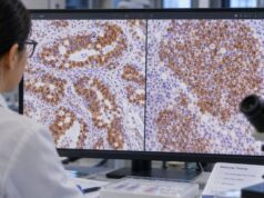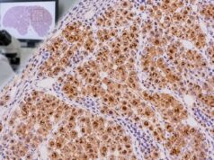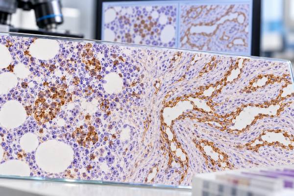
CD34 immunohistochemistry (IHC) detects the CD34 protein in tissue and is used mainly to identify hematopoietic precursor cells, blood-vessel endothelium, and a range of tumors that share CD34 expression. In bone marrow, it is especially useful for highlighting blasts and estimating their distribution when leukemia or a myelodysplastic neoplasm is being evaluated. In soft-tissue pathology, CD34 can support vascular differentiation and can also be positive in nonvascular tumors such as solitary fibrous tumor, dermatofibrosarcoma protuberans, and many gastrointestinal stromal tumors. Because normal endothelial cells and marrow progenitors are naturally positive, CD34 is sensitive but not specific for cancer. A “CD34-positive” result therefore does not name a diagnosis on its own. Pathologists interpret the stain by asking which cells are positive, how many are present, whether staining is diffuse or focal, and whether the morphology and companion markers fit the suspected disease.
- CD34 positivity usually identifies immature hematopoietic cells or endothelial/stromal differentiation; it does not automatically mean leukemia.
- In bone marrow, CD34 IHC can help estimate and map blasts, but blast percentage must be integrated with morphology, flow cytometry, genetics, and current classification criteria.
- Vascular endothelial cells normally stain for CD34, providing an internal control and helping outline blood vessels.
- Many nonvascular tumors can be CD34 positive, so vascular tumors require correlation with markers such as ERG and CD31.
- There is no single universal CD34-positive percentage that diagnoses a tumor; the relevant cutoff depends on the specific hematologic or surgical-pathology question.
Table of Contents
- What CD34 IHC detects
- CD34 in bone marrow and leukemia
- CD34 in vascular tumors
- Other CD34-positive tumors
- How positive staining is interpreted
- Common pitfalls and false impressions
- What happens after the result
- Practical CD34 interpretation scenarios
What CD34 IHC detects
CD34 is a transmembrane glycoprotein expressed by early hematopoietic progenitor cells and by vascular endothelial cells. It is also present in several stromal cell populations. IHC places an antibody against CD34 on a fixed tissue section, producing visible staining where the protein is present. The result preserves tissue architecture, which is an important advantage over tests that analyze dissociated cells.
In a bone marrow core biopsy, a pathologist can see whether CD34-positive immature cells are scattered normally, increased, or clustered abnormally. In a soft-tissue biopsy, the stain may highlight tumor cells, normal vessels, or both. Those are very different findings. The cell type must therefore be identified from the microscopic context.
CD34 IHC does not directly measure circulating stem cells. Stem-cell collections for transplant are usually quantified with flow cytometry, which counts CD34-positive cells in blood or an apheresis product. Tissue IHC answers a different question: where are CD34-expressing cells within a biopsy, and what pattern do they form?
A related distinction matters in leukemia. CD34 is a marker of immaturity in many—but not all—acute leukemias. Some leukemic blasts are CD34 negative, and several genetically defined acute myeloid leukemias can be diagnosed under modern classification systems without relying on CD34 expression. Thus, the stain supports blast identification but does not replace leukemia flow cytometry, cytogenetics, or molecular testing.
CD34 in bone marrow and leukemia
Normal adult marrow contains a small population of CD34-positive progenitor cells. They are generally scattered rather than forming large sheets. In a marrow involved by acute leukemia or certain myelodysplastic neoplasms, CD34-positive blasts may become more numerous and may form abnormal clusters or aggregates. That architectural information can be valuable when aspirate material is dilute, hemodilute, fibrotic, or otherwise difficult to assess.
The stain is especially helpful when a pathologist wants to estimate the proportion and distribution of blasts on a core biopsy. International standardization guidance emphasizes that the method and readout should be validated because fixation, decalcification, antibody clone, tissue quality, and counting approach can all influence the apparent result. A pathologist may describe the percentage of marrow cellularity represented by CD34-positive blasts, but the number should be interpreted as one component of the full marrow assessment.
CD34 positivity is not required for acute leukemia
A common misconception is that blasts must be CD34 positive. They do not. Acute myeloid leukemia, acute lymphoblastic leukemia, and other immature neoplasms have variable CD34 expression. Some AML subtypes are characteristically often negative or only partially positive. Conversely, increased CD34-positive cells can occur in myelodysplastic neoplasms and other marrow disorders without meeting criteria for acute leukemia.
Modern classifications integrate morphology with disease-defining genetic findings. In selected genetically defined AML categories, a rigid 20% blast threshold is no longer universally required in the WHO system, while other classification frameworks may use different thresholds. This is why a pathology report should be read together with the molecular and cytogenetic sections rather than by treating CD34 as a stand-alone blast counter.
CD34 is also useful in marrow staging and follow-up, but residual or regenerating progenitors can complicate interpretation after chemotherapy or transplant. When minimal residual disease is the clinical question, highly sensitive flow cytometry, PCR, or next-generation sequencing is usually more informative than routine CD34 IHC alone. The role of IHC is architectural and contextual.
CD34 in vascular tumors
Endothelial cells lining normal blood vessels usually express CD34, so vascular channels are strongly highlighted in many tissue sections. This makes CD34 a useful endothelial marker and also provides an internal positive control. Benign and malignant vascular tumors can be positive, including many hemangiomas, epithelioid hemangioendotheliomas, and angiosarcomas.
However, CD34 is not the most specific proof of endothelial differentiation. Other markers, particularly CD31 and the nuclear transcription factor ERG, are generally more specific in difficult cases. A malignant spindle or epithelioid tumor that is CD34 positive should not be called angiosarcoma unless its morphology and broader vascular panel support that diagnosis. Some vascular neoplasms may also lose CD34 expression, so a negative result does not exclude them.
The exact panel depends on morphology. For a vasoformative lesion, pathologists may combine CD34 with ERG, CD31, FLI1, or factor VIII-related antigen. For Kaposi sarcoma, HHV-8 testing is central. For a lesion with lymphatic differentiation, D2-40/podoplanin may be useful; the D2-40 IHC test highlights lymphatic endothelium but has its own important nonvascular applications.
Recent reviews of vascular tumors emphasize that diagnosis increasingly combines morphology, IHC, and molecular alterations. This matters because several vascular tumors have characteristic gene fusions or mutations that can confirm an otherwise difficult diagnosis. CD34 remains useful, but it is one layer of evidence rather than a universal vascular “yes/no” test.
Other CD34-positive tumors
CD34 expression is common in a variety of mesenchymal tumors that are not endothelial. Important examples include solitary fibrous tumor, dermatofibrosarcoma protuberans, spindle cell lipoma, and many GISTs. Some peripheral nerve sheath, fibroblastic, and other soft-tissue lesions can also show staining.
This broad expression explains why tumor location and architecture matter so much. A CD34-positive spindle-cell tumor of the pleura or deep soft tissue may raise solitary fibrous tumor, for which nuclear STAT6 is a much more specific confirmatory marker. A dermal spindle-cell lesion may require distinction between dermatofibrosarcoma protuberans and dermatofibroma using morphology, CD34 distribution, and sometimes molecular testing. A gastrointestinal spindle-cell tumor may be CD34 positive but is assessed with KIT and DOG1 if GIST is suspected; CD117 IHC is often part of that workup.
CD34 is also expressed in normal stromal dendritic/interstitial cells in several organs. These background cells can create apparent positivity near a tumor and must not be mistaken for tumor-cell staining. Careful comparison with the hematoxylin and eosin slide helps determine whether the brown reaction is actually in neoplastic cells.
The practical message is that CD34 is a pattern-recognition marker. It narrows or redirects a differential diagnosis only when the pathologist knows which compartment is staining and what the tumor looks like. Its wide distribution makes it versatile, but the same versatility limits specificity.
How positive staining is interpreted
Most pathology reports interpret CD34 in relation to the diagnostic question rather than by a universal score. The report may state that tumor cells are positive, that only background vessels are positive, or that a certain approximate percentage of marrow cells is highlighted. The distinction is crucial.
| Finding | Typical interpretation | Important next context |
|---|---|---|
| Scattered CD34-positive marrow cells | Can be a normal progenitor pattern | Overall cellularity, morphology, clinical setting |
| Increased or clustered CD34-positive marrow blasts | Supports an abnormal immature-cell population | Flow cytometry, genetics, formal blast assessment |
| Tumor cells and vascular channels strongly positive | May support a CD34-positive mesenchymal or vascular tumor | Specific lineage markers and morphology |
| Only internal blood vessels positive | Stain worked but tumor is CD34 negative | Use alternative markers for the suspected diagnosis |
| Focal weak tumor staining | Often nonspecific or nonexclusive | Do not over-weight without corroborating markers |
In marrow, percentages require particular care. The denominator may be all nucleated marrow cells, overall cellularity on the core, or another defined population depending on laboratory practice. Patchy disease can also produce local areas with a higher blast concentration than the average. That is one reason standardized terminology and explicit reporting methods improve reproducibility.
In solid tumors, intensity alone rarely has a therapeutic cutoff. CD34 does not function like ER, HER2, or PD-L1 scoring systems where specific thresholds can directly affect treatment eligibility. Its main value is diagnostic.
Common pitfalls and false impressions
The most common error is equating CD34 positivity with one disease. In bone marrow, positive cells are not automatically leukemic. In soft tissue, positivity is not automatically vascular. And in GIST, positivity is supportive but not specific.
Technical variables can also change results. Decalcification of marrow cores may reduce antigenicity. Overfixation, underfixation, tissue exhaustion, necrosis, and suboptimal antigen retrieval can produce weaker staining. Internal vascular staining helps determine whether a negative tumor result is technically believable.
Another pitfall is counting endothelial cells as blasts in marrow. CD34 strongly labels small vessels, and these can be prominent in hypercellular or neoplastic marrows. A trained observer distinguishes endothelial morphology and vessel architecture from individual hematopoietic cells. Similarly, stromal CD34-positive cells can be mistaken for tumor cells in some soft-tissue biopsies.
Sampling is also important. A tiny biopsy may not represent the full tumor. Heterogeneous lesions can contain positive and negative regions, and a marrow core may miss focal disease. When the clinical and morphologic picture strongly conflicts with the stain, repeating the stain, testing another block, or using orthogonal methods may be appropriate.
Finally, CD34 status generally does not tell how aggressive a solid tumor is. Prognosis depends on the specific diagnosis, grade, stage, molecular alterations, and other factors. The presence or absence of CD34 should not be interpreted as a universal good- or bad-risk sign.
What happens after the result
The next step depends entirely on why CD34 was ordered. In a marrow with suspected leukemia or myelodysplasia, the pathologist integrates the stain with aspirate morphology, flow cytometry, chromosome analysis, fluorescence in situ hybridization, and molecular sequencing. If an acute leukemia is diagnosed, the genetic subtype and measurable residual disease strategy become more important for treatment planning than CD34 positivity alone.
If a vascular tumor is suspected, a broader endothelial panel and, in selected entities, molecular confirmation may follow. For a CD34-positive spindle-cell tumor, site-specific markers such as STAT6, KIT/DOG1, SOX10, or others may be chosen. This targeted approach avoids wasting tissue on large unfocused panels.
Patients reviewing a pathology report can ask three practical questions: Which cells are CD34 positive? What diagnosis does that support? What other tests confirm the diagnosis? Those questions usually clarify the result far better than asking whether “CD34 is high.” Tissue IHC is not a blood concentration and does not have a normal numeric range.
When a report says “CD34 highlights approximately X% blasts,” the percentage should be interpreted in the context of the complete bone marrow diagnosis. If the percentage appears near a diagnostic threshold, classification may depend on the marrow aspirate, defining genetic alterations, or the laboratory’s validated method. A hematopathologist may also compare multiple compartments because aspirate and core estimates can differ when disease is patchy or the aspirate is diluted.
For rare soft-tissue tumors, expert pathology review can be useful when morphology and IHC are discordant or when a molecularly defined entity is being considered. The goal is not to accumulate more positive stains but to arrive at a coherent diagnosis in which morphology, immunophenotype, and molecular findings agree.
Practical CD34 interpretation scenarios
A bone marrow biopsy with suspected acute leukemia illustrates why architecture matters. Suppose the aspirate is hemodilute and contains too few marrow particles for a reliable blast count. On the core biopsy, CD34 may highlight clusters of immature cells that are not obvious on routine staining. That finding can support increased blasts and help the hematopathologist estimate their distribution, but the final diagnosis still depends on the complete immunophenotype and genetic data. If the blasts are CD34 negative, morphology, flow cytometry, MPO, TdT, CD117, and disease-defining molecular abnormalities may still establish acute leukemia.
The same stain behaves very differently in a spindle-cell soft-tissue tumor. Diffuse CD34 positivity in a pleural or deep soft-tissue spindle tumor raises entities such as solitary fibrous tumor, but nuclear STAT6 is the more specific confirmatory test. In a dermal spindle-cell proliferation, diffuse CD34 may support dermatofibrosarcoma protuberans, while a peripheral rim or patchy pattern may be seen in other fibrohistiocytic lesions. Here, the shape and location of the tumor are at least as important as the stain.
A vascular lesion creates a third scenario. CD34 may strongly outline tumor channels, but the pathologist still asks whether those cells are genuinely endothelial. ERG and CD31 can provide stronger lineage confirmation, and the tumor’s growth pattern—such as vasoformation, atypia, necrosis, and infiltrative behavior—helps distinguish a benign vascular lesion from angiosarcoma. A lesion that is CD34 positive but ERG and CD31 negative should prompt reconsideration of a nonvascular CD34-positive tumor.
CD34 can also be useful when the result is negative. If the internal blood vessels stain strongly but the spindle tumor does not, the pathologist knows the assay worked and can place real weight on tumor negativity. In contrast, if both vessels and tumor fail to stain, the test is technically noninformative and may need repetition. This internal-control principle is a basic but important part of IHC interpretation.
Specimen processing deserves attention in marrow. Decalcification is necessary to soften bone but can damage antigens. Laboratories validate their protocols so that CD34 remains interpretable, yet a heavily decalcified or poorly fixed core may still underperform. When aspirate flow cytometry and core IHC appear discordant, technical quality, sampling differences, and hemodilution should be considered before assuming the biology changed.
Finally, CD34 should not be confused with a transplant stem-cell count. A report that says “CD34-positive blasts comprise approximately 8% of marrow cellularity” is a histologic estimate in a diagnostic biopsy. A report that says “CD34+ cells/kg” in an apheresis product is a flow-cytometric measurement used for transplantation. The same antigen is being measured for different purposes, with different methods and clinical meanings.
A final distinction is between describing a cell population and diagnosing a disease. CD34 can estimate or highlight immature hematopoietic cells in bone marrow, but the percentage of CD34-positive cells is not automatically identical to the blast percentage used for classification. Some blasts lack CD34, and some normal progenitors express it. Pathologists therefore reconcile CD34 IHC with the aspirate differential, flow cytometry, morphology, and genetics. In vascular lesions, the same principle applies in a different way: CD34 can confirm endothelial or fibroblastic differentiation in the right context, but it does not establish benign versus malignant behavior. Growth pattern, atypia, mitotic activity, necrosis, and more specific markers determine the diagnosis. This explains why the same “CD34 positive” phrase can appear in reports for very different tissues without carrying the same clinical meaning.
References
- Clinical Applications of Bone Marrow CD34 Immunohistochemistry (BM CD34 IHC) Assay: International Council for Standardization in Hematology (ICSH) Guidelines. 2025 (Guideline)
- Updates in Immunohistochemistry for Hematopoietic and Lymphoid Neoplasms 2024 (Review)
- Fifth edition WHO classification: myeloid neoplasms 2025 (Review)
- The 5th edition of the World Health Organization Classification of Haematolymphoid Tumours: Myeloid and Histiocytic/Dendritic Neoplasms 2022 (Review)
- Cutaneous vascular tumors: an updated review 2023 (Review)
- Vascular tumors of intermediate malignancy: An update 2024 (Review)
Disclaimer
CD34 IHC is a tissue-based diagnostic test and cannot diagnose leukemia, a vascular tumor, or another neoplasm by itself. Individual results should be interpreted by the pathology and clinical teams together with morphology, flow cytometry, genetics, imaging, and other relevant findings.