CD117 immunohistochemistry (IHC) is a tissue stain that detects the KIT receptor protein on or within cells. Pathologists use it most often when evaluating a possible gastrointestinal stromal tumor (GIST), identifying mast cells in bone marrow or other tissues, and working through selected germ cell, myeloid, melanocytic, and other tumor differentials. A positive CD117 stain does not by itself prove a specific cancer. Its meaning depends on the tumor’s shape, location, staining pattern, other IHC markers, and—when GIST or systemic mastocytosis is suspected—molecular findings such as KIT or PDGFRA mutations. In GIST, strong diffuse CD117 staining is common, but a small minority of genuine GISTs can be weak or negative. In mast cell disorders, CD117 is highly sensitive for mast cells but does not distinguish normal/reactive mast cells from neoplastic ones. The most useful interpretation therefore comes from a panel rather than from CD117 alone.
- CD117-positive usually means the cells express KIT protein, not that a KIT mutation is automatically present.
- Most GISTs are CD117 positive, but DOG1 and molecular testing help resolve CD117-negative or unusual cases.
- Mast cells normally express CD117, so positivity in bone marrow must be interpreted with morphology and markers such as tryptase, CD25, CD2, or CD30.
- There is no universal “positive percentage” cutoff for all CD117 IHC uses; intensity, distribution, cellular localization, and the diagnostic question matter.
- A positive stain is not a treatment decision by itself; mutation testing is often needed when targeted KIT/PDGFRA therapy is being considered.
Table of Contents
- What the CD117 IHC test shows
- Normal expression and staining pattern
- CD117 in gastrointestinal stromal tumor
- CD117 in mast cell disorders
- Other tumors that can be positive
- How pathologists interpret CD117 results
- Limitations, follow-up, and next tests
- Practical CD117 interpretation scenarios
What the CD117 IHC test shows
CD117 is another name for the KIT receptor, a transmembrane receptor tyrosine kinase encoded by the KIT gene. In normal biology, KIT signaling helps regulate several cell populations, including mast cells, hematopoietic progenitors, melanocytes, germ cells, and interstitial cells of Cajal. IHC uses an antibody against the KIT protein on a formalin-fixed tissue section, allowing a pathologist to see which cells contain the target and where staining is located.
The test answers a protein-expression question. It does not directly sequence the KIT gene. That distinction matters because a tumor can express KIT protein without carrying an activating KIT mutation, and some tumors driven by a KIT mutation can show variable staining. In a suspected GIST, a CD117-positive result supports the diagnosis when the microscopic appearance and anatomic setting are compatible. It is especially useful when paired with DOG1 IHC for GIST, which is also highly sensitive and can remain positive in some KIT-negative GISTs.
CD117 can also be used as a cell-identification stain. In bone marrow, for example, mast cells generally stain strongly, and immature myeloid cells may also express CD117. That makes the stain useful, but it also creates a common interpretive trap: seeing CD117-positive cells does not automatically mean mastocytosis or leukemia. The pathologist must ask which cells are staining, whether they form abnormal aggregates, what their shape is, and whether the wider immunophenotype is clonal or expected.
Unlike blood tests, CD117 IHC has no serum reference range. Reports may describe a stain as negative, focal, patchy, diffuse, weak, moderate, or strong. Some laboratories may also record a percentage of positive tumor cells, but the significance of that percentage varies by tumor type and is not standardized as a single universal diagnostic cutoff.
Normal expression and staining pattern
Normal internal controls are important because they help show that the stain worked. Mast cells are a classic internal positive control and may be visible as scattered strongly positive cells in many tissues. Interstitial cells of Cajal in the gastrointestinal tract also express KIT. Bone marrow progenitor cells can be positive, while mature lymphocytes are generally not a major CD117-positive population.
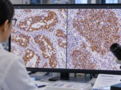
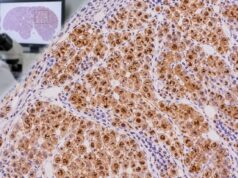
In GIST, CD117 staining is typically cytoplasmic with membranous accentuation. Some cases show a dot-like or paranuclear Golgi-type pattern. The exact appearance can vary with antibody clone, antigen retrieval, fixation, tumor differentiation, and mutation subtype. A pathologist does not diagnose GIST merely because brown staining is present; the staining must occur in the cells that make up the lesion and must fit the expected morphology.
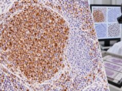
In mast cells, staining is often strong and membranous/cytoplasmic, making even relatively inconspicuous or hypogranulated mast cells easier to identify than with routine hematoxylin and eosin alone. This can be particularly helpful in a bone marrow biopsy where abnormal mast cells are spindle shaped or where the infiltrate is small.
Interpretation also depends on the background. Poor fixation can reduce antigen detection and produce a false-negative or weak result. Endogenous pigment, necrosis, crush artifact, and nonspecific background may complicate interpretation. Good IHC practice therefore uses appropriate controls and correlates staining with viable, morphologically intact tumor.
A useful practical distinction is lineage support versus mutation status. CD117 is primarily a lineage/differentiation marker in diagnostic IHC. It tells the pathologist that KIT protein is present. A DNA or RNA assay answers a separate question about genetic alteration. Those two results can be related but are not interchangeable.
CD117 in gastrointestinal stromal tumor
GIST is the tumor most strongly associated with CD117 in routine surgical pathology. Most GISTs arise in the stomach or small intestine, although they can occur elsewhere in the gastrointestinal tract and, rarely, in nearby soft tissues. Microscopically they may be spindle-cell, epithelioid, or mixed. Strong diffuse KIT expression in a compatible mesenchymal tumor is highly supportive of GIST.
Modern diagnosis usually combines morphology with CD117 and DOG1 rather than relying on either stain in isolation. If a spindle-cell tumor in the stomach is strongly positive for both CD117 and DOG1, while markers supporting smooth muscle or neural differentiation are absent or limited, the profile strongly favors GIST. If CD117 is negative, however, GIST is not excluded. DOG1 may still be positive, and molecular testing may identify a PDGFRA, KIT, or other defining alteration.
Other stains help address the differential diagnosis. Desmin and smooth muscle actin are considered when leiomyoma or leiomyosarcoma is in the differential; S100 and SOX10 can support neural tumors; and SDHB IHC may help identify succinate dehydrogenase-deficient GIST. The relationship between CD117 and CD34 also needs context: many GISTs express CD34, but CD34 is less specific and is shared by numerous vascular and soft-tissue tumors. The separate CD34 IHC test is therefore best viewed as complementary rather than interchangeable.
CD117 positivity does not predict imatinib sensitivity by itself
A common misunderstanding is that a positive CD117 stain automatically means a patient will respond to a KIT inhibitor. Treatment sensitivity depends much more on the tumor’s molecular driver and mutation site. For example, many KIT-mutant GISTs respond well to imatinib, but certain mutations have different sensitivity, and the common PDGFRA D842V mutation is resistant to imatinib while being targetable with other agents such as avapritinib. Conversely, KIT protein expression can occur in tumors that are not biologically dependent on activating KIT signaling.
For that reason, mutation testing is important in GIST when systemic therapy, adjuvant treatment, resistance evaluation, or an unusual phenotype makes genotype clinically relevant. The IHC result helps establish the tumor type; the molecular result can help refine biology and therapy.
CD117 in mast cell disorders
Mast cells normally express abundant KIT, so CD117 is a sensitive way to highlight them in tissue. In systemic mastocytosis, bone marrow may contain multifocal dense mast-cell aggregates, often with spindle-shaped or atypical forms. CD117 makes these cells easier to see, but it does not establish clonality because normal mast cells are also strongly positive.
The diagnosis of systemic mastocytosis uses a combination of morphology, immunophenotype, molecular findings, and clinical/laboratory data. Tryptase is another highly useful mast-cell marker. Aberrant expression of CD25, CD2, or CD30 can support a neoplastic mast-cell population in the right setting, and sensitive molecular testing frequently looks for KIT D816V or another activating KIT variant. The exact diagnostic criteria have evolved, so the final classification should follow the current hematopathology framework rather than a single stain.
CD117 can also highlight mast cells in reactive conditions. Allergic inflammation, chronic inflammatory disorders, healing tissue, and other benign states may contain increased mast cells. A numerical increase alone is not enough to call mastocytosis. Architecture matters: abnormal dense aggregates, cytologic atypia, associated marrow findings, and clonal markers carry more weight.
In acute myeloid leukemia and other myeloid neoplasms, blasts can express CD117. In that setting the stain is interpreted with other myeloid markers, flow cytometry, blast percentage, morphology, cytogenetics, and molecular testing. CD117 is therefore not a “mast cell only” marker. Its strength is sensitivity across several KIT-expressing cell types; its limitation is the same lack of absolute specificity.
Other tumors that can be positive
CD117 positivity occurs beyond GIST and mast-cell disease. The differential varies with site and morphology, but recognized examples include seminoma and related germ-cell tumors, adenoid cystic carcinoma, some acute myeloid leukemias, subsets of melanoma, and variable subsets of ovarian, pulmonary, thyroid, and other neoplasms. This broad expression is why the phrase “CD117-positive tumor” is descriptive, not a final diagnosis.
The staining pattern and the rest of the panel are critical. A testicular mass with large clear cells and strong membranous CD117 expression raises a very different diagnostic question from a spindle-cell gastric mass with diffuse KIT and DOG1 positivity. Likewise, a small blue-cell tumor that expresses CD117 may need an entirely different panel than either of those examples.
Pathologists usually start with morphology and broad lineage markers, then use more specific stains. For an undifferentiated tumor, keratins can support epithelial differentiation, CD45 can support hematolymphoid origin, and S100/SOX10 may support melanocytic or neural differentiation. CD117 then contributes within that framework.
This explains why isolated weak CD117 staining should be treated cautiously. Weak or focal reactivity is more likely to be nonspecific or diagnostically nonexclusive than a strong, diffuse pattern in the expected cell population. Even strong staining, however, must be reconciled with the tumor’s architecture and other markers.
How pathologists interpret CD117 results
A CD117 report may contain several layers of information. The first is simply whether the target cells are positive or negative. The second is the extent of staining—focal versus diffuse, for example. The third is intensity, and the fourth is localization, such as membranous, cytoplasmic, or dot-like. The pathologist then integrates those features with the histologic diagnosis.
| Finding | What it may support | What else is usually needed |
|---|---|---|
| Strong diffuse CD117 in a GI spindle/epithelioid tumor | GIST | DOG1, morphology, and often KIT/PDGFRA molecular testing |
| CD117-positive mast-cell aggregates in marrow | Mast-cell proliferation | Tryptase, CD25/CD2/CD30, KIT testing, diagnostic criteria |
| CD117-positive blasts | Immature myeloid phenotype | Flow cytometry, MPO and other markers, genetics, blast assessment |
| Focal or weak staining in an otherwise discordant tumor | Potential nonspecific or nonexclusive expression | Broader lineage- and site-specific panel |
| CD117-negative suspected GIST | GIST still possible | DOG1, SDHB when indicated, molecular testing |
A “positive” result should therefore be read together with the final diagnosis line and comment. If a report says “CD117 positive” but the diagnosis is not GIST, that is not contradictory. The stain may have been one piece of a larger panel or may simply document an expression pattern.
For patients, the most useful questions are often: What diagnosis is the pathologist favoring? Which other markers support it? Is molecular testing needed? Does the stain affect treatment, or is it mainly diagnostic? These questions are more informative than focusing on a percentage alone.
Limitations, follow-up, and next tests
CD117 IHC has several limitations. It is not specific to one tumor, not all tumors of a given type are positive, technical factors can change staining, and protein expression does not equal mutation status. Small biopsies can also miss heterogeneous areas, while necrosis or poor fixation may reduce staining quality.
When GIST is the leading diagnosis, the usual next steps depend on the case. DOG1 can confirm lineage, while molecular testing for KIT and PDGFRA is often important for therapy and for unusual or negative-staining tumors. SDHB IHC may be used when an SDH-deficient GIST is suspected. Risk assessment after resection also incorporates tumor site, size, mitotic rate, and rupture—not CD117 intensity.
When a mast-cell disorder is suspected, the workup is different. The pathologist may use tryptase and aberrant mast-cell markers, while the laboratory performs sensitive KIT mutation testing. Serum tryptase and clinical findings may contribute, but tissue morphology remains central. If leukemia is suspected, flow cytometry and molecular/cytogenetic testing usually provide far more classification detail than CD117 alone.
The key practical point is that CD117 works best as a context marker. It is highly useful when the diagnostic question is well formed and the correct companion tests are used. It becomes misleading when treated as a stand-alone cancer label.
A patient should discuss the full pathology report with the treating clinician, especially if the report uses wording such as “supports,” “compatible with,” “focal,” “equivocal,” or “requires molecular correlation.” Those phrases often signal that the IHC result is part of a probabilistic diagnosis rather than a binary answer. If the tumor is rare, unusual, or treatment depends on a specific molecular subtype, expert pathology review may also be appropriate.
Practical CD117 interpretation scenarios
A few common scenarios show why the same positive stain can mean different things. In a gastric submucosal spindle-cell mass, diffuse CD117 and DOG1 positivity is highly supportive of GIST because the site and morphology create a high pretest probability. The pathologist may then use molecular testing to define KIT or PDGFRA status, especially if systemic therapy is relevant. In contrast, CD117 positivity in an ovarian carcinoma or melanoma does not make that tumor a GIST; the tumor’s epithelial or melanocytic phenotype has already established a different lineage.
A bone marrow biopsy creates another kind of problem. Scattered CD117-positive mast cells can be completely normal, and CD117-positive myeloid precursors are expected at low levels. Concern rises when mast cells form abnormal dense aggregates, show spindle-cell morphology, or carry an aberrant phenotype. Likewise, an increase in CD117-positive blasts matters only when their identity and proportion are supported by the marrow morphology and flow-cytometric or molecular findings. A pathologist may use the stain to map the abnormal cells while avoiding a diagnosis based only on the color reaction.
Negative staining also has different implications by setting. If a classic-appearing GIST is CD117 negative, the result prompts DOG1 and often molecular testing rather than immediate rejection of the diagnosis. If both CD117 and DOG1 are negative, the pathologist rechecks morphology, considers technical quality, and expands the differential to smooth-muscle, neural, fibroblastic, and other mesenchymal tumors. Conversely, a strongly CD117-positive lesion with completely incompatible morphology should trigger the same kind of diagnostic reassessment.
Specimen type can influence confidence. A large resection shows architecture and tumor heterogeneity better than a tiny needle core. Fine-needle aspiration or a small cell block can establish an immunophenotype, but the limited material may need to be conserved for molecular testing. When treatment choices depend on genotype, laboratories often prioritize an efficient IHC panel and preserve enough tissue for sequencing.
Patients sometimes see “c-KIT positive” and assume it is the same as “KIT mutation positive.” Those statements are not equivalent. IHC detects protein; sequencing detects a DNA alteration. A useful report or oncology discussion should state which type of test was performed and what decision it informs. This distinction prevents both overestimating the meaning of a positive stain and overlooking the value of molecular testing in a confirmed GIST or mast-cell disorder.
References
- 2023 GEIS Guidelines for gastrointestinal stromal tumors 2023 (Guideline)
- SEOM-GEIS clinical guideline for gastrointestinal stromal tumors (2022) 2023 (Guideline)
- Pathologic diagnosis and molecular features of gastrointestinal stromal tumors: a mini-review 2024 (Review)
- Systemic mastocytosis: current status and challenges in 2024. 2025 (Review)
- How I diagnose systemic mastocytosis 2024 (Review)
- Standards of Pathology in the Diagnosis of Systemic Mastocytosis: Recommendations of the EU-US Cooperative Group 2022 (Position Statement)
Disclaimer
CD117 IHC is a pathology test that must be interpreted with the tissue appearance, other stains, and—when relevant—molecular results. This article is educational and cannot determine the meaning of an individual pathology report or replace advice from a pathologist, oncologist, hematologist, or other qualified clinician.