
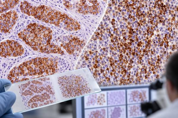
CD45 immunohistochemistry (IHC), also called leukocyte common antigen or LCA staining, detects a protein expressed on most nucleated blood and immune cells. Pathologists use it as a broad lineage marker when they need to decide whether an abnormal tissue mass is hematolymphoid rather than epithelial, melanocytic, or mesenchymal. Strong staining in a suspicious cell population can support lymphoma or another blood-cell neoplasm, but CD45 does not identify the exact lymphoma subtype. It can also be weak or absent in some hematologic tumors, including classic Hodgkin lymphoma, many plasma-cell neoplasms, and some very immature or anaplastic neoplasms. A useful CD45 result therefore opens a diagnostic pathway rather than ending it. Pathologists combine the stain with morphology and more specific markers such as CD20, PAX5, CD3, CD30, ALK, MPO, TdT, or plasma-cell markers, as well as flow cytometry and molecular tests when needed.
- CD45-positive tumor cells usually support hematolymphoid origin, but they do not identify a specific lymphoma or leukemia subtype.
- CD45 is normally present on many leukocytes, so background lymphocytes provide a useful internal positive control.
- A CD45-negative malignant tumor is more likely to be nonhematolymphoid, but important lymphomas can be negative or only weakly positive.
- Classic Hodgkin lymphoma often shows absent or weak CD45 in Hodgkin/Reed–Sternberg cells, even though surrounding reactive leukocytes stain strongly.
- The final diagnosis depends on a marker panel, tissue architecture, clinical setting, and often flow cytometry or genetic testing.
Table of Contents
- What CD45 IHC tests
- Why CD45 helps identify tumor origin
- CD45 in lymphoma and leukemia
- What positive and negative staining mean
- Marker panels used with CD45
- Pitfalls and limitations
- Reading the report and next steps
- Practical CD45 interpretation scenarios
What CD45 IHC tests
CD45 is a transmembrane protein tyrosine phosphatase encoded by PTPRC. It is expressed in most nucleated hematopoietic cells and exists in multiple isoforms associated with different leukocyte populations and activation states. In surgical pathology, a pan-CD45 antibody is typically used to answer a broad question: do the cells in this lesion belong to the hematolymphoid system?
That broad role is why CD45 is often included early in the workup of a poorly differentiated or undifferentiated malignancy. A biopsy may show sheets of large atypical cells without obvious glands, keratinization, melanin, or other defining features. A small screening panel can separate major lineages. Cytokeratin positivity supports carcinoma, SOX10 or S100 may support melanoma or certain neural tumors, and CD45 positivity supports lymphoma or another hematolymphoid process.
CD45 staining is usually membranous, sometimes with accompanying cytoplasmic reactivity. Normal lymphocytes in the same section should stain, which provides an internal control. If those background cells fail to stain, a negative tumor result may not be technically reliable.
The test is not a blood count and has no normal concentration or serum range. Pathologists generally describe whether the abnormal cells are positive or negative and may comment on strength or distribution. The diagnostic value lies in the identity of the stained cells rather than a universal percentage cutoff.
Why CD45 helps identify tumor origin
When cancer appears in a lymph node, soft tissue, bone, skin, or another organ, the first challenge may be determining what kind of tumor it is. A metastasis from carcinoma and a high-grade lymphoma can look similar, especially in a small or crushed biopsy. CD45 is valuable because broad leukocyte expression contrasts with most carcinomas, melanomas, and sarcomas.
For example, a diffuse large-cell tumor that is strongly CD45 positive and pancytokeratin negative raises hematolymphoid neoplasia. The next stains can then focus on B-cell, T-cell, myeloid, or other lineages. If the same tumor is keratin positive and CD45 negative, carcinoma becomes more likely. This stepwise method preserves tissue and reduces the chance of ordering an unfocused panel.
CD45 is particularly helpful in lymph nodes, but location alone never determines lineage. Lymph nodes commonly contain metastatic carcinomas and melanomas, while lymphomas can involve almost any extranodal organ. A gastric, skin, lung, or brain biopsy can therefore require the same basic lineage question.
The marker is also useful in mixed specimens. Normal inflammatory cells may be CD45 positive around a CD45-negative carcinoma, sharply outlining the nonhematolymphoid tumor. Conversely, a lymphoma can contain many reactive cells, so the pathologist must confirm that the atypical cells themselves express CD45 rather than simply noting that the tissue contains positive leukocytes.
CD45 in lymphoma and leukemia
Most mature B-cell and T-cell lymphomas express CD45. However, the intensity can vary, and the stain does not distinguish benign from malignant lymphocytes. Normal small lymphocytes, reactive immunoblasts, and lymphoma cells may all be positive. Architecture, cytology, clonality, and subtype-specific markers are therefore essential.
Once hematolymphoid origin is established, lineage markers are usually added. CD20 IHC and PAX5 help identify B-cell differentiation; CD3 IHC supports T-cell lineage. Other markers—such as CD10, BCL6, MUM1, BCL2, cyclin D1, SOX11, CD30, ALK, and EBV-related testing—are selected according to morphology and the suspected entity.
Acute leukemias and myeloid neoplasms can also express CD45, often with intensity that differs from mature lymphocytes. In flow cytometry, the combination of CD45 intensity and side scatter is routinely used to help locate blast populations. Tissue IHC is less quantitative but can still help establish hematopoietic lineage. Myeloperoxidase, CD34, CD117, TdT, lysozyme, and lineage-specific markers may be needed to classify immature neoplasms.
Important CD45-negative or weakly positive exceptions
Classic Hodgkin lymphoma is the best-known exception. The neoplastic Hodgkin/Reed–Sternberg cells are typically CD30 positive and usually negative or only weak for CD45, while surrounding reactive lymphocytes are strongly positive. This pattern is diagnostically useful. Some plasma-cell neoplasms also show absent or reduced CD45, and certain anaplastic large-cell lymphomas may be weak or negative. Very immature acute leukemias can show weak expression as well.
These exceptions mean a negative CD45 stain does not absolutely exclude lymphoma. When the morphology suggests hematolymphoid disease, the pathologist uses additional markers rather than abandoning the possibility based on one result.
What positive and negative staining mean
A CD45-positive result means the antibody detected leukocyte common antigen in the stained cells. The key question is whether those are the abnormal cells. A report may specify “tumor cells are positive for CD45” or “CD45 highlights background lymphocytes only.” Those statements have opposite implications.
| Pattern | What it suggests | Typical next step |
|---|---|---|
| Diffuse CD45 positivity in atypical cells | Hematolymphoid lineage | B-, T-, myeloid, or other lineage markers |
| Atypical cells CD45 negative; lymphocytes positive | Consider carcinoma, melanoma, sarcoma, or CD45-negative hematologic exception | Use morphology-directed lineage panel |
| Large atypical cells CD30 positive and CD45 negative/weak | Can fit classic Hodgkin lymphoma in the correct setting | PAX5, MUM1, EBER and differential-diagnosis markers |
| Weak CD45 in blast-like cells | Possible immature hematopoietic neoplasm | Flow cytometry, CD34/TdT/MPO and genetics |
There is no universal rule that stronger CD45 means a more aggressive lymphoma. Staining intensity can reflect cell type, isoform, fixation, antibody performance, or differentiation state. Prognosis is determined by the specific diagnosis, stage, genetic abnormalities, proliferation, patient factors, and treatment response.
Likewise, a “positive” result is not equivalent to “cancer positive.” Reactive lymph nodes are full of CD45-positive cells. The test is useful because it labels leukocytes; malignancy is determined by whether those cells form an abnormal clonal proliferation with the expected morphologic and molecular features.
Marker panels used with CD45
The most efficient IHC panel is driven by the differential diagnosis. For a high-grade undifferentiated tumor, a pathologist may begin with CD45, broad-spectrum cytokeratin, and a melanocytic marker. If CD45 is positive, the panel narrows toward hematopathology.
For suspected B-cell lymphoma, common markers include CD20, PAX5, CD10, BCL6, MUM1, BCL2, cyclin D1, and Ki-67. Certain patterns prompt molecular studies for MYC, BCL2, or BCL6 rearrangements. For a small B-cell lymphoma, cyclin D1 and SOX11 can be important when mantle cell lymphoma is considered; the cyclin D1 IHC test is closely linked to CCND1 dysregulation but must still be interpreted in a panel.
For T- and NK-cell neoplasms, CD2, CD3, CD5, CD7, CD4, CD8, CD30, ALK, cytotoxic markers, CD56, and EBV testing may be selected. Loss of a normally expressed T-cell antigen can support an abnormal phenotype, but no single loss pattern is diagnostic.
For acute leukemia in tissue, markers such as CD34, TdT, MPO, CD117, PAX5, CD79a, cytoplasmic CD3, and others may help define lineage. Flow cytometry is often preferred when fresh material is available because it can evaluate many antigens simultaneously and quantify abnormal populations.
The goal of the panel is internal consistency. A convincing diagnosis should explain the morphology, CD45 status, lineage markers, and clinical context rather than depend on one unexpected stain.
Pitfalls and limitations
The first pitfall is background staining. Almost every tissue biopsy contains some leukocytes, so CD45 positivity somewhere on the slide is expected. Only staining in the cells of interest answers the diagnostic question. This is especially important in carcinomas with a heavy lymphoid infiltrate and in lymph nodes partly replaced by metastasis.
The second pitfall is assuming CD45 negativity rules out hematologic malignancy. Classic Hodgkin lymphoma, plasma-cell neoplasms, some anaplastic lymphomas, and some precursor neoplasms can be negative or weak. The morphology may justify additional hematolymphoid markers even after a negative stain.
Technical issues include fixation, decalcification, necrosis, crush artifact, and low tumor volume. Bone specimens can be especially challenging after decalcification. If internal lymphocytes stain appropriately, the technical performance is more reassuring; if they do not, repeat testing or another block may be needed.
Another limitation is that CD45 cannot prove clonality. A florid reactive immune response may contain sheets or clusters of CD45-positive cells. Flow cytometry, immunoglobulin or T-cell receptor gene studies, EBV testing, cytogenetics, or sequencing may be needed depending on the suspected process.
Finally, some nonhematolymphoid tumors can show unexpected or aberrant marker expression. A pathologist resolves discordant staining by looking for a coherent multiparameter pattern rather than accepting one surprising result at face value.
Reading the report and next steps
A CD45 result is most useful when read within the pathology diagnosis and comment. If the report says “neoplastic cells are positive for CD45,” the next sentence often names the lineage or lists additional stains. If it says “CD45 highlights numerous background lymphocytes,” the marker may simply be documenting the inflammatory component around a nonhematolymphoid tumor.
If lymphoma is diagnosed, additional testing may define the exact entity and clinically relevant features. Flow cytometry can establish a clonal B-cell population or an aberrant T-cell phenotype. FISH can detect rearrangements. Molecular sequencing may identify mutations with diagnostic, prognostic, or treatment relevance. The current WHO and International Consensus classifications intentionally combine these layers because modern lymphoma diagnosis is no longer purely morphologic.
For a patient, practical questions include: Are the tumor cells themselves CD45 positive? Which lineage is favored? Which markers establish the subtype? Is flow cytometry or genetic testing pending? Does the diagnosis require a larger biopsy because architecture is important? These questions help translate the stain into a clinical plan.
A small needle biopsy may occasionally establish lineage but not provide enough tissue for full lymphoma classification. In that situation, the pathologist may recommend a larger core or excisional biopsy rather than ordering every possible stain on limited material. Preserving tissue for flow cytometry and molecular studies can be more valuable than adding numerous low-yield IHC tests.
CD45 is therefore best thought of as a highly useful gateway marker. It can rapidly shift the diagnostic pathway toward or away from hematolymphoid disease, but accurate classification comes from the evidence that follows.
Practical CD45 interpretation scenarios
Consider a liver biopsy containing sheets of large malignant cells. If the cells are strongly CD45 positive and broad-spectrum keratin negative, metastatic carcinoma becomes less likely and a hematolymphoid process moves to the front of the differential. The pathologist may then use PAX5 and CD20 for B-cell lineage, CD3 for T-cell lineage, and additional markers based on cell size and morphology. A strong B-cell phenotype might lead to lymphoma-specific studies, while MPO or other myeloid markers could redirect the workup toward a myeloid sarcoma.
A lymph-node biopsy can present the opposite problem. A metastatic carcinoma may be surrounded by dense CD45-positive lymphocytes, making the slide appear extensively positive at low power. At higher power, however, the malignant epithelial nests are CD45 negative and keratin positive. This is why pathology reports distinguish “tumor cells positive” from “background lymphocytes positive.” The location of staining matters as much as the existence of staining.
Classic Hodgkin lymphoma demonstrates why a negative result can be informative rather than simply disappointing. The large Hodgkin/Reed–Sternberg cells are typically CD30 positive and weak or negative for CD45, whereas the surrounding reactive leukocytes are strongly CD45 positive. In the correct morphology, that contrast supports the diagnosis. Other stains, including weak PAX5, MUM1, and EBV studies in selected cases, help separate classic Hodgkin lymphoma from anaplastic large cell lymphoma, diffuse large B-cell lymphoma, and reactive mimics.
In bone marrow, CD45 staining is usually too broad to count blasts accurately because most hematopoietic cells express the antigen. Flow cytometry uses relative CD45 intensity together with light scatter and multiple antigens to isolate suspicious populations. Tissue IHC is more useful for determining lineage in an infiltrate and for showing which cells are hematopoietic within the preserved architecture.
A difficult case may also contain a genuinely CD45-negative hematologic tumor. Plasma-cell myeloma is a common example in which neoplastic plasma cells may lose or reduce CD45. Strong CD138 expression, light-chain restriction, marrow morphology, and plasma-cell flow cytometry can establish the diagnosis despite negative CD45. The same principle applies to other known exceptions: one broad marker should not overrule a coherent alternative phenotype.
The practical way to use CD45 is therefore sequential. First determine whether the atypical cells express it. Second establish lineage with more specific markers. Third classify the neoplasm using morphology, immunophenotype, genetics, and clinical data. This sequence explains why CD45 remains valuable even though modern hematopathology uses increasingly sophisticated molecular tests: it answers an early, high-impact question quickly while preserving a logical path to the final diagnosis.
A final practical issue is specimen triage. When lymphoma is suspected before biopsy, sending fresh tissue for flow cytometry can preserve information that paraffin IHC cannot recreate. If all tissue is fixed, IHC can still classify many lymphomas, but some light-chain, surface-antigen, and population-level relationships are easier to assess by flow. Communication between the procedural team and pathology laboratory therefore improves diagnostic yield. A result that remains indeterminate after limited tissue is not necessarily a laboratory failure; repeating a well-chosen stain on another block may help, but sometimes the biologically important answer requires architecture from a larger biopsy or material suitable for genetics.
A useful practical point is that CD45 can also guide specimen triage. When a fresh lymph node or mass shows a CD45-positive atypical population, preserving viable tissue for flow cytometry, cytogenetics, or molecular testing may be more informative than adding a large number of stains to a limited block. This is especially important when the differential includes leukemia or lymphoma that requires immunophenotypic or genetic classification.
CD45 interpretation also depends on recognizing the background cells. Almost every solid-tumor biopsy contains normal lymphocytes, and those cells can stain strongly even when the malignant cells are completely CD45 negative. A report should therefore identify which population is positive rather than simply recording that staining is present somewhere on the slide. This distinction is especially important in tiny biopsies, necrotic tumors, and specimens after therapy, where viable tumor may be sparse. When the atypical cells are convincingly CD45 positive, the stain redirects the workup toward hematolymphoid disease; when only background lymphocytes label, it instead provides a useful internal control showing that the antibody worked. Careful localization prevents a technically successful stain from being given the wrong biologic interpretation.
References
- Updates in Immunohistochemistry for Hematopoietic and Lymphoid Neoplasms 2024 (Review)
- The 5th edition of the World Health Organization Classification of Haematolymphoid Tumours: Lymphoid Neoplasms 2022 (Review)
- The International Consensus Classification of Mature Lymphoid Neoplasms: a report from the Clinical Advisory Committee 2022 (Position Statement)
- Update from the 5th Edition of the World Health Organization Classification of Head and Neck Tumors: Hematolymphoid Proliferations and Neoplasia 2022 (Review)
- Classification and diagnostic evaluation of nodal T- and NK-cell lymphomas 2023 (Review)
- The 5th Edition of the World Health Organization Classification of Hematolymphoid Tumors 2022 (Review)
Disclaimer
CD45 IHC is a diagnostic pathology tool, not a stand-alone cancer test. The meaning of an individual result depends on which cells stain, the tissue appearance, other immunostains, and any flow-cytometric or molecular findings, and should be reviewed with the treating clinical and pathology teams.





