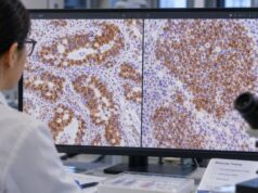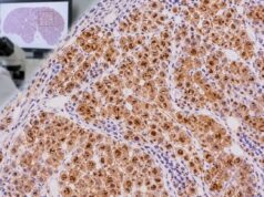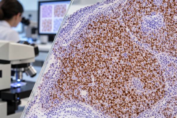
Cyclin D1 immunohistochemistry (IHC) detects a cell-cycle protein that is strongly associated with mantle cell lymphoma (MCL) when it is abnormally expressed in B lymphocytes. Most conventional MCLs carry the t(11;14) translocation involving CCND1 and IGH, which drives cyclin D1 overexpression and produces characteristic nuclear staining. In the right morphologic and immunophenotypic setting, diffuse nuclear cyclin D1 is a powerful diagnostic clue. It is not completely specific, however: cyclin D1 can be positive in plasma-cell neoplasms, hairy cell leukemia, and several nonhematologic tumors, while a small minority of genuine MCLs are cyclin D1 negative. Pathologists therefore interpret cyclin D1 with B-cell markers, CD5, SOX11, morphology, flow cytometry, and—when needed—FISH or molecular testing. A positive stain supports a diagnosis; it does not replace the full lymphoma classification.
- Diffuse nuclear cyclin D1 in a CD5-positive B-cell lymphoma strongly supports mantle cell lymphoma.
- Most MCLs have a CCND1 rearrangement, usually t(11;14), but rare MCLs are cyclin D1 negative and may require SOX11 and molecular studies.
- Cyclin D1 can be positive in tumors other than MCL, so the lineage and morphology must match.
- The stain is interpreted as nuclear tumor-cell expression; cytoplasmic or background staining is not equivalent.
- Cyclin D1 intensity or percentage is not a stand-alone prognosis score; Ki-67, morphology, TP53 status, stage, and clinical factors are more important for risk.
Table of Contents
- What cyclin D1 IHC detects
- Why cyclin D1 marks mantle cell lymphoma
- How positive staining is interpreted
- Cyclin D1-negative mantle cell lymphoma
- Other cyclin D1-positive neoplasms
- Marker panels and confirmatory tests
- Report meaning, limitations, and next steps
- Practical cyclin D1 interpretation scenarios
What cyclin D1 IHC detects
Cyclin D1 is a regulatory protein that helps cells move from the G1 phase of the cell cycle into DNA synthesis. The CCND1 gene encodes it. In normal lymphoid tissue, most mature B cells do not show the strong diffuse nuclear cyclin D1 expression that characterizes conventional MCL.
IHC detects the protein on a fixed tissue section. A true positive result is primarily nuclear because cyclin D1 acts in cell-cycle regulation within the nucleus. The pathologist evaluates whether the atypical lymphoid cells stain, how diffuse the staining is, and whether the pattern fits the microscopic architecture.
The test is commonly performed on lymph node, bone marrow, gastrointestinal, spleen, or other biopsies when a small B-cell lymphoma is suspected. MCL can involve many extranodal sites, so the stain is not limited to lymph nodes. In marrow, staining can help highlight otherwise subtle involvement, although architecture and other B-cell markers remain important.
Cyclin D1 IHC is not a blood concentration and has no normal numeric range. The clinical question is whether an abnormal cell population has convincing nuclear overexpression. In many cases, a simple positive/negative interpretation is enough; in others, the distribution helps identify in situ or focal involvement.
Why cyclin D1 marks mantle cell lymphoma
The hallmark genetic event in most MCL is t(11;14)(q13;q32), which places CCND1 under the influence of immunoglobulin heavy-chain regulatory elements. This causes excess cyclin D1 production and contributes to abnormal cell-cycle progression. IHC provides a practical tissue-level surrogate for that molecular event in most cases.
A classic MCL phenotype includes mature B-cell markers such as CD20 and PAX5, frequent CD5 coexpression, and cyclin D1 positivity, often with CD23 absent or only weak. The cells may be small to medium sized with irregular nuclear contours. Growth patterns can be diffuse, nodular, mantle-zone, or mixed. Blastoid and pleomorphic variants have larger or more aggressive cytology.
The diagnosis is not made from cyclin D1 alone. A small B-cell lymphoma with CD5 expression also raises chronic lymphocytic leukemia/small lymphocytic lymphoma. Cyclin D1 strongly favors MCL in that differential, but other features—including CD23, LEF1, SOX11, flow cytometry, and FISH—may be needed in unusual cases.
Modern classifications also recognize biologic subtypes of MCL, including conventional and leukemic non-nodal forms. SOX11 expression, IGHV mutation status, morphology, genomic alterations, and clinical pattern can differ between them. The SOX11 test can be especially informative in difficult or cyclin D1-negative cases.
How positive staining is interpreted
The expected MCL pattern is nuclear staining in the neoplastic B cells. Diffuse convincing nuclear positivity in a morphologically compatible B-cell lymphoma is much more informative than rare weak cells. Background histiocytes, endothelial cells, or epithelial structures may have unrelated staining depending on the tissue and assay.
| Finding | Typical implication | Useful follow-up |
|---|---|---|
| Diffuse nuclear cyclin D1 in a CD20+/CD5+ small B-cell lymphoma | Strongly supports MCL | SOX11 and/or CCND1 FISH when confirmation is needed |
| Cyclin D1 negative but MCL morphology/phenotype persists | Rare cyclin D1-negative MCL possible | SOX11, CCND2/CCND3-related studies, expert review |
| Cyclin D1 positive plasma-cell neoplasm | Can reflect CCND1 dysregulation outside MCL | Plasma-cell markers and myeloma workup |
| Weak/focal staining without a coherent B-cell phenotype | Potential nonspecific or non-MCL expression | Interpret with lineage panel and morphology |
Cyclin D1 staining is not the same as Ki-67. Cyclin D1 is a diagnostic/proliferative pathway marker; Ki-67 estimates the fraction of cells actively cycling and is more directly used as a proliferation index. In MCL, a high Ki-67 index and blastoid or pleomorphic morphology are associated with higher-risk biology, while simply being cyclin D1 positive is expected for the diagnosis.
Cyclin D1-negative mantle cell lymphoma
A small subset of MCL lacks cyclin D1 overexpression and the usual CCND1 rearrangement. These cases can be difficult because the stain that normally anchors the diagnosis is absent. Some harbor rearrangements involving CCND2 or CCND3 instead.
SOX11 is particularly useful in this setting. Many conventional MCLs express SOX11, including a substantial proportion of cyclin D1-negative cases. Strong nuclear SOX11 in a morphologically and immunophenotypically compatible B-cell neoplasm can provide important support, although SOX11 is not absolutely exclusive to MCL.
Molecular or cytogenetic testing may be required to establish the diagnosis. The CCND1/IGH fusion test can confirm the canonical rearrangement in typical cases, while broader testing may be needed when CCND1 is not involved.
This is an example of why negative IHC should not automatically overrule a strong clinicopathologic pattern. Instead, discordance prompts the pathologist to ask whether technical failure, unusual biology, or an alternative lymphoma better explains the findings.
Other cyclin D1-positive neoplasms
Cyclin D1 is not exclusive to MCL. Plasma-cell myeloma can show strong cyclin D1, particularly in cases with t(11;14). Hairy cell leukemia may express cyclin D1, usually in a different clinical, morphologic, and immunophenotypic setting. A variety of epithelial tumors—including subsets of breast, head and neck, and other carcinomas—can also overexpress cyclin D1.
The distinction is usually straightforward once lineage is established. A keratin-positive carcinoma is not MCL simply because cyclin D1 is positive. A plasma-cell neoplasm expressing CD138 and light-chain restriction is interpreted through the plasma-cell framework. Likewise, a B-cell process with the classic MCL phenotype carries much more diagnostic weight than cyclin D1 expression in isolation.
Within hematopathology, the pathologist also considers whether the cells are truly neoplastic. Rare positive non-neoplastic cells or technical background should not be overcalled. Internal morphology is important because the brown nuclear signal must line up with the atypical population seen on the H&E slide.
Because cyclin D1 dysregulation can occur through different mechanisms, the presence of protein does not always identify the exact genetic event. FISH or sequencing provides direct genetic information when that distinction matters.
Marker panels and confirmatory tests
A typical MCL workup may include CD20 or PAX5 to confirm B-cell lineage, CD5, cyclin D1, SOX11, CD23, LEF1, and a proliferation marker such as Ki-67. The exact panel varies with specimen and differential diagnosis. CD45 IHC may be used earlier when hematolymphoid origin itself is uncertain.
Flow cytometry can detect a clonal B-cell population and characterize surface markers, often showing CD5-positive B cells with light-chain restriction. Flow findings should match the tissue because some MCLs have atypical phenotypes.
FISH for CCND1/IGH or a CCND1 break-apart assay can confirm the canonical rearrangement. Molecular testing may add information about TP53 and other abnormalities that influence prognosis or treatment planning. Current classifications increasingly integrate genetics with morphology and immunophenotype rather than treating them as separate silos.
The Ki-67 IHC test deserves separate attention in MCL because proliferation has prognostic value. A Ki-67 percentage should be assessed in representative tumor and reported using an appropriate method. Cyclin D1 and Ki-67 answer different questions and should not be confused.
Report meaning, limitations, and next steps
A report that says “cyclin D1 positive” is most informative when it also specifies the cell population and the favored diagnosis. For example, “the atypical B cells show diffuse nuclear cyclin D1 expression” is strong evidence in a compatible MCL workup. If a report simply lists cyclin D1 among many stains, the final diagnostic comment explains its weight.
Technical limitations include fixation, decalcification, small sample size, and variable antibody performance. Bone marrow specimens can be challenging after decalcification. When staining is unexpectedly negative, internal controls and repeat testing on another block may help.
Sampling can also matter. MCL may involve marrow or gastrointestinal biopsies focally, and a small biopsy can underestimate disease. Conversely, an in situ mantle cell neoplasia pattern may show cyclin D1-positive B cells restricted to mantle zones without the features of overt lymphoma. Clinical and morphologic context prevents overdiagnosis.
For patients, useful questions include whether the diagnosis is conventional MCL, leukemic non-nodal MCL, or another subtype; whether CCND1 rearrangement was confirmed; whether SOX11 and Ki-67 were assessed; and whether TP53 or other molecular testing is indicated. These features can matter more for prognosis and management than the simple fact that cyclin D1 is positive.
If the IHC pattern is atypical or the diagnosis has major treatment implications, hematopathology review at an experienced center can be valuable. The goal is a diagnosis in which morphology, flow cytometry, IHC, and genetics tell the same biological story.
Practical cyclin D1 interpretation scenarios
A lymph-node biopsy containing a monotonous small B-cell proliferation is the classic scenario. If flow cytometry shows a CD5-positive clonal B-cell population and the tissue shows diffuse nuclear cyclin D1, mantle cell lymphoma becomes highly likely. The pathologist may still confirm the underlying CCND1 rearrangement, particularly when morphology or the immunophenotype is atypical. This is a high-value use of IHC because the stain links a visible protein pattern to a well-characterized genetic pathway.
A different challenge arises when the lymphoid cells look like MCL but cyclin D1 is negative. Rather than excluding the diagnosis immediately, the pathologist checks technical controls, reviews CD5 and other B-cell markers, and may order SOX11. Strong nuclear SOX11 can support cyclin D1-negative MCL, after which molecular studies can look for alternative cyclin D family rearrangements. This pathway matters because such cases may otherwise be misclassified as another small B-cell lymphoma.
Cyclin D1 positivity in a plasma-cell neoplasm demonstrates the need to establish lineage before naming the disease. Plasma cells may show strong nuclear cyclin D1, often associated with t(11;14), but they express plasma-cell markers such as CD138 and show light-chain restriction rather than a conventional mature B-cell phenotype. The same protein abnormality can therefore participate in different diseases. The rest of the immunophenotype tells the pathologist which biological context applies.
Bone marrow can be another source of uncertainty. MCL may infiltrate marrow in nodular, interstitial, paratrabecular, or diffuse patterns. Cyclin D1 can make small deposits more conspicuous, but decalcification can weaken staining. If a marrow core is negative while a known nodal MCL is strongly cyclin D1 positive, the pathologist considers sampling and technical factors before concluding there is no marrow involvement. Flow cytometry may detect a small clonal population that is not obvious morphologically.
The stain also has no stand-alone threshold for treatment. Nearly all conventional MCLs are cyclin D1 positive, so stronger staining does not mean a patient needs more aggressive therapy. Risk assessment uses features such as Ki-67, blastoid or pleomorphic morphology, TP53 abnormalities, clinical stage, performance status, LDH, and other disease-specific variables. Cyclin D1 establishes identity much more than severity.
A common patient question is whether “cyclin D1 positive” means a gene mutation was found. Usually it does not. IHC demonstrates protein overexpression. In MCL, that overexpression is commonly driven by a chromosomal rearrangement rather than a point mutation. FISH can directly demonstrate the CCND1/IGH rearrangement, while sequencing answers still other genetic questions. Keeping protein, chromosome, and sequence tests separate helps make sense of a modern pathology report.
Finally, small biopsies require tissue stewardship. Once a convincing MCL phenotype is established, indiscriminate additional IHC may consume tissue needed for FISH or sequencing. A focused panel that answers lineage, MCL identity, proliferation, and key differentials is often better than a very large panel. In difficult cases, expert hematopathology review can help decide which ancillary test is most likely to resolve the remaining uncertainty.
Another practical distinction is between proving mantle cell lineage and estimating how aggressively the lymphoma may behave. Cyclin D1 is central to lineage confirmation in a typical case, but its staining intensity is not a substitute for proliferation assessment, morphology, stage, or clinical risk. Ki-67 is commonly reported because the proliferation index can add prognostic information, and blastoid or pleomorphic morphology signals more aggressive biology. A strongly cyclin D1-positive tumor can therefore still range from relatively indolent clinical behavior to a high-risk lymphoma depending on the broader findings.
The leukemic non-nodal form of mantle cell lymphoma deserves special attention. These cases may involve blood, bone marrow, and spleen with little lymph-node enlargement and can show low or absent SOX11 expression despite retaining cyclin D1 and the CCND1 rearrangement. That pattern should not be mistaken for evidence against mantle cell lymphoma. Conversely, a cyclin D1-positive small B-cell population discovered incidentally should not be labeled overt lymphoma without considering the amount and distribution of disease, morphology, and clinical findings.
Small biopsies can create another challenge. Crush artifact, scant tumor, decalcification of bone marrow, or uneven fixation may weaken nuclear staining. When morphology and other markers strongly suggest mantle cell lymphoma but cyclin D1 is unexpectedly negative, the pathologist may repeat the stain on another block, add SOX11, or request cytogenetic or molecular confirmation. This targeted escalation is more reliable than forcing a diagnosis from one technically limited result.
For patients, the most useful questions are whether the complete B-cell phenotype supports mantle cell lymphoma, whether a CCND1 rearrangement was demonstrated when needed, whether SOX11 or Ki-67 adds clinically useful information, and whether the sample was adequate. These details explain both the confidence of the diagnosis and which findings matter beyond simply calling cyclin D1 “positive.”
Cyclin D1 also needs to be interpreted in the correct cellular compartment. The diagnostically expected pattern is nuclear staining in the neoplastic lymphoid cells. Background endothelial cells, histiocytes, or other non-neoplastic elements should not be mistaken for tumor positivity. When only scattered questionable cells label, correlation with the B-cell markers and architecture is essential.
Cyclin D1 can also be positive in non-lymphoid tumors, so its meaning depends on first establishing what kind of cells are being stained. Breast carcinomas, some squamous carcinomas, and other solid tumors can express cyclin D1 through mechanisms unrelated to mantle cell lymphoma. In a lymph node replaced by an epithelial malignancy, nuclear cyclin D1 therefore does not convert that tumor into a lymphoma. Broad lineage markers and morphology come first. Within a confirmed B-cell proliferation, the combination of cyclin D1 with CD5, SOX11, B-cell markers, and CCND1 rearrangement testing is what creates the characteristic mantle cell lymphoma profile. This hierarchy prevents a biologically common cell-cycle protein from being mistaken for a disease-specific antigen.
References
- Updates in Immunohistochemistry for Hematopoietic and Lymphoid Neoplasms 2024 (Review)
- The 5th edition of the World Health Organization Classification of Haematolymphoid Tumours: Lymphoid Neoplasms 2022 (Review)
- The International Consensus Classification of Mature Lymphoid Neoplasms: a report from the Clinical Advisory Committee 2022 (Position Statement)
- SOX11+ Large B-Cell Neoplasms: Cyclin D1-Negative Blastoid/Pleomorphic Mantle Cell Lymphoma or Large B-Cell Lymphoma? 2024
- Mantle Cell Lymphoma Under the Scope of Personalized Medicine: Perspective and Directions 2024 (Review)
- Mantle Cell Lymphoma 2024 (Review)
Disclaimer
Cyclin D1 IHC is one component of lymphoma diagnosis and cannot establish mantle cell lymphoma by itself in every case. Results should be interpreted by a pathologist or hematopathologist together with morphology, immunophenotyping, genetic studies, and the clinical picture.