
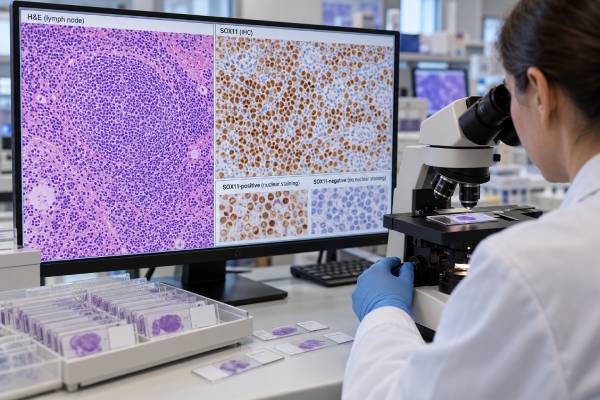
A SOX11 test is mainly a pathology test used to help identify and classify mantle cell lymphoma (MCL). SOX11 is a transcription factor that is abnormally expressed in most conventional mantle cell lymphomas, including many uncommon cases that do not show the usual cyclin D1 overexpression. Pathologists usually assess SOX11 by immunohistochemistry on a lymph node, bone marrow, or other tissue biopsy and look for nuclear staining in the abnormal lymphoid cells. A positive result can strongly support MCL when the cell appearance and other markers fit, but SOX11 should never be interpreted alone. Most MCL is established by a combination of morphology, B-cell markers, cyclin D1, and evidence of a CCND1 rearrangement such as t(11;14). Some SOX11-negative cases are genuine MCL, particularly leukemic non-nodal forms that can behave more indolently. Conversely, rare non-MCL tumors can express SOX11. The practical meaning therefore comes from the entire pathology panel, not simply from whether the stain says positive or negative.
- Positive SOX11 nuclear staining strongly supports conventional mantle cell lymphoma when the morphology and B-cell immunophenotype are compatible.
- SOX11 is especially helpful in cyclin D1-negative suspected MCL, where alternative cyclin D2 or cyclin D3 abnormalities may be present.
- A negative SOX11 test does not rule out MCL; leukemic non-nodal MCL is often SOX11-negative or weak.
- SOX11 is primarily a diagnostic and biologic marker, not a stand-alone treatment-selection test and is not equivalent to TP53 or Ki-67 risk assessment.
- The result is usually based on tissue immunohistochemistry, so there is no normal blood “range” or numeric cutoff that applies across all laboratories.
Table of Contents
- What SOX11 Is and How It Is Tested
- How SOX11 Fits the Diagnosis of Mantle Cell Lymphoma
- What Positive SOX11 Staining Means
- What Negative SOX11 Staining Means
- SOX11, Cyclin D1, and CCND1 Testing
- Prognosis and Treatment Meaning
- Common Interpretation Problems and Next Steps
What SOX11 Is and How It Is Tested
SOX11 is a transcription factor—a protein that helps regulate which genes a cell turns on and off. Mature B cells normally do not show the strong nuclear SOX11 expression seen in most conventional mantle cell lymphomas. That abnormal expression makes SOX11 useful to hematopathologists as part of an immunohistochemistry panel.
The usual SOX11 test is performed on a tissue section from a lymph node, bone marrow core, gastrointestinal biopsy, spleen, or another involved site. A pathologist applies an antibody that binds SOX11 and then examines where the stain appears. In MCL, the diagnostically useful pattern is nuclear staining in the abnormal lymphoid cells. Cytoplasmic background staining is not interpreted the same way.
Unlike a blood chemistry test, SOX11 immunohistochemistry does not have a universal reference interval such as 0–10 units. Reports may describe staining as positive or negative and may sometimes note strength or percentage, but laboratories use different antibodies, platforms, and interpretive thresholds. The pathologist also evaluates whether the cells that stain are the same population that looks abnormal under the microscope.
SOX11 is not generally ordered as an isolated test by a patient or clinician. It is added when the pathologic pattern suggests MCL or when a B-cell lymphoma needs further classification. It often sits alongside CD20, PAX5, CD5, cyclin D1, CD23, LEF1, Ki-67, and other stains, with fluorescence in situ hybridization (FISH) or molecular studies when needed.
Because the diagnosis rests on an integrated panel, a SOX11 result should be read in the pathology report’s final interpretation rather than as a stand-alone laboratory flag.
How SOX11 Fits the Diagnosis of Mantle Cell Lymphoma
Mantle cell lymphoma is a mature B-cell lymphoma most often driven by a chromosomal rearrangement that places CCND1 near an immunoglobulin enhancer. The classic t(11;14)(q13;q32) rearrangement leads to cyclin D1 overexpression, and most MCL cases are cyclin D1-positive.
A typical diagnosis combines several layers of evidence:
- lymph node or tissue architecture and cell morphology;
- a mature B-cell phenotype, commonly including CD20 and other B-cell markers;
- frequent coexpression of CD5;
- cyclin D1 overexpression and/or a CCND1 rearrangement;
- SOX11 expression in most conventional cases; and
- clinical and molecular findings that help resolve unusual presentations.
SOX11 becomes particularly valuable when the usual pattern is incomplete. Some MCL lacks cyclin D1 expression and does not have a detectable CCND1 rearrangement. These rare tumors may instead involve CCND2 or CCND3 and can be difficult to distinguish from other small B-cell lymphomas. Strong nuclear SOX11 expression in the right morphologic and immunophenotypic setting can provide an important clue that the tumor belongs in the mantle cell lymphoma family.
For a conventional cyclin D1-positive case, SOX11 is often supportive rather than essential. The cyclin D1 test for mantle cell lymphoma and a CCND1/IGH fusion test can demonstrate the defining cell-cycle abnormality more directly.
Current lymphoma classifications also recognize that MCL is biologically heterogeneous. Conventional nodal MCL and leukemic non-nodal MCL can differ in SOX11 expression, IGHV mutation patterns, genomic complexity, and clinical behavior. This is why “SOX11-negative” should not be translated into “not MCL” without considering the subtype.
What Positive SOX11 Staining Means
A positive SOX11 result means that the pathologist detected the protein in the nuclei of the tested cells at a level and pattern considered abnormal. In an appropriate mature B-cell neoplasm, this strongly supports mantle cell lymphoma.
The result has the most diagnostic weight when several features agree. For example, a CD20-positive, CD5-positive B-cell lymphoma with compatible morphology, cyclin D1 expression, and nuclear SOX11 staining has a classic MCL profile. In that setting, SOX11 reinforces a diagnosis already supported by other markers.
SOX11 is even more useful when cyclin D1 is absent. A cyclin D1-negative B-cell neoplasm with strong nuclear SOX11 expression may prompt additional studies for cyclin D2/D3 dysregulation and careful review for cyclin D1-negative MCL. Recent pathologic studies show that SOX11-positive blastoid or pleomorphic B-cell tumors without CCND1 rearrangement can still have molecular and clinical features of mantle cell lymphoma.
Positive staining does not mean all of the following:
- It does not by itself prove t(11;14).
- It does not tell the total tumor burden.
- It does not provide a stage.
- It does not determine whether treatment is required immediately.
- It does not independently establish a high-risk TP53-mutated tumor.
SOX11 expression also is not absolutely exclusive to MCL. It can occur in some other neoplasms, including certain non-hematologic tumors and uncommon hematolymphoid settings. The practical specificity is highest when the pathologist limits interpretation to the correct morphologic and immunophenotypic differential diagnosis.
The wording “positive” may also vary between laboratories. Some reports specify diffuse strong nuclear staining; others simply state “SOX11: positive.” If the significance is unclear, the final diagnosis and comment section of the pathology report usually explains how the stain was used.
What Negative SOX11 Staining Means
A negative SOX11 result means the abnormal cells did not show diagnostically significant nuclear staining with the assay used. It does not exclude mantle cell lymphoma.
The most important exception is leukemic non-nodal mantle cell lymphoma. This biologic subtype often involves blood, bone marrow, and spleen with little or no lymph-node disease. It is frequently SOX11-negative and tends to have more mutated IGHV genes and, in many cases, a more indolent course than conventional SOX11-positive MCL. Some patients can initially be observed rather than treated if they are asymptomatic and meet appropriate clinical criteria.
A negative result also requires technical context. Tissue fixation, decalcification of bone marrow samples, low tumor content, antibody performance, and staining quality can affect immunohistochemistry. Pathologists use internal controls and the rest of the panel to decide whether a negative stain is reliable.
When MCL remains suspected despite negative SOX11, other evidence can establish the diagnosis. Cyclin D1 overexpression or a CCND1 rearrangement is highly informative. In cyclin D1-negative disease, molecular or FISH studies may investigate CCND2 or CCND3 rearrangements, while morphology and the broader immunophenotype are reviewed carefully.
A SOX11-negative result therefore changes the diagnostic probability rather than closing the case. The right question is not simply “Is SOX11 negative?” but “Does the entire profile still fit a recognized SOX11-negative MCL subtype, or is another B-cell lymphoma a better explanation?”
SOX11, Cyclin D1, and CCND1 Testing
SOX11, cyclin D1 immunohistochemistry, and CCND1 rearrangement testing are related but they measure different biology.
| Test | What it detects | Main diagnostic role |
|---|---|---|
| SOX11 immunohistochemistry | Nuclear SOX11 protein expression | Supports conventional MCL and helps identify many cyclin D1-negative cases |
| Cyclin D1 immunohistochemistry | Overexpression of cyclin D1 protein | Key routine marker for classic MCL |
| CCND1 FISH | CCND1 rearrangement, commonly involving IGH | Genetic confirmation when morphology/staining is uncertain |
| Ki-67 | Proportion of proliferating tumor cells | Helps assess biologic aggressiveness; not an identity marker like SOX11 |
| TP53 testing | TP53 mutation and/or related abnormalities | Important high-risk biologic information in MCL |
In a straightforward case, cyclin D1 positivity and a compatible B-cell phenotype may make the diagnosis clear without needing every possible marker. In an atypical case, the pathologist may layer tests in stages to avoid both underdiagnosis and unnecessary testing.
The distinction matters because SOX11 expression is not a substitute for a genetic demonstration of CCND1 rearrangement. Likewise, a CCND1 rearrangement identifies the classic driver but does not describe proliferation rate, TP53 status, stage, or symptoms. Each marker adds a separate piece of the biologic picture.
Flow cytometry may also support classification by demonstrating a clonal B-cell population and its surface-marker pattern. A lymphoma flow cytometry panel can be especially useful in blood, marrow, or fresh tissue, but flow cytometry cannot replace the tissue architecture and immunohistochemistry needed in many lymphoma diagnoses.
Prognosis and Treatment Meaning
SOX11 has biologic associations with MCL subtype, but it should not be treated as a simple good-versus-bad prognostic switch. Conventional SOX11-positive MCL often has a more nodal presentation and more aggressive biology than leukemic non-nodal, SOX11-negative MCL. However, prognosis within either group depends on many other factors.
Modern risk assessment considers clinical features such as age, performance status, lactate dehydrogenase, and white blood cell count through the Mantle Cell Lymphoma International Prognostic Index. Pathologists also evaluate morphology and Ki-67. Blastoid or pleomorphic morphology and a high proliferation index generally signal more aggressive disease. TP53 abnormalities are particularly important high-risk features and may influence treatment strategy or clinical-trial consideration.
SOX11 does not by itself select a specific approved drug. Treatment decisions are based on whether treatment is needed, patient age and fitness, symptoms, stage and distribution, high-risk biology, prior therapy, and patient preference. Some asymptomatic patients with indolent leukemic non-nodal MCL can be monitored initially. Others with symptomatic conventional MCL need systemic treatment.
Research continues to clarify whether SOX11 itself can become a therapeutic vulnerability. Laboratory work has shown that SOX11 participates in oncogenic transcriptional programs and interacts with chromatin-remodeling machinery such as SMARCA4. Those findings are scientifically important but should not be mistaken for a routine SOX11-directed treatment currently chosen from an immunohistochemistry result.
For an individual report, a more useful prognostic discussion includes SOX11 status together with Ki-67, morphology, TP53, MIPI, disease distribution, and treatment response rather than assigning a prognosis from SOX11 alone.
Common Interpretation Problems and Next Steps
The most common mistake is treating SOX11 as a binary cancer test. It is a marker used inside a differential diagnosis, and its value depends on which cells stain and what the rest of the specimen shows.
Another pitfall is equating SOX11 positivity with cyclin D1 positivity. The two often occur together in conventional MCL, but they are not the same marker and they can separate in diagnostically important cases. Cyclin D1-negative, SOX11-positive MCL is a recognized situation. SOX11-negative, cyclin D1-positive MCL also occurs, particularly in leukemic non-nodal disease.
Small biopsies create additional challenges. A limited sample may not show the full architecture, and crush artifact or decalcification can reduce stain quality. If the pathology and clinical presentation disagree, a larger tissue sample or additional molecular testing may be needed.
After a SOX11 result, next steps can include:
- Confirming the final integrated lymphoma diagnosis rather than interpreting the stain in isolation.
- Reviewing cyclin D1 and CCND1 rearrangement results.
- Adding CCND2/CCND3 studies when cyclin D1-negative MCL remains likely.
- Assessing Ki-67, morphology, and TP53-related risk features when appropriate.
- Completing clinical staging with blood tests, imaging, and marrow evaluation according to the presentation.
Patients should ask what role SOX11 played in the final diagnosis: Was it confirmatory, did it resolve a cyclin D1-negative case, or did it help recognize a leukemic non-nodal subtype? That answer is much more informative than the word “positive” alone and keeps the marker connected to the actual clinical decision.
The site and pattern of disease can be as informative as the stain itself. Conventional SOX11-positive MCL commonly presents with lymph-node enlargement and may involve marrow, spleen, blood, or gastrointestinal tract. Leukemic non-nodal MCL more often shows blood, marrow, and spleen involvement with little bulky nodal disease. Recognizing that pattern helps a hematopathologist understand why a genuine MCL might be SOX11-negative.
SOX11 should also be separated conceptually from markers used to estimate aggressiveness. Ki-67 measures the fraction of tumor cells actively cycling and is commonly reported as a proliferation index. TP53 mutation or disruption identifies a particularly high-risk molecular subgroup. Blastoid or pleomorphic morphology reflects a more aggressive histologic variant. SOX11 status can correlate with biologic subtype, but it is not a substitute for any of those measurements.
A pathology report may sometimes describe heterogeneous or weak staining rather than a simple positive/negative result. In that situation, the pathologist considers whether staining is truly nuclear, whether internal controls worked, how much viable tumor is present, and whether the result fits cyclin D1 and genetic studies. Borderline staining should not be forced into a definitive diagnosis if stronger molecular evidence points elsewhere.
For patients, the most useful way to discuss SOX11 is to ask how it changed the final diagnosis. In a classic cyclin D1-positive case it may be confirmatory. In a cyclin D1-negative case it may be a key clue that prompts CCND2 or CCND3 testing. In a SOX11-negative leukemic presentation it may help explain a distinct biologic subtype rather than excluding MCL.
SOX11 is also not a routine blood monitoring marker after treatment. Although research assays have measured SOX11 RNA, standard response assessment relies on clinical evaluation, imaging when appropriate, blood and marrow findings, and validated molecular or flow-based MRD approaches in selected settings. A tissue SOX11 stain that was positive at diagnosis is not usually repeated serially to measure whether treatment is working.
When an outside pathology review is requested, sending the original tissue block or unstained slides can be more useful than repeating a single marker. Expert hematopathology review can reassess architecture, cyclin D1, SOX11, Ki-67, and genetic findings together, which is especially valuable in cyclin D1-negative, blastoid, or otherwise atypical cases.
References
- The 5th edition of the World Health Organization Classification of Haematolymphoid Tumours: Lymphoid Neoplasms 2022 (Classification)
- The International Consensus Classification of Mature Lymphoid Neoplasms: a report from the Clinical Advisory Committee 2022 (Consensus)
- Mantle Cell Lymphoma Under the Scope of Personalized Medicine: Perspective and Directions 2024 (Review)
- SOX11+ Large B-Cell Neoplasms: Cyclin D1-Negative Blastoid/Pleomorphic Mantle Cell Lymphoma or Large B-Cell Lymphoma? 2024
- The SOX11:SMARCA4 complex is a driver of oncogenic transcriptional programs in mantle cell lymphoma 2025
- The pathologic diagnosis of mantle cell lymphoma 2021 (Review)
Disclaimer
SOX11 is a specialized pathology marker and cannot diagnose or exclude mantle cell lymphoma by itself. Interpretation depends on tissue morphology, other immunostains, genetic studies, and the clinical presentation. Treatment and prognosis should be based on the integrated diagnosis and risk assessment rather than SOX11 status alone.





