A BCL2 rearrangement test looks for a structural change involving the BCL2 gene, most often the classic IGH::BCL2 fusion created by t(14;18)(q32;q21). This abnormality is a hallmark of conventional, or classic, follicular lymphoma and is found in most typical cases, but it is not present in every follicular lymphoma. Testing is usually performed on lymph node or other tumor tissue with fluorescence in situ hybridization (FISH), sometimes supplemented by chromosome analysis or sequencing. A positive result supports a diagnosis of classic follicular lymphoma when the biopsy also shows the expected follicular growth pattern, germinal-center B-cell phenotype, and other pathologic features. A negative result does not rule follicular lymphoma out, because a meaningful minority of cases are BCL2-rearrangement negative and may belong to biologically distinct subgroups. The result should therefore be interpreted as one component of an integrated hematopathology diagnosis rather than as a stand-alone cancer test.
- A positive BCL2 rearrangement usually means an abnormal BCL2 gene fusion is present, commonly IGH::BCL2 from t(14;18).
- About 85% of classic follicular lymphomas harbor IGH::BCL2, while roughly 10%–15% lack the classic rearrangement.
- FISH is the most common test, using either BCL2 break-apart probes or probes designed to detect an IGH::BCL2 fusion.
- BCL2 protein staining and BCL2 gene rearrangement are not the same test; one measures protein expression and the other detects a DNA-level structural abnormality.
- A positive result does not diagnose follicular lymphoma by itself, because the same rearrangement can occur in precursor lesions and other B-cell lymphomas.
Table of Contents
- What a BCL2 rearrangement means
- How BCL2 FISH testing works
- What a positive FISH result means
- Negative and atypical results
- Role in follicular lymphoma diagnosis
- BCL2 rearrangement beyond follicular lymphoma
- How to read the report and plan follow-up
What a BCL2 rearrangement means
BCL2 encodes a protein that helps cells resist programmed cell death, or apoptosis. In classic follicular lymphoma, a chromosome rearrangement places BCL2 next to regulatory elements from the immunoglobulin heavy-chain gene IGH. The usual event is t(14;18)(q32;q21), producing an IGH::BCL2 rearrangement. This change can lead to persistent BCL2 expression in germinal-center B cells that would normally be more tightly controlled.
The rearrangement is strongly associated with classic follicular lymphoma because that disease arises from germinal-center B cells. Most classic cases are positive, and the molecular finding fits with the familiar immunophenotype of CD10 and BCL6 expression together with abnormal BCL2 expression in neoplastic follicles.
The terms BCL2 rearrangement, IGH::BCL2 fusion, and t(14;18) are related but not always interchangeable. A BCL2 break-apart FISH assay can show that BCL2 is rearranged without proving that IGH is the partner. A dual-fusion FISH assay is designed specifically to demonstrate an IGH::BCL2 fusion. Modern sequencing can sometimes identify the exact partner and breakpoint.
The gene result is different from a BCL2 IHC test. Immunohistochemistry measures BCL2 protein in cells, while FISH evaluates the DNA structure. A tumor can show BCL2 protein without a detectable BCL2 rearrangement, and uncommon technical or biologic situations can produce the reverse pattern.
How BCL2 FISH testing works
FISH uses fluorescently labeled DNA probes that bind to selected chromosome regions. It can be performed on nuclei from formalin-fixed, paraffin-embedded biopsy tissue, fresh tissue, bone marrow, or another validated specimen. The laboratory chooses the assay based on the diagnostic question and available material.
BCL2 break-apart FISH
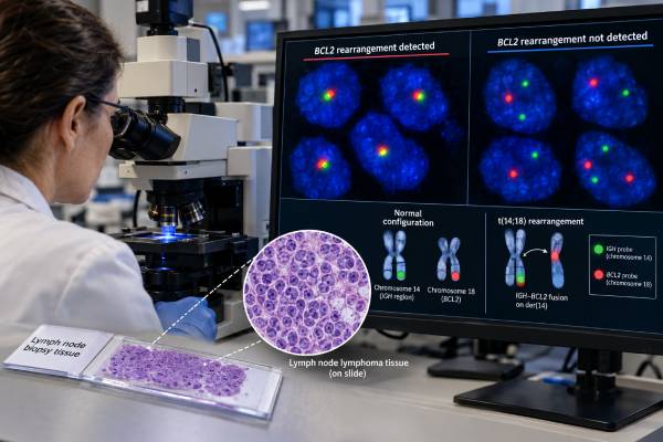
A break-apart assay places two differently colored probes on opposite sides of the BCL2 locus. In cells without a rearrangement, the colors remain together or nearly overlap. If BCL2 has been disrupted by a translocation, the probes separate. The laboratory counts a defined number of tumor-cell nuclei and compares the percentage with a validated cutoff.
A positive break-apart result answers the question “Is BCL2 rearranged?” It usually cannot answer “Which gene is fused to BCL2?” without additional testing.
IGH/BCL2 dual-fusion FISH
A dual-fusion assay uses probes for both IGH on chromosome 14 and BCL2 on chromosome 18. The classic translocation brings the loci together, producing a fusion signal pattern. This approach directly targets the common IGH::BCL2 event and can be particularly useful when classic follicular lymphoma is suspected.
FISH has advantages over conventional chromosome analysis because the cells do not need to be actively dividing. It also works on many routinely processed tissue samples. However, fixation, decalcification, tissue age, nuclear truncation, and low tumor content can reduce signal quality. Laboratories therefore use controls, specimen-specific criteria, and established positivity thresholds.
There is usually no fasting or medication preparation for the patient. The main practical requirement is adequate tumor material. If a small needle biopsy has already been exhausted by histology, flow cytometry, and other molecular tests, the pathologist may need to prioritize studies or request additional tissue.
What a positive FISH result means
A positive BCL2 FISH result means that enough analyzed cells show a signal pattern consistent with a BCL2 rearrangement to exceed the laboratory’s cutoff. If a dual-fusion assay was used, the report may specifically state that IGH::BCL2 is detected.
In a typical follicular lymphoma biopsy, this result strongly supports classic follicular lymphoma. It fits especially well when the tissue shows back-to-back or abnormal follicles composed mainly of centrocytes and centroblasts, with germinal-center markers such as CD10 and BCL6. A CD10 IHC result can help establish germinal-center differentiation, but no single immunostain replaces the full morphologic assessment.
A positive result is usually reported qualitatively as detected/positive rather than as a blood concentration. Some laboratories include the percentage of nuclei with an abnormal pattern. That percentage is not the same as a tumor stage, and small differences between percentages should not be treated as a direct measure of how aggressive the lymphoma is.
The laboratory may also describe the signal pattern in technical language, such as one or two fusion signals plus separate copies of the normal probes. Extra signals can occur because tumor cells sometimes have additional copies of chromosome regions or more complex rearrangements. These patterns are interpreted according to the probe design and laboratory validation; they are not reliably decoded by counting colors on the report without that context.
When both break-apart and dual-fusion probes are used, they can answer complementary questions. A positive BCL2 break-apart assay with a negative IGH/BCL2 dual-fusion assay may indicate that BCL2 is rearranged to a less common partner, although technical factors must also be considered. Conversely, a classic dual-fusion pattern provides direct evidence for the common IGH::BCL2 event.
The result also does not tell whether the lymphoma is localized or widespread. Staging requires clinical evaluation, imaging, laboratory studies, and sometimes marrow assessment. Likewise, a BCL2 rearrangement does not by itself determine whether treatment is needed immediately; some patients with low-burden follicular lymphoma can be observed, while others require therapy based on symptoms, tumor burden, organ effects, pace of disease, and other clinical factors.
One important nuance is that the t(14;18) event can be found at very low frequency in otherwise healthy people and can also be present in in situ follicular B-cell neoplasia, a microscopic precursor-type lesion. Therefore, the molecular abnormality must be located within an appropriate pathologic process before it is used to classify a lymphoma.
Negative and atypical results
A negative BCL2 rearrangement result does not exclude follicular lymphoma. Roughly 10%–15% of follicular lymphomas lack the classic t(14;18), and current classifications recognize that BCL2-rearrangement-negative follicular lymphomas are heterogeneous. Some have distinctive clinical sites, morphology, immunophenotypes, or alternative genetic changes.
A negative result should prompt the pathologist to ask whether the case still looks like classic follicular lymphoma or whether another entity fits better. Factors can include age, nodal versus extranodal location, follicular architecture, cytology, expression of CD10, BCL6, BCL2, MUM1, and other markers, and the results of additional cytogenetic or sequencing studies.
Technical false negatives are also possible. A rearrangement may have an unusual breakpoint outside the region effectively covered by a particular probe, although modern clinical probes are designed to capture the common events. Low tumor content or poor signal quality can also make testing less sensitive. If the result conflicts with strong morphologic evidence, a different FISH strategy or a sequencing-based assay may be considered.
An equivocal result generally means the abnormal signal percentage falls near the laboratory cutoff or the specimen quality prevents confident interpretation. The report may recommend repeat testing on another block or specimen. “Equivocal” should not be converted by the reader into either “positive” or “negative” without the pathologist’s interpretation.
BCL2 protein staining can remain positive even when the rearrangement is absent. Conversely, staining intensity can vary with technical factors and tumor biology. Gene-level and protein-level results answer different questions and are most useful when interpreted together.
Role in follicular lymphoma diagnosis
Follicular lymphoma is diagnosed by integrating architecture, cytology, immunophenotype, and genetics. BCL2 rearrangement is one of the strongest genetic clues for classic follicular lymphoma, but the diagnosis is not a FISH-only diagnosis.
The pathologist first determines whether the lymph node or tissue has a neoplastic follicular pattern. Tumor follicles often lack the normal polarization and tingible-body macrophages seen in reactive germinal centers. The cells usually express B-cell markers and germinal-center markers such as CD10 and BCL6. BCL2 expression within abnormal follicles supports the diagnosis because normal reactive germinal centers are typically BCL2 negative or weak compared with mantle-zone cells.
The molecular result becomes particularly helpful when the morphology is limited, the BCL2 protein stain is difficult to interpret, or the differential diagnosis includes a reactive process. It can also help classify unusual follicular proliferations when combined with other findings.
Current lymphoma classifications place greater emphasis on recognizing forms of follicular lymphoma that do not fit the classic BCL2-rearranged pattern. This avoids forcing every follicular B-cell neoplasm into a single category. Examples include certain pediatric-type follicular lymphomas and other BCL2-rearrangement-negative variants with different molecular features and clinical behavior.
Clinical location can be especially helpful in a BCL2-negative case. Nodal disease in an adult with conventional follicular morphology raises different possibilities from a localized lesion in a child or young adult, or from a process centered in the duodenum. The pathologist may therefore use the patient’s age and anatomic site as real diagnostic data rather than treating them as background details. Molecular findings such as mutations in epigenetic regulators, STAT6-related pathways, or other recurrent genes can provide additional support in selected difficult cases, but broad sequencing is not required for every routine follicular lymphoma.
Another distinction is in situ follicular B-cell neoplasia. In this lesion, BCL2-rearranged B cells are confined to otherwise preserved germinal centers and may be discovered incidentally. It is not managed like overt follicular lymphoma unless there is separate evidence of clinically significant lymphoma. This illustrates why molecular positivity alone cannot define tumor burden or stage.
Grade and transformation are separate issues. A BCL2-positive FISH result does not by itself determine histologic grade, and it does not prove that a follicular lymphoma has transformed to a large B-cell lymphoma. Those determinations require examination of cell size, growth pattern, proliferation, and clinical context.
This also means that a newly positive or newly negative laboratory result should not be used alone to declare progression. Tumor heterogeneity, sampling, and assay sensitivity can affect what is detected in a particular specimen. Clinical progression is established from the overall disease picture, not from a single FISH line item.
BCL2 rearrangement beyond follicular lymphoma
BCL2 rearrangements also matter in aggressive B-cell lymphomas. A large B-cell lymphoma may carry BCL2 rearrangement, sometimes together with MYC rearrangement. The meaning is different from an isolated BCL2 rearrangement in otherwise typical follicular lymphoma.
When the biopsy has large-cell morphology or other high-grade features, laboratories may perform a broader FISH workup that includes MYC, BCL2, and sometimes BCL6. A double-hit lymphoma FISH panel is used to identify clinically important combinations of rearrangements in appropriate high-grade B-cell tumors. The terminology for these lymphomas has evolved in recent classification systems, so the final diagnosis should follow the integrated pathology report rather than older shorthand alone.
BCL2 rearrangement can also persist when a previously diagnosed follicular lymphoma transforms into diffuse large B-cell lymphoma. In that setting, finding the same rearrangement may support a clonal relationship, but it is not sufficient by itself to prove transformation or lineage continuity.
This broader context explains why the specimen type and diagnostic question matter. “BCL2 rearrangement detected” is not a complete diagnosis. The same molecular event can contribute to different final classifications depending on the cells in which it occurs and the architecture of the tumor.
How to read the report and plan follow-up
Start with the final diagnosis, then read the FISH section. Useful report details include the assay type, whether the result was positive or negative, the percentage of abnormal nuclei if provided, and whether the test detected a generic BCL2 rearrangement or a specific IGH::BCL2 fusion.
Questions worth asking the hematology or pathology team include:
- Was BCL2 tested with a break-apart probe, an IGH/BCL2 dual-fusion probe, or sequencing?
- Does the result support classic follicular lymphoma, or is the case considered a BCL2-rearrangement-negative variant?
- Do the morphology and markers such as CD10, BCL6, and BCL2 protein agree with the FISH result?
- Was MYC or BCL6 testing needed because of large-cell or high-grade features?
- Is the specimen adequate, or did fixation, decalcification, or low tumor content limit the test?
- Does the result change the final tumor classification, prognosis discussion, or treatment plan?
Follow-up is based on the lymphoma diagnosis and clinical situation rather than serial BCL2 FISH percentages. Patients with follicular lymphoma are typically monitored with history, examination, blood tests, and imaging when clinically indicated. A new biopsy may be recommended if disease behavior changes substantially or transformation is suspected.
For a newly diagnosed patient, the FISH result may therefore have its greatest value at the classification stage. Once the diagnosis is established, clinicians generally do not repeat BCL2 FISH at every visit. If a new specimen is obtained later, testing may be repeated when the lineage is uncertain, the disease has transformed morphologically, or a broader molecular reassessment is needed. The decision is based on what new information the test could change, not simply on the passage of time.
A good report should also make limitations visible. Phrases such as “insufficient nuclei,” “suboptimal hybridization,” “below the validated cutoff,” or “cannot exclude a rearrangement outside the probe region” change how confidently a negative result can be used. When the final diagnosis and the molecular result seem inconsistent, asking whether the case was reviewed by a hematopathologist can be more useful than focusing on the FISH number alone.
Urgent medical assessment is appropriate for severe shortness of breath, rapidly enlarging masses, new neurologic symptoms, major bleeding, or signs of infection during treatment. More gradual symptoms such as persistent fevers, drenching night sweats, weight loss, worsening fatigue, or steadily enlarging nodes should also be reported to the treating team.
References
- The 5th edition of the World Health Organization Classification of Haematolymphoid Tumours: Lymphoid Neoplasms 2022 (Review)
- The International Consensus Classification of Mature Lymphoid Neoplasms: a report from the Clinical Advisory Committee 2022 (Position Statement)
- Bridging clinicopathologic features and genetics in follicular lymphoma: Towards enhanced diagnostic accuracy and subtype differentiation 2024 (Review)
- Diagnosis and Classification of Follicular Lymphoma and Related Entities 2025 (Review)
- Mature B, T and NK-cell, plasma cell and histiocytic/dendritic cell neoplasms: classification according to the World Health Organization and International Consensus Classification 2024 (Review)
Disclaimer
BCL2 rearrangement testing is a specialized pathology test and cannot diagnose follicular lymphoma by itself. Results must be interpreted with the biopsy appearance, immunophenotype, clinical findings, and other molecular studies. Discuss the complete pathology report and any treatment implications with a hematologist or oncologist.