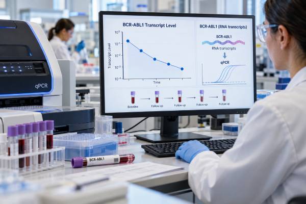
The BCR-ABL1 quantitative PCR test is the main blood test used to monitor molecular response in chronic myeloid leukemia (CML). It measures the amount of BCR::ABL1 messenger RNA made by leukemia cells and reports the result on the standardized International Scale, usually as BCR::ABL1% IS. After treatment with a tyrosine kinase inhibitor, the goal is for this percentage to fall over time. Key response levels include ≤10% at 3 months, ≤1% at 6 months, and ≤0.1% at 12 months in the 2025 European LeukemiaNet framework, although treatment decisions depend on the trend, adherence, dose changes, side effects, and individual clinical circumstances. A result of ≤0.1% is called major molecular response, while deeper responses such as MR4 and MR4.5 indicate progressively lower measurable disease. An “undetectable” result is reassuring only when the sample has enough control-gene transcripts to prove that the assay was sufficiently sensitive.
- BCR::ABL1 quantitative PCR reports leukemia transcript level as a percentage on the International Scale (IS).
- Favorable milestones are ≤10% at 3 months, ≤1% at 6 months, and ≤0.1% at 12 months, interpreted with prior results rather than as isolated cutoffs.
- Major molecular response (MMR) is BCR::ABL1 ≤0.1% IS; MR4 is ≤0.01%, MR4.5 is ≤0.0032%, and MR5 is ≤0.001% when assay quality is adequate.
- Testing is generally recommended at least every 3 months until MMR is achieved and confirmed, then about every 4–6 months when the response remains stable.
- “Not detected” does not mean every leukemia cell is gone; it means BCR::ABL1 was below that specimen’s validated detection limit.
Table of Contents
- What the test measures
- International Scale and molecular response levels
- When testing is done
- How to interpret rising and falling results
- Undetectable results and deep molecular response
- Unexpected results, atypical transcripts, and resistance
- Using results for treatment decisions
What the test measures
CML is driven by the BCR::ABL1 fusion gene, usually created by the Philadelphia chromosome translocation t(9;22). The fusion gene produces abnormal RNA and a continuously active tyrosine kinase protein. Quantitative reverse-transcription PCR, usually abbreviated RT-qPCR, converts BCR::ABL1 RNA into complementary DNA and then measures it during amplification.
The test does not count leukemia cells one by one. Instead, it compares BCR::ABL1 transcript copies with copies of a reference gene such as ABL1, BCR, or GUSB. The relationship is converted to the International Scale so that results from properly standardized laboratories can be compared with established CML response thresholds.
This monitoring test is different from the initial BCR-ABL1 diagnostic test. At diagnosis, testing establishes that the fusion is present and identifies the transcript type. During treatment, quantitative PCR repeatedly measures how much of the transcript remains.
About 98% of patients with CML express the standard e13a2 and/or e14a2 BCR::ABL1 transcripts, historically called b2a2 and b3a2, which encode the p210 BCR::ABL1 protein. A small minority have atypical transcripts that may not be measured accurately by standard International Scale assays. Knowing the transcript at diagnosis prevents a falsely reassuring monitoring result later.
No fasting is required. The test is commonly performed on peripheral blood, which makes repeated monitoring much easier than repeated bone marrow biopsies. Consistency matters: results are most interpretable when samples are handled by a laboratory with validated International Scale calibration and adequate quality controls.
International Scale and molecular response levels
The International Scale (IS) anchors BCR::ABL1 measurements to a common reference. At the standardized baseline used to create the system, the value is 100% IS. Response depths are expressed as progressively lower percentages and, equivalently, as log reductions from that baseline.
| Response level | BCR::ABL1 on International Scale | Approximate reduction from standardized baseline |
|---|---|---|
| Equivalent to complete cytogenetic response | ≤1% IS | About 2-log reduction |
| Major molecular response (MMR, MR3) | ≤0.1% IS | 3-log reduction |
| MR4 | ≤0.01% IS | 4-log reduction |
| MR4.5 | ≤0.0032% IS | 4.5-log reduction |
| MR5 | ≤0.001% IS | 5-log reduction |
These labels describe depth of molecular response, not separate disease stages. A patient can move from 1% to 0.1% to 0.01% as therapy suppresses the CML clone more deeply.
The 2025 European LeukemiaNet recommendations retain familiar milestone values while emphasizing trends and clinical context. At 3 months, ≤10% is favorable; at 6 months, ≤1% is favorable; at 12 months, ≤0.1% is favorable. Values above these thresholds may fall into warning or unfavorable categories depending on the level and whether an abnormal result is confirmed.
A single percentage should therefore never be read without its date and prior values. A result of 0.2% can have very different implications in someone steadily falling from 5% than in someone who had been stable at 0.02% and is now rising repeatedly.
It is also useful to distinguish response depth from response speed. Two patients may eventually reach the same MMR level even if one gets there earlier. Early milestones help estimate the likelihood of resistance and later deep response, but clinicians increasingly use the whole trajectory rather than treating every deadline as an automatic pass-or-fail event. That is why a confirmatory sample can be important when a value sits near a decision threshold.
Percentage changes can look dramatic when the numbers are already very small. Moving from 0.02% to 0.04% is a twofold increase, but both values remain below the 0.1% MMR boundary. By contrast, a sustained rise from 0.05% to 0.5% crosses a clinically meaningful response category. This is one reason CML teams focus on repeatable category changes and trends, not percentage arithmetic alone.
When testing is done
A quantitative BCR::ABL1 level is usually obtained at diagnosis or soon after treatment begins to establish a molecular reference. During tyrosine kinase inhibitor therapy, molecular monitoring is recommended at least every 3 months until MMR is achieved and confirmed. Once a patient remains in stable MMR or a deeper response, intervals of about 4–6 months are generally reasonable.
The schedule becomes especially intensive after a planned TKI discontinuation because most molecular relapses occur relatively early. Treatment-free remission protocols therefore use close PCR surveillance and a predefined trigger for restarting therapy, commonly loss of MMR. Patients considering discontinuation need access to a reliable laboratory and must be willing to complete testing on schedule; treatment-free remission is a monitored strategy, not simply stopping medication.
More frequent testing may be needed when:
- BCR::ABL1 values are rising or fluctuating unexpectedly;
- treatment has been interrupted or the dose substantially reduced;
- adherence is uncertain;
- a treatment change has recently occurred;
- resistance is suspected;
- the patient is being evaluated for treatment-free remission; or
- a TKI has been stopped and close molecular surveillance is required.
Blood counts are also followed, especially early in treatment, but blood counts and molecular response answer different questions. A normal white blood cell count can occur while measurable BCR::ABL1 remains, which is why molecular testing continues even when the complete blood count looks normal.
Bone marrow cytogenetics is much less sensitive than qPCR for routine molecular follow-up. It still has a role when there is evidence of resistance, suspected progression, unusual transcript biology, or a need to look for additional chromosome abnormalities. FISH may also help in selected cases with rare transcripts that cannot be tracked reliably by standard qPCR.
Patients do not need to time the test around meals. The practical priority is to obtain the sample at the recommended interval and, when possible, avoid unnecessary switching between laboratories with different assay systems unless results are clearly standardized to the IS.
How to interpret rising and falling results
The most important pattern is a sustained decline in BCR::ABL1 after treatment starts. Early molecular response shows that the TKI is suppressing the BCR::ABL1-positive clone. Reaching MMR by 12 months is a favorable target and increases the likelihood of later achieving a deep molecular response.
A result above a milestone does not automatically mean treatment has failed. The 2025 recommendations specifically caution against switching therapy based on one number alone. Clinicians consider whether the value was confirmed, whether the overall curve is still falling, whether treatment was held for toxicity, and whether the patient was able to take the prescribed medication consistently.
For example, BCR::ABL1 >10% at 3 months is concerning. If it remains >10% on a repeat sample within roughly 1–3 months, it falls into an unfavorable category. At 6 months, >10% is considered established resistance in the ELN framework, while >1% to 10% is a warning range. At 12 months, >1% is unfavorable, although individual circumstances can justify a more nuanced decision.
A rise after a previously good response deserves attention, especially a loss of MMR. Possible explanations include missed doses, drug interactions that reduce exposure, dose interruption, biologic resistance, or progression. The team may repeat qPCR sooner, review medication adherence and interactions, perform BCR::ABL1 kinase-domain mutation testing, and obtain cytogenetic studies when appropriate.
Small changes near very low levels can reflect normal analytic variation. A change from 0.006% to 0.009%, for example, remains within MR4 territory and should not be interpreted like a large, sustained rise across response categories. Trends become more convincing when the increase is repeated and clinically coherent.
A common source of confusion is the phrase “one-log change.” A 1-log increase means roughly a tenfold rise, such as 0.01% to 0.1%; a 1-log reduction means roughly a tenfold fall. Because assay variation is greater at very low transcript levels, a laboratory may recommend confirmation before a small numerical shift is labeled significant. The clinical meaning also depends on whether the change causes loss of MMR or another established response.
Laboratories sometimes report both the current IS value and the molecular response label. If the two appear inconsistent, the numeric International Scale result and the laboratory’s validated interpretation should be reviewed rather than recalculating the response from a consumer portal. Rounding, detection-limit rules, and control-gene requirements can affect the displayed label.
Undetectable results and deep molecular response
An undetectable BCR::ABL1 result is encouraging, but the phrase “not detected” requires a sensitivity statement. PCR can only detect what is present in the amount of RNA actually tested. If the specimen contains too few control-gene transcripts, the assay may not be sensitive enough to document MR4.5 or MR5 even though no fusion transcript was amplified.
For this reason, deep response definitions include minimum reference-gene requirements. With ABL1 as the control gene, MR4.5 generally requires at least about 32,000 ABL1 control transcripts when BCR::ABL1 is undetectable; MR5 requires about 100,000. Laboratories may use GUSB instead, with different validated transcript requirements.
A report may therefore say something like “BCR::ABL1 not detected; sensitivity consistent with MR4.5.” That is more informative than “negative” alone. It means no transcript was detected within a sample that had enough amplifiable material to support that depth of response.
Deep molecular response is important because sustained MR4 or deeper is one prerequisite considered when evaluating selected patients for treatment-free remission. It does not guarantee that stopping therapy will be successful, and it does not mean the disease has been eradicated. Very small numbers of CML cells may persist below the assay’s detection limit.
This is one way BCR::ABL1 monitoring functions as a highly sensitive minimal residual disease test for leukemia, although CML uses its own standardized molecular terminology and International Scale.
Unexpected results, atypical transcripts, and resistance
A result that does not fit the clinical picture should be investigated before major decisions are made. One of the most important causes of an apparently false-negative result is an atypical BCR::ABL1 transcript. Standard IS assays are optimized for e13a2/e14a2. Patients with rare fusion transcripts may require a personalized qPCR assay, digital PCR, FISH, or cytogenetic monitoring.
Pre-analytic problems can also affect results. Delayed sample processing can degrade RNA. Different reference genes, assay platforms, conversion factors, and laboratory sensitivities can create small differences even when laboratories report on the IS. Large unexpected shifts after a laboratory change should be reviewed with that possibility in mind.
When a genuine rise suggests resistance, the next test may be BCR::ABL1 kinase-domain mutation analysis rather than simply repeating the same qPCR indefinitely. Certain mutations affect sensitivity to specific TKIs. The clinical team integrates the mutation, prior therapies, side effects, cardiovascular risk, disease phase, and available drugs when choosing the next treatment.
Cytogenetic testing may also be repeated to look for additional chromosome abnormalities, especially with loss of response or suspected progression. These changes can provide information that transcript level alone cannot.
The key distinction is that qPCR tells how much measurable BCR::ABL1 is present, while resistance testing asks why the disease may no longer be responding as expected.
The distinction also prevents overtesting. A stable patient with a deep response does not need kinase-domain sequencing at every routine visit. Mutation analysis becomes useful when the response pattern raises a real question about resistance, while qPCR remains the repeated surveillance tool for long-term CML follow-up.
Using results for treatment decisions
BCR::ABL1 qPCR is powerful because it connects a laboratory number to a longitudinal treatment strategy. Clinicians use it to confirm that first-line therapy is working, identify warning signs early, decide when resistance testing is needed, document deep response, and monitor patients attempting treatment-free remission.
The result is never the only factor. Before changing an effective TKI, the team weighs the transcript trend against adverse effects, other medical conditions, treatment goals, pregnancy plans when relevant, adherence, and the risks of an alternative drug. A more potent TKI may produce deeper responses but can also have a different toxicity profile.
For patients whose main goal is durable disease control, maintaining a stable response may be more important than pursuing the deepest possible number at any cost. For others, achieving and sustaining MR4 or deeper may matter because they hope to attempt treatment-free remission later. The same PCR result can therefore lead to different reasonable discussions depending on the patient’s goals and health profile.
A practical way to review each report is to ask four questions:
- What is the current BCR::ABL1% IS?
- What was the previous value, and is the overall trend falling, stable, or rising?
- Which response category applies—above 10%, ≤10%, ≤1%, MMR, MR4, MR4.5, or deeper?
- Was the assay sensitive enough for the response depth being claimed?
Contact the treating team promptly if a report notes loss of a previous molecular response, a major confirmed rise, a newly detected resistant mutation, or progression-related findings. Do not stop, skip, or change a TKI based on a portal result without medical guidance; interruptions themselves can cause BCR::ABL1 to rise and complicate interpretation.
References
- 2025 European LeukemiaNet recommendations for the management of chronic myeloid leukemia 2025 (Guideline)
- European LeukemiaNet laboratory recommendations for the diagnosis and management of chronic myeloid leukemia 2023 (Guideline)
- Chronic myeloid leukemia: 2025 update on diagnosis, therapy, and monitoring 2024 (Review)
- Standardization of molecular monitoring of CML: results and recommendations from the European treatment and outcome study 2022 (Review)
- Comparative analysis of BCR::ABL1 p210 mRNA transcript quantification and ratio to ABL1 control gene converted to the International Scale by chip digital PCR and droplet digital PCR for monitoring patients with chronic myeloid leukemia 2024 (Comparative Study)
Disclaimer
BCR::ABL1 quantitative PCR results must be interpreted as a trend by the clinician managing CML. Do not change or stop a tyrosine kinase inhibitor based on a single laboratory value without medical guidance. Assay sensitivity, transcript type, treatment interruptions, and other clinical factors can materially change the meaning of a result.