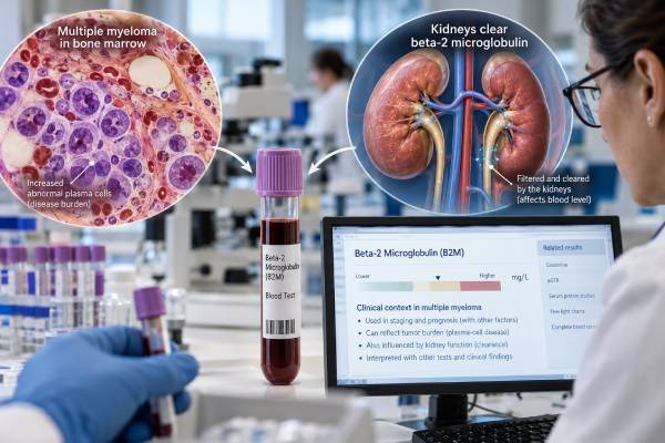
A beta-2 microglobulin test measures a small protein in blood that is used as part of staging and prognosis in multiple myeloma. In myeloma, a higher serum beta-2 microglobulin level often reflects a larger amount of disease, but the result is not a pure tumor-burden measurement. Beta-2 microglobulin is cleared largely by the kidneys, so reduced kidney function can raise the level even when the plasma-cell burden has not changed. This dual influence is especially important in myeloma because kidney injury can itself be caused by the disease. The test is therefore most useful as one component of a larger evaluation that includes serum albumin, lactate dehydrogenase, cytogenetic findings, kidney tests, monoclonal protein studies, bone marrow findings, imaging, and clinical features. It is central to the original International Staging System and still contributes to newer staging systems, but it should not be used by itself to diagnose myeloma or judge whether treatment is working.
- Beta-2 microglobulin is a prognostic and staging marker in multiple myeloma, not a stand-alone diagnostic test.
- Higher levels can reflect greater myeloma burden, poorer kidney filtration, or both, so creatinine and estimated kidney function are essential context.
- In the original ISS, stage I requires beta-2 microglobulin below 3.5 mg/L with albumin at least 3.5 g/dL; stage III uses beta-2 microglobulin at least 5.5 mg/L.
- There is no single “cancer normal range” that applies to every laboratory; reference intervals and units should be read directly from the report.
- Modern risk assessment adds cytogenetic abnormalities and LDH to clinical stage because beta-2 microglobulin alone cannot capture the full biology of myeloma.
Table of Contents
- What beta-2 microglobulin measures
- Why beta-2 microglobulin rises in myeloma
- How beta-2 microglobulin is used for myeloma staging
- How kidney function changes the result
- What high and low results mean
- How the test fits with other myeloma markers
- How to interpret the result in practice
What beta-2 microglobulin measures
Beta-2 microglobulin, often written β2-microglobulin or B2M, is a small protein associated with major histocompatibility complex class I molecules on the surface of most nucleated cells. As cells turn over, beta-2 microglobulin is released into the circulation. The kidneys filter it from the blood and normally reabsorb and break down much of the filtered protein in the proximal tubules.
A serum beta-2 microglobulin test therefore sits at the intersection of cell turnover and kidney clearance. Increased production can raise the blood concentration, while impaired filtration can do the same. This is why an elevated result is not specific for cancer. Chronic kidney disease, inflammation, infection, immune activation, and several lymphoid malignancies can produce elevations.
In multiple myeloma, the test has long been valuable because the level correlates with overall disease burden and clinical outcome across large patient populations. It is easy to measure from blood, and when combined with serum albumin it forms the original International Staging System, or ISS. Newer staging systems add tumor genetics and lactate dehydrogenase because the biology of myeloma cannot be represented fully by two serum proteins.
The laboratory report generally gives a numeric concentration, often in milligrams per liter. Reference intervals vary by method and laboratory. A number marked “high” means it exceeds that laboratory’s expected interval; it does not automatically assign a myeloma stage. Staging uses specific thresholds and other required variables.
Beta-2 microglobulin is also different from the multiple myeloma test panel used to establish and characterize a monoclonal protein. It does not identify an M protein, determine immunoglobulin type, or prove that plasma cells are clonal.
Why beta-2 microglobulin rises in myeloma
In active myeloma, a higher beta-2 microglobulin level often accompanies a greater mass of malignant plasma cells and more advanced disease. Myeloma cells and the surrounding immune environment contribute to protein turnover, so the serum concentration tends to rise as disease burden increases. This population-level relationship is strong enough to have made beta-2 microglobulin one of the most durable prognostic markers in the disease.
However, “tumor burden marker” is an incomplete description. Myeloma frequently affects the kidneys through filtered monoclonal free light chains, dehydration, high calcium, infection, medications, amyloid, or other mechanisms. When glomerular filtration falls, beta-2 microglobulin is cleared less efficiently and the serum concentration rises. A high value may therefore represent both more myeloma and less kidney clearance at the same time.
This overlap is clinically meaningful. Two patients with the same beta-2 microglobulin concentration may have very different combinations of plasma-cell burden and renal impairment. That does not make the marker useless; it explains why the test predicts outcome but is not a direct cell count. Kidney dysfunction is itself an important complication and adverse clinical feature in myeloma, so the combined signal still carries prognostic information.
Inflammation can add another layer. Beta-2 microglobulin can increase during immune activation and in other hematologic cancers. For this reason, an isolated elevation in someone without established myeloma should not be treated as evidence of the disease. Myeloma diagnosis instead requires clonal plasma cells or a plasmacytoma plus myeloma-defining events, assessed with marrow, monoclonal protein testing, imaging, and other clinical data.
Similarly, a declining beta-2 microglobulin after treatment can be encouraging, but it is not the preferred stand-alone response measure. The level may fall because kidney function improves, because tumor burden falls, or because both occur. Response assessment relies more directly on monoclonal protein measurements, free light chains when appropriate, bone marrow and measurable residual disease in selected settings, and imaging when indicated.
How beta-2 microglobulin is used for myeloma staging
The original ISS divides newly diagnosed multiple myeloma into three stages using only serum beta-2 microglobulin and albumin. It was designed as a simple, reproducible prognostic system rather than a map of where cancer is physically located.
| ISS stage | Beta-2 microglobulin | Albumin |
|---|---|---|
| I | Below 3.5 mg/L | At least 3.5 g/dL |
| II | Does not meet stage I or stage III criteria | Any value consistent with the stage II definition |
| III | At least 5.5 mg/L | Any value |
These cutoffs are staging thresholds, not laboratory reference ranges. A patient can have beta-2 microglobulin above the lab’s normal interval yet still be below the ISS stage III threshold. Conversely, staging cannot be assigned correctly without the full required information.
The Revised ISS, or R-ISS, retained the ISS backbone and added serum lactate dehydrogenase plus selected high-risk chromosome abnormalities. This was an important step because two patients with similar beta-2 microglobulin and albumin can have myeloma with very different genetic risk.
The Second Revision of the ISS, called R2-ISS, further refined prognostic grouping. It uses weighted contributions from ISS stage, elevated LDH, del(17p), t(4;14), and chromosome 1q gain or amplification. In the original R2-ISS model, ISS stage II contributes 1 point and ISS stage III contributes 1.5 points, showing that the beta-2 microglobulin/albumin framework remains relevant while no longer being treated as the whole risk model.
A staging system predicts outcomes across groups; it does not determine an individual person’s exact survival. Modern treatment, age, frailty, kidney recovery, cytogenetics, depth of response, access to therapies, and many other factors can shift an individual’s course substantially. A stage should therefore be understood as a risk category used to guide clinical discussion, trial stratification, and treatment planning, not as a countdown.
How kidney function changes the result
Kidney function is one of the most important modifiers of beta-2 microglobulin. The protein is small enough to pass through the glomerular filtration barrier. Healthy proximal tubules then reabsorb and metabolize most of it. When filtration is reduced, serum beta-2 microglobulin accumulates.
This means a high result should be read alongside serum creatinine, estimated glomerular filtration rate, calcium, hydration status, and the clinical cause of kidney dysfunction. In myeloma, clinicians may also examine serum free light chains and the kappa/lambda ratio, urine protein studies, and sometimes kidney biopsy findings when the mechanism of renal injury is uncertain.
A common misunderstanding is that kidney dysfunction makes the beta-2 microglobulin result “invalid.” It does not. Renal impairment is part of what the measured concentration reflects and is one reason the marker is prognostic. The limitation is more specific: a clinician cannot convert a beta-2 microglobulin number into a precise estimate of tumor mass without accounting for kidney clearance.
Changes over time require the same caution. Suppose beta-2 microglobulin falls from a markedly elevated level after treatment. If creatinine also improves substantially, part of the fall may reflect restored clearance. If the monoclonal protein and free light chains simultaneously drop, marrow disease improves, and kidney function is stable, the decrease is more clearly aligned with lower disease burden. The most reliable interpretation comes from the pattern across tests.
Urine beta-2 microglobulin testing is a different application and is not the basis of myeloma ISS staging. Urinary concentrations can reflect proximal tubular handling and are used in selected renal evaluations. When people discuss beta-2 microglobulin as a myeloma stage marker, they generally mean the serum test.
What high and low results mean
A high serum beta-2 microglobulin value in a person with confirmed myeloma generally signals a less favorable prognostic profile, but it does not identify the cause of the elevation by itself. The first interpretive question is whether the result is being used for initial staging, general risk assessment, or longitudinal follow-up. The second is what the kidney function and other myeloma markers show at the same time.
For staging, values below 3.5 mg/L and at or above 5.5 mg/L are especially important because they help define ISS stage I and III, respectively. Values between those thresholds commonly contribute to stage II, but albumin and the exact ISS definition still matter. Current R-ISS or R2-ISS classification adds other variables.
A result below the laboratory’s upper reference limit is generally reassuring with respect to this marker, but it does not exclude myeloma. Some patients with early or lower-burden disease have beta-2 microglobulin within the reference interval. Diagnosis depends on evidence of a clonal plasma-cell disorder and myeloma-defining events, not on beta-2 microglobulin being high.
There is also no useful target of “as low as possible” outside context. Very low serum values are not typically a clinical problem. The concern in myeloma is an elevated concentration and what it implies when combined with the rest of the evaluation.
Laboratory units deserve attention. Myeloma staging thresholds are commonly expressed in mg/L. If a report uses a different unit, clinicians should not compare the raw number directly with ISS cutoffs without conversion. Likewise, a patient’s serial measurements are easiest to interpret when performed with a consistent method and laboratory, although standardized clinical interpretation can accommodate changes when necessary.
The size of a change matters less than whether it aligns with other objective evidence. Beta-2 microglobulin is best treated as a contextual prognostic marker rather than a day-to-day tumor meter.
How the test fits with other myeloma markers
No single blood test captures all of multiple myeloma. Beta-2 microglobulin contributes mainly to staging and prognosis. Other tests answer different questions.
Serum protein electrophoresis and immunofixation identify and characterize monoclonal immunoglobulin in many patients. The serum protein electrophoresis test can quantify an M-spike when it is measurable. Serum free light chains are particularly important in light-chain myeloma and in patients whose monoclonal protein is not well measured by electrophoresis. Quantitative immunoglobulins show immunoglobulin levels but do not replace immunofixation for defining clonality.
Bone marrow aspiration and biopsy determine the percentage and appearance of plasma cells and provide material for flow cytometry and genetic testing. FISH on enriched plasma cells can detect high-risk abnormalities such as del(17p), t(4;14), and chromosome 1q gain or amplification that influence modern risk classification. LDH provides another measure associated with aggressive biology and is included in revised staging systems.
Imaging identifies lytic bone lesions, focal marrow disease, plasmacytomas, and other structural complications. Calcium, hemoglobin, creatinine, and related tests evaluate classic organ damage. Together, these findings distinguish asymptomatic precursor conditions such as monoclonal gammopathy of undetermined significance or smoldering myeloma from active multiple myeloma that meets treatment criteria.
For response monitoring, clinicians usually prioritize the marker that directly represents the patient’s secreted monoclonal protein, along with marrow or imaging assessment when required. Beta-2 microglobulin may improve as treatment succeeds, but its dependence on renal function makes it a secondary rather than definitive response marker.
This division of labor explains why a patient can have a favorable drop in M protein while beta-2 microglobulin remains elevated because chronic kidney disease persists. It also explains why beta-2 microglobulin can improve rapidly after hydration and kidney recovery even before the full antimyeloma response is known.
How to interpret the result in practice
A useful interpretation starts with the exact number, unit, and laboratory reference interval, then asks whether the person has a confirmed plasma-cell disorder. In someone being evaluated for possible myeloma, beta-2 microglobulin can contribute prognostic information but cannot confirm the diagnosis. In someone with newly diagnosed myeloma, it should be paired with albumin and the other inputs needed for the staging system the clinical team uses.
Next, review kidney function from the same time point. A beta-2 microglobulin of 7 mg/L means something different in a patient with normal filtration than in a patient with severe acute kidney injury. Both are clinically important, but the contribution from tumor burden cannot be assumed to be equal. The direction of creatinine and estimated filtration over time can help explain why beta-2 microglobulin changes.
Then place the result beside cytogenetics and LDH. A lower ISS stage does not neutralize a high-risk plasma-cell genetic profile, and a higher beta-2 microglobulin level does not tell the clinician which chromosome abnormalities are present. R-ISS and R2-ISS were created specifically to combine these different dimensions of risk.
For patients comparing reports, it is also important to distinguish stage from response. A person generally keeps the staging label assigned at diagnosis for prognostic description; clinicians do not usually “restage” the disease every time beta-2 microglobulin changes during therapy as though the original stage were a dynamic response score. Treatment response has its own criteria.
Questions worth asking the hematology team include: Was this value drawn before treatment? What was my creatinine or estimated kidney function at the same time? Which staging system is being used? What are my LDH and FISH findings? Which laboratory marker will be used to track treatment response? These questions help separate prognosis from diagnosis and monitoring.
An unexpectedly high beta-2 microglobulin result should not prompt self-directed conclusions about disease progression. Infection, inflammation, kidney injury, dehydration, and other changes can alter the level. A clinician can determine whether repeat testing, kidney evaluation, or a broader myeloma reassessment is appropriate based on the full clinical picture.
References
– Multiple myeloma: 2024 update on diagnosis, risk-stratification, and management 2024 – Second Revision of the International Staging System (R2-ISS) for Overall Survival in Multiple Myeloma: A European Myeloma Network (EMN) Report Within the HARMONY Project 2022 – Diagnosis and Management of Multiple Myeloma: A Review 2022 – Allocation and validation of the second revision of the International Staging System in the ICARIA-MM and IKEMA studies 2024 – A Real-World International Staging System (RW-ISS) for patients with newly diagnosed multiple myeloma 2025
Disclaimer
This article is for general education and does not replace evaluation by a hematologist or other qualified clinician. Beta-2 microglobulin can be affected by kidney function and other medical conditions, so a result should never be interpreted as a direct measure of myeloma burden without the rest of the clinical data. Treatment and monitoring decisions should be based on the complete myeloma assessment.