
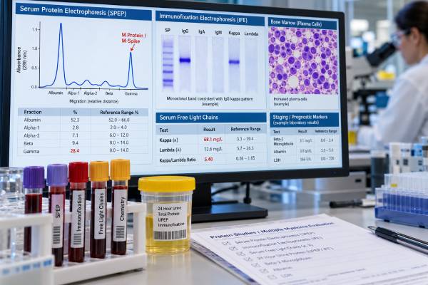
A multiple myeloma test panel combines several blood, urine, bone marrow, and imaging studies because no single test can diagnose or stage myeloma reliably. The core laboratory workup usually includes serum protein electrophoresis (SPEP), serum immunofixation, serum free light chains, quantitative immunoglobulins, a complete blood count, creatinine, calcium, albumin, and beta-2 microglobulin. Urine protein studies, bone marrow examination, fluorescence in situ hybridization (FISH), and whole-body imaging are added as needed to confirm the plasma-cell disorder, identify myeloma-defining events, and estimate prognosis.
The panel answers several separate questions: Is there a monoclonal protein? What immunoglobulin type is it? How much is present? Are free kappa or lambda light chains abnormal? Has myeloma caused anemia, kidney dysfunction, hypercalcemia, or bone disease? What percentage of marrow plasma cells are clonal, and do they carry high-risk genetic abnormalities? The diagnosis comes from the combined pattern rather than one abnormal number.
- Main screening tests: SPEP, serum immunofixation, and serum free light chains are commonly used together to detect monoclonal proteins.
- What M protein means: An M spike shows a monoclonal immunoglobulin, but it can occur in MGUS and smoldering myeloma as well as active multiple myeloma.
- Free light-chain warning: A markedly abnormal involved/uninvolved ratio can be diagnostically important, but kidney function and assay-specific reference ranges affect interpretation.
- Staging tests: Beta-2 microglobulin, albumin, LDH, and myeloma FISH abnormalities contribute to modern risk stratification.
- Diagnosis requires context: Active myeloma requires clonal plasma cells or plasmacytoma plus a myeloma-defining event, not merely a positive monoclonal protein test.
Table of Contents
- What is in a multiple myeloma test panel
- M protein and SPEP
- Immunofixation and immunoglobulin type
- Serum free light chains
- Bone marrow, imaging, and FISH
- Staging and prognosis
- How to interpret the whole panel
What is in a multiple myeloma test panel
A useful myeloma panel is not one branded bundle. It is a set of complementary tests chosen to detect a clonal plasma-cell process and determine whether it meets criteria for active disease.
Common tests include:
- complete blood count for anemia and other cytopenias;
- creatinine and estimated kidney function;
- serum calcium;
- total protein and albumin;
- SPEP to identify and quantify an M spike;
- serum immunofixation to type the monoclonal protein;
- serum free kappa and lambda light chains with a ratio;
- quantitative IgG, IgA, and IgM;
- urine protein electrophoresis and urine immunofixation in selected settings;
- beta-2 microglobulin and LDH for prognostic assessment;
- bone marrow aspirate and biopsy;
- plasma-cell FISH for cytogenetic risk; and
- whole-body low-dose CT, PET/CT, or MRI to evaluate bone and focal disease.
A serum protein electrophoresis test and a serum immunofixation test are closely related but not interchangeable. SPEP is better for seeing and measuring a discrete protein peak, while immunofixation is more sensitive for confirming and typing a small monoclonal immunoglobulin.
The panel is interpreted against diagnostic criteria. Active myeloma generally requires at least 10% clonal bone marrow plasma cells or a biopsy-proven plasmacytoma plus one or more myeloma-defining events. Those events include attributable CRAB features—hypercalcemia, renal impairment, anemia, or lytic bone lesions—and certain validated biomarkers of near-term progression.
M protein and SPEP
M protein, also called monoclonal protein, paraprotein, or M spike, is an immunoglobulin produced by one clone of plasma cells. SPEP separates serum proteins according to electrical properties and displays their distribution. A narrow peak in the gamma or beta region can indicate a monoclonal protein.
The report often quantifies the M spike in g/dL. The size is useful for monitoring, but there is no single M-protein cutoff that by itself proves active myeloma. A person with MGUS can have a small M protein for years without organ damage, while a patient with light-chain myeloma may have little or no measurable intact M spike.
M protein also changes slowly in some settings because circulating immunoglobulin has a biological half-life. After treatment, the tumor burden may fall before the serum protein fully clears. This is one reason response assessment uses more than SPEP alone.
A dedicated M protein blood test interpretation is most useful for understanding how the spike is quantified over time.
When SPEP is normal but myeloma is still possible
SPEP can miss disease dominated by free light chains or very small monoclonal proteins. If symptoms suggest a plasma-cell disorder—such as unexplained anemia, renal injury, hypercalcemia, bone pain, or recurrent fractures—serum free light chains and immunofixation remain important even when SPEP does not show a large spike.
Rare nonsecretory or oligosecretory myeloma produces little measurable protein. In those cases marrow, imaging, and MRD approaches carry more weight.
Immunofixation and immunoglobulin type
Immunofixation answers what type of monoclonal protein is present. It identifies heavy chains such as IgG, IgA, or IgM and light chains such as kappa or lambda. A result might read “IgG kappa monoclonal protein” or “free lambda monoclonal light chain.”
This typing matters for diagnosis and follow-up. Once the original clone is known, later immunofixation can determine whether the same protein persists.
Immunofixation is more sensitive than SPEP for small monoclonal proteins but is not ideal for precise quantification. The two tests therefore work together: SPEP measures, immunofixation identifies.
A positive immunofixation result is not synonymous with myeloma. MGUS, smoldering myeloma, Waldenström macroglobulinemia, AL amyloidosis, and other monoclonal gammopathies can also produce a monoclonal band. The immunoglobulin class can help narrow the differential—for example, IgM monoclonal proteins raise different diagnostic questions from typical IgG or IgA myeloma.
Urine immunofixation remains useful when light-chain excretion or AL amyloidosis is a concern. A urine immunofixation test can identify monoclonal free light chains in urine even when the serum pattern is subtle.
Serum free light chains
Plasma cells normally produce immunoglobulin heavy chains and light chains. Small amounts of unbound, or “free,” kappa and lambda light chains circulate in blood. Myeloma can overproduce one light-chain type, causing the involved chain to rise and the kappa/lambda ratio to become abnormal.
A serum free light-chain test reports:
- free kappa concentration;
- free lambda concentration; and
- the kappa/lambda ratio.
If kappa is the monoclonal involved chain, the ratio may become high. If lambda is involved, the ratio may become low. Laboratories use assay-specific reference intervals, so the report’s range should be used rather than a memorized universal number.
Myeloma-defining free light-chain biomarker
One recognized myeloma-defining biomarker is an involved/uninvolved free light-chain ratio of 100 or greater, provided the involved free light chain is at least 100 mg/L and the other diagnostic requirements are met. This criterion is intended to identify a group at sufficiently high risk that waiting for organ damage would be unsafe.
That threshold should not be applied casually to every abnormal ratio. Kidney impairment raises both kappa and lambda because renal clearance falls and can widen the ratio modestly. Infection, inflammation, assay variation, and monoclonal gammopathies below the myeloma threshold can also produce abnormal results.
For monitoring, the difference between involved and uninvolved free light chains may be useful in light-chain or oligosecretory disease. Trends are generally more informative than isolated values.
Bone marrow, imaging, and FISH
Blood tests can detect the products and consequences of myeloma, but marrow and imaging show the disease more directly.
Bone marrow examination
A marrow aspirate and biopsy estimate the percentage of plasma cells and determine whether they are clonal. Flow cytometry or immunohistochemistry may establish light-chain restriction and abnormal antigen expression. Marrow involvement can be patchy, so the percentage should be interpreted with imaging and clinical data.
A clonal marrow plasma-cell percentage of 60% or greater is itself a myeloma-defining biomarker when diagnostic conditions are satisfied. Lower percentages can still represent active myeloma if another defining event is present.
Imaging
Modern imaging looks for lytic bone lesions and focal marrow abnormalities. Whole-body low-dose CT is commonly used for skeletal assessment; PET/CT can show metabolically active lesions and extramedullary disease; MRI is particularly sensitive for marrow focal lesions.
More than one focal MRI lesion of appropriate size is another recognized myeloma-defining biomarker in the correct setting.
FISH and molecular risk
Plasma-cell FISH assesses recurrent chromosome abnormalities such as del(17p), t(4;14), t(14;16), t(14;20), and 1q gain/amplification. These findings do not establish the diagnosis alone, but they help define biological risk and guide treatment intensity.
Because plasma cells may be a minority of total marrow cells, many laboratories enrich CD138-positive plasma cells before FISH to improve sensitivity.
Staging and prognosis
Myeloma stage is not based on M-protein size alone. The traditional International Staging System uses serum beta-2 microglobulin and albumin. The Revised ISS adds LDH and high-risk cytogenetics. The newer R2-ISS further refines risk by incorporating chromosome 1q abnormalities and weighted risk features.
A beta-2 microglobulin test for myeloma is important but must be interpreted with kidney function because impaired renal clearance can raise beta-2 microglobulin independently of tumor burden.
| Test or feature | What it contributes | Important limitation |
|---|---|---|
| Beta-2 microglobulin | Tumor burden and prognostic information | Also rises with kidney dysfunction |
| Albumin | Part of ISS staging | Nonspecific and influenced by systemic illness |
| LDH | Identifies more aggressive biology when elevated | Nonspecific marker of tissue turnover |
| FISH | Detects high-risk chromosome abnormalities | Requires adequate clonal plasma-cell sampling |
| MRD | Measures depth of treatment response | Usually a post-treatment marker, not baseline stage |
Stage and risk are related but not identical. A patient can have a lower stage but high-risk cytogenetics, or a higher beta-2 microglobulin partly because of renal dysfunction. Hematologists therefore interpret the full pattern rather than using one staging label as a complete prognosis.
How to interpret the whole panel
The easiest way to read a myeloma workup is to divide it into five questions.
- Is there a monoclonal plasma-cell process? SPEP, immunofixation, free light chains, marrow clonality, and sometimes urine studies answer this.
- Does it meet criteria for active myeloma? Look for CRAB features or validated myeloma-defining biomarkers.
- How much disease is present? M protein, free light chains, marrow percentage, and imaging estimate burden in different ways.
- What is the biological risk? FISH, LDH, beta-2 microglobulin, albumin, and stage contribute.
- How well is treatment working? Serial SPEP, free light chains, immunofixation, imaging, and myeloma MRD testing measure response.
Several common mistakes are worth avoiding. An M spike is not automatically myeloma. A normal calcium does not exclude myeloma. A near-normal SPEP does not exclude light-chain disease. A high beta-2 microglobulin may partly reflect kidney dysfunction. And a normal free light-chain ratio does not eliminate every plasma-cell neoplasm.
If the initial panel is abnormal, useful follow-up questions include: What is the monoclonal protein type? What is the clonal marrow plasma-cell percentage? Are there lytic or focal lesions? Is kidney injury attributable to the plasma-cell disorder? What FISH abnormalities are present? Does the patient meet MGUS, smoldering myeloma, or active myeloma criteria?
The central principle is that myeloma diagnosis is integrated. Protein studies detect what the clone secretes, marrow measures the clone itself, imaging shows its tissue effects, and staging tests estimate risk. The complete panel is more reliable than any individual result.
How kidney function changes protein test interpretation
Kidney function can complicate the free light-chain result because the kidneys normally clear free kappa and lambda chains from blood. When filtration falls, both light chains can rise and the ratio may shift modestly even without a malignant plasma-cell clone. The pattern is different from a strongly skewed ratio caused by one clonal light-chain type, but borderline results should be interpreted with creatinine, estimated glomerular filtration rate, immunofixation, and the clinical setting.
Kidney injury can also be a consequence of myeloma. A patient with a rapidly rising involved free light chain, acute kidney dysfunction, and a compatible monoclonal process may need urgent hematology evaluation because light-chain cast nephropathy can threaten kidney function. The distinction between “kidney disease raising the light chains” and “light chains causing kidney disease” requires the whole pattern rather than one number.
Why serial results should be compared using the same method
Myeloma monitoring depends heavily on change over time. A fall in M protein or involved free light chain can indicate response, while a confirmed rise may signal biochemical progression before symptoms appear. Small differences between laboratories or assay platforms can occur, so longitudinal testing is easiest to interpret when the same laboratory method is used consistently.
Immunofixation is more sensitive than routine electrophoresis for identifying a small monoclonal protein, but it is not ideal for quantifying how much protein is present. SPEP is better for measuring a discrete serum M spike when one is measurable. Free light-chain assays are especially important in light-chain or oligosecretory disease, where SPEP may show little or no M spike.
MGUS, smoldering myeloma, and active myeloma are separated by the whole picture
The presence of a monoclonal protein does not by itself establish active myeloma. MGUS usually has a smaller clone and no myeloma-defining organ damage. Smoldering myeloma has a larger clonal burden but still lacks myeloma-defining events. Active myeloma is diagnosed when the clonal plasma-cell disorder is accompanied by qualifying CRAB organ damage—hypercalcemia, renal impairment, anemia, or bone lesions—or by validated biomarker-defined events that predict a very high risk of progression.
Because those categories have very different management, the test panel must be paired with marrow percentage, imaging, kidney function, calcium, hemoglobin, and the relevant biomarkers. The safest interpretation is a diagnostic category, not simply “M protein positive.”
Treatment can create confusing monoclonal bands
Some therapeutic monoclonal antibodies used in myeloma can appear on serum protein studies and interfere with interpretation of immunofixation or a small M spike. Laboratories may use drug-specific reflex methods or mass-spectrometry approaches in selected settings to separate a treatment antibody from the patient’s original monoclonal protein. This issue becomes most important when the disease protein is already very low and the team is trying to determine whether a deep response has been reached.
The original isotype at diagnosis is useful context. If a new faint band has a different migration pattern or immunoglobulin type, the laboratory may consider therapy interference or oligoclonal immune recovery rather than assuming immediate relapse.
Baseline documentation is especially valuable before treatment starts. Recording the original M-protein quantity, heavy- and light-chain type, involved free light chain, marrow plasma-cell percentage, imaging findings, and cytogenetic risk creates a reference point for later response assessment. Without that baseline, a faint post-treatment band or an abnormal light-chain value can be harder to interpret accurately.
The same baseline also helps distinguish a true biochemical relapse from normal assay variation, kidney-related light-chain changes, or a new small band unrelated to the original clone.
References
- Multiple myeloma: 2024 update on diagnosis, risk-stratification, and management 2024 (Review)
- Diagnosis and Management of Multiple Myeloma: A Review 2022 (Review)
- Second Revision of the International Staging System (R2-ISS) for Overall Survival in Multiple Myeloma: A European Myeloma Network (EMN) Report Within the HARMONY Project 2022
- Performance Characteristics and Limitations of the Available Assays for the Detection and Quantitation of Monoclonal Free Light Chains and New Emerging Methodologies 2024 (Review)
- International Myeloma Working Group updated criteria for the diagnosis of multiple myeloma 2014 (Guideline)
- Molecular Diagnostics of Plasma Cell Neoplasms 2023 (Review)
Disclaimer
This article is for general educational purposes and is not a substitute for diagnosis or treatment by a hematologist. Monoclonal proteins and abnormal free light chains can occur in disorders other than active myeloma, and kidney function can alter several results. Seek prompt medical evaluation for severe weakness, confusion, dehydration, reduced urine output, new neurologic symptoms, or severe bone pain.





