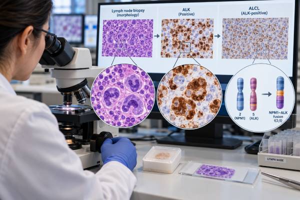
An ALK test helps determine whether an anaplastic large cell lymphoma (ALCL) belongs to the ALK-positive subtype, a biologically distinct form of systemic T-cell lymphoma. The test looks for abnormal activation of the ALK gene or its protein product, usually caused by a gene fusion. In most cases, the first clue comes from ALK immunohistochemistry on a lymph node or other biopsy, with molecular testing such as fluorescence in situ hybridization (FISH) used when confirmation or clarification is needed. A positive result supports classification as ALK-positive ALCL when the tumor also has the expected morphology and strong CD30 expression. It is not a stand-alone cancer diagnosis, because ALK abnormalities occur in other tumors and the staining pattern must be interpreted in context. The distinction matters because ALK-positive and ALK-negative systemic ALCL differ in age distribution, molecular biology, prognosis, and sometimes treatment planning.
- A positive ALK result in the right lymphoma pattern supports ALK-positive systemic ALCL, a recognized subtype of mature T-cell lymphoma.
- Most ALK-positive ALCL is driven by an ALK gene fusion, most often NPM1::ALK from t(2;5)(p23;q35).
- ALK immunohistochemistry is commonly the first test, while ALK break-apart FISH or another molecular method can confirm rearrangement in selected cases.
- ALK negativity does not rule out ALCL; systemic ALK-negative ALCL is a separate diagnosis that still requires characteristic morphology and strong CD30 expression.
- No special fasting or medication preparation is usually needed, because testing is generally performed on biopsy tissue, bone marrow, or another previously collected specimen.
Table of Contents
- What the ALK test detects
- How ALK testing is done
- What a positive ALK result means
- Negative, equivocal, and unusual results
- How ALK affects tumor classification
- Clinical meaning and follow-up
- Questions to ask about the report
What the ALK test detects
The ALK test evaluates abnormal activity of the anaplastic lymphoma kinase (ALK) gene in tumor cells. ALK is a receptor tyrosine kinase gene located on chromosome 2. Normal mature lymphocytes do not usually show the strong ALK protein expression seen in ALK-positive ALCL. In this lymphoma, a chromosome rearrangement places the kinase portion of ALK next to another gene, creating a fusion protein that sends continuous growth and survival signals.
The classic rearrangement is t(2;5)(p23;q35), which creates an NPM1::ALK fusion. NPM1 is the most common fusion partner, but it is not the only one. Other ALK partners can produce different intracellular locations of the fusion protein and therefore different staining patterns on immunohistochemistry. The biologic result is similar: the ALK kinase is switched on abnormally.
ALK is only one part of the diagnostic picture. Systemic ALCL is usually characterized by large atypical lymphoid cells, often including so-called hallmark cells with horseshoe- or kidney-shaped nuclei, and strong, usually uniform expression of CD30. A pathologist may pair ALK testing with a CD30 test, T-cell markers, epithelial markers, and other stains to determine what lineage the abnormal cells represent.
An ALK-positive result is therefore best thought of as a classification marker rather than a general tumor marker measured in the blood. It helps answer a focused question: does this CD30-positive large-cell lymphoma belong to the ALK-positive ALCL category?
How ALK testing is done
ALK testing is usually performed on tissue from a lymph node, extranodal mass, skin lesion, or another biopsy site. Bone marrow may also be assessed when clinically indicated. The exact laboratory approach depends on the specimen, the appearance of the tumor, and whether the initial result is straightforward.
ALK immunohistochemistry
Immunohistochemistry (IHC) uses an antibody to show ALK protein in fixed tissue. It is fast, widely available, and highly useful for ALCL. The pathologist evaluates both whether the tumor cells stain and where the staining appears inside the cell.
NPM1::ALK often produces both nuclear and cytoplasmic staining because the NPM1 partner can shuttle between these compartments. Other ALK fusions may produce cytoplasmic, membranous, granular, or other non-nuclear patterns. A characteristic pattern can suggest a fusion partner, but IHC usually does not identify the exact partner by itself. Readers who want more detail on the protein-based method may also find the ALK IHC test useful for understanding how staining is interpreted across tumor types.
ALK FISH
Fluorescence in situ hybridization (FISH) examines the ALK locus at the DNA level. A break-apart probe places differently colored probes on opposite sides of the ALK breakpoint region. In a non-rearranged cell, the signals remain together or overlap. When ALK is rearranged, the signals separate beyond the laboratory’s validated cutoff.
A break-apart FISH result confirms that ALK is rearranged but usually does not name the partner gene. A targeted dual-fusion assay or sequencing-based fusion test can be used when the exact partner matters or when the case is unusual.
RNA- or DNA-based molecular testing
Reverse-transcription PCR, targeted RNA sequencing, or broader next-generation sequencing can identify specific ALK fusions. RNA-based testing is particularly useful because it can demonstrate the expressed fusion transcript, including less common partners. These methods are not required in every typical ALK-positive ALCL, but they can resolve discrepancies between morphology, IHC, and FISH.
There is usually no special patient preparation for ALK testing. Fasting does not affect the result, and common medicines do not change the tumor’s underlying fusion. The important pre-analytic issues are specimen quality, adequate viable tumor, fixation, and choosing a block or cell sample with enough abnormal cells for the requested assay.
Laboratories also use validated cutoffs and internal controls rather than judging a few isolated signals. For FISH, the report should state whether the proportion of abnormal nuclei exceeds the laboratory’s reference threshold. For IHC, the pathologist compares tumor-cell staining with the expected cellular and background pattern. These safeguards matter because crushed tissue, necrosis, overlapping nuclei, and non-neoplastic cells can make a small specimen difficult to read.
When more than one method is performed, the results should be biologically consistent. Strong, characteristic ALK IHC with a matching rearrangement is straightforward. A discordant case deserves review of the slides, specimen quality, probe design, and the possibility of an uncommon alteration. A negative break-apart FISH result does not automatically override convincing molecular evidence from a validated fusion assay, and an isolated weak IHC signal should not outweigh the total pathology.
What a positive ALK result means
A positive ALK result means that the tumor shows abnormal ALK protein expression or an ALK gene rearrangement, depending on the method used. In a lesion with the expected morphology and strong CD30 expression, this finding strongly supports ALK-positive anaplastic large cell lymphoma.
The wording of the result matters. Common report phrases include:
- “ALK positive by immunohistochemistry” — tumor cells show a staining pattern considered abnormal and compatible with ALK expression.
- “ALK rearrangement detected” — FISH demonstrates separation of ALK break-apart signals above the validated threshold.
- “NPM1::ALK fusion detected” — a molecular assay identifies the specific fusion transcript or genomic event.
- “t(2;5) detected” — cytogenetic or targeted molecular testing identifies the classic chromosome translocation associated with NPM1::ALK.
A positive result does not mean that ALK is elevated throughout the blood, and it does not provide a numerical “high” or “low” level like a serum biomarker. It is primarily a qualitative or pattern-based tumor finding.
It also does not establish ALCL without the rest of the pathology. ALK rearrangements occur in other neoplasms, including certain lung cancers, inflammatory myofibroblastic tumors, and rare ALK-positive large B-cell lymphomas. Those diseases have different cell lineages and different morphologic and immunophenotypic profiles. Pathologists therefore integrate ALK with CD30, leukocyte markers, T-cell markers, B-cell or plasma-cell markers when needed, cytokeratins, clinical site, and histology.
A useful way to read the report is to separate three questions: Was ALK detected? Is the abnormal cell population truly a lymphoma? Does the overall pattern meet criteria for ALK-positive ALCL? Only the combined answer determines the final classification.
Negative, equivocal, and unusual results
An ALK-negative result does not automatically make the biopsy benign and does not exclude anaplastic large cell lymphoma. Systemic ALK-negative ALCL is a separate lymphoma category. It shares strong CD30 expression and characteristic large-cell morphology with ALK-positive ALCL but lacks ALK rearrangement and ALK protein expression.
A negative result can also occur for technical reasons. Poor fixation, very small amounts of viable tumor, decalcified tissue, low RNA quality, or a test not designed to detect an unusual fusion may reduce sensitivity. If the morphology is highly suggestive but the result is unexpected, the pathologist may repeat IHC on another block or use FISH or sequencing as an orthogonal method.
Equivocal IHC can occur when staining is weak, focal, or difficult to distinguish from background. In that setting, a clear molecular result can be valuable. Conversely, an apparently positive molecular signal should be checked against the tumor’s lineage. An ALK rearrangement in a B-cell or non-hematologic neoplasm has a different meaning from the same finding in classic ALCL morphology.
The differential diagnosis may include Hodgkin lymphoma, peripheral T-cell lymphoma not otherwise specified, other CD30-positive T-cell lymphomas, ALK-positive large B-cell lymphoma, and metastatic or mesenchymal tumors. A broader lymphoma flow cytometry panel can help define abnormal B-, T-, or NK-cell populations in suitable fresh specimens, although flow cytometry is not a substitute for tissue architecture and ALK IHC in ALCL.
Another source of confusion is the distinction between systemic ALCL and primary cutaneous ALCL. Primary cutaneous ALCL is generally ALK negative and is defined by its clinical distribution as well as pathology. ALK positivity in a skin lesion should therefore prompt careful assessment for systemic disease rather than being interpreted as a routine feature of primary cutaneous ALCL.
How ALK affects tumor classification
Modern lymphoma classification treats ALK-positive systemic ALCL as a distinct disease entity. The diagnosis is not simply “T-cell lymphoma with ALK.” It reflects a recognizable combination of morphology, CD30 expression, immunophenotype, and ALK-driven biology.
Systemic ALCL is broadly separated into ALK-positive and ALK-negative forms. This division is clinically meaningful because the groups differ in typical age, genetics, and outcome. ALK-positive ALCL occurs more often in children, adolescents, and younger adults, although it can occur at any age. ALK-negative ALCL is more common in older adults and is itself molecularly heterogeneous.
The tumor cells in ALCL may lose one or more conventional T-cell antigens, so some cases appear “null” by routine immunostains even though molecular and biologic evidence supports T-cell origin. Strong CD30 is a defining feature, while epithelial membrane antigen can be positive in many cases. ALK expression then identifies the ALK-positive subgroup.
The exact fusion partner usually does not create a separate routine ALCL category. NPM1::ALK is the dominant fusion, while other partners are less common. The staining pattern can offer a clue to the partner, but treatment and classification generally focus first on whether the lymphoma is ALK positive and on its clinical stage and risk features.
Classification also depends on excluding mimics. ALK-positive large B-cell lymphoma, for example, is a rare aggressive B-cell neoplasm with plasmablastic features and a different marker profile. It should not be called ALK-positive ALCL simply because ALK is present. Likewise, an ALK-rearranged non-hematologic tumor is classified according to its own tissue lineage.
This is why pathology reports often use integrated wording such as “findings support ALK-positive anaplastic large cell lymphoma” rather than treating the ALK result as an isolated diagnosis.
Clinical meaning and follow-up
ALK status provides important context for prognosis, but it is not the only factor that predicts outcome. ALK-positive ALCL generally has a more favorable prognosis than ALK-negative systemic ALCL when broad groups are compared, especially in younger patients. However, age, stage, performance status, disease bulk, extranodal involvement, laboratory findings, and treatment response remain important. An older patient with ALK-positive disease may not have the same expected course as a child or young adult with otherwise favorable features.
The test result may also influence therapeutic reasoning. Standard systemic therapy for CD30-positive peripheral T-cell lymphomas can include regimens incorporating the CD30-directed antibody-drug conjugate brentuximab vedotin. ALK inhibitors have activity in relapsed or refractory ALK-positive ALCL and may be considered in selected clinical settings. The exact choice depends on age, prior treatment, disease status, local approvals, and specialist judgment rather than on the ALK result alone.
ALK testing is usually a diagnostic and classification test, not the main routine method for monitoring disease burden during treatment. Follow-up relies on clinical assessment, blood counts and chemistry tests, imaging when appropriate, and sometimes bone marrow evaluation. In specialized settings, a known fusion may be measurable with a sensitive molecular assay, but this is not interchangeable with standard response assessment for every patient.
Staging is a separate step from ALK classification. Once systemic ALCL is diagnosed, clinicians determine where the lymphoma is present using history, physical examination, laboratory tests, and imaging, with marrow assessment in selected patients. A person can have ALK-positive disease at an early or advanced stage; the ALK label describes the tumor’s biology, not how far it has spread. Likewise, a positive ALK result does not by itself indicate that treatment is working or failing.
The distinction between prognostic and predictive information is also useful. ALK status is prognostic because it helps describe expected behavior when considered with age and other clinical factors. It can be predictive in selected settings because an ALK-driven tumor may be vulnerable to ALK-directed drugs. Those roles are related but not identical, and neither should be reduced to a simple “good” or “bad” result.
If a lymphoma returns after treatment, a new biopsy may be important when feasible. Relapsed disease can differ in morphology or marker expression, and treatment planning may require confirmation that the recurrent lesion represents the same disease. Additional molecular testing may also look for changes that affect targeted treatment options.
Patients should seek prompt medical evaluation for rapidly enlarging lymph nodes, persistent unexplained fever, drenching night sweats, significant unintentional weight loss, shortness of breath, new severe pain, or other concerning symptoms. These symptoms are not specific to ALCL, but they warrant clinical assessment in someone being evaluated or followed for lymphoma.
Questions to ask about the report
An ALK report is easiest to understand when it is read together with the final pathology diagnosis. Useful questions include:
- Was ALK tested by IHC, FISH, sequencing, or more than one method?
- If IHC was positive, what staining pattern was seen, and was it considered typical for ALCL?
- Was an ALK rearrangement confirmed, and was the fusion partner identified?
- Did the tumor show strong CD30 expression and the expected ALCL morphology?
- Were B-cell, T-cell, and other lineage markers used to exclude an ALK-positive mimic?
- Is the final diagnosis systemic ALK-positive ALCL, or is additional staging needed to distinguish systemic from skin-limited disease?
- Does the pathology result change treatment options, clinical-trial eligibility, or the need for additional molecular testing?
A pathology result should not be interpreted from a single marker in isolation. The most reliable diagnosis comes from integrating the biopsy appearance, immunophenotype, molecular findings, clinical presentation, and staging studies.
References
- The 5th edition of the World Health Organization Classification of Haematolymphoid Tumours: Lymphoid Neoplasms 2022 (Review)
- The International Consensus Classification of Mature Lymphoid Neoplasms: a report from the Clinical Advisory Committee 2022 (Position Statement)
- Classification and diagnostic evaluation of nodal T- and NK-cell lymphomas 2022 (Review)
- ALK-positive anaplastic large cell lymphoma in adults 2023 (Review)
- Anaplastic Large Cell Lymphoma: Molecular Pathogenesis and Treatment 2022 (Review)
Disclaimer
ALK testing must be interpreted by a hematopathologist together with the biopsy findings and other laboratory results. A positive or negative ALK result alone cannot diagnose or exclude lymphoma, determine stage, or select treatment. Discuss the complete pathology report and treatment implications with the treating hematology or oncology team.