
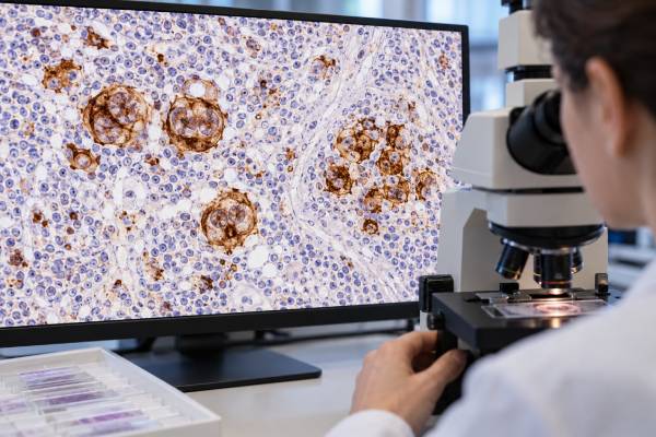
A CD30 test looks for CD30 protein on abnormal lymphoid cells and is especially important in the diagnosis of classic Hodgkin lymphoma and anaplastic large cell lymphoma. CD30 can also be expressed in other T-cell and B-cell lymphomas and in activated, noncancerous immune cells, so a positive stain is not a cancer diagnosis by itself. In tissue, pathologists usually evaluate CD30 by immunohistochemistry and pay attention to which cells stain, how strongly they stain, and whether staining is diffuse or limited to a subset. In classic Hodgkin lymphoma, the large Hodgkin/Reed-Sternberg cells are typically CD30-positive, often with strong membrane and Golgi-region staining. In anaplastic large cell lymphoma, tumor cells usually show strong CD30 expression as well. CD30 also matters therapeutically because it is the target of brentuximab vedotin and is being studied with other CD30-directed approaches. The result therefore has to be interpreted with morphology, the complete immunophenotype, disease subtype, and treatment setting.
- Strong CD30 staining supports certain lymphoma diagnoses but is not specific to one disease. Classic Hodgkin lymphoma and anaplastic large cell lymphoma are key examples.
- There is no universal “normal CD30 level.” Pathology reports describe the percentage, intensity, distribution, and cellular pattern of staining rather than a blood reference range.
- Reactive immune cells can be CD30-positive. Infection, immune activation, and inflammatory conditions can produce scattered positive cells.
- CD30 expression may guide targeted therapy. Its treatment significance depends on the lymphoma subtype, approved indication, prior therapy, and overall clinical context.
- A negative or low result does not automatically exclude lymphoma. Sampling, tumor heterogeneity, technical factors, and disease subtype can affect expression.
Table of Contents
- What CD30 Is and Why It Is Tested
- How CD30 Testing Is Done
- What Positive CD30 Staining Means
- CD30 in Hodgkin Lymphoma
- CD30 in T-Cell Lymphomas
- CD30 and Targeted Treatment
- Limitations and Follow-Up
What CD30 Is and Why It Is Tested
CD30 is an activation-associated cell-surface receptor that becomes strongly expressed in several important lymphoma types. It belongs to the tumor necrosis factor receptor family and is also known as TNFRSF8. In healthy immune biology, CD30 can appear on activated T cells and B cells. In lymphoma, abnormal cells may express it persistently or at much higher levels.
The marker is useful because the pattern of CD30 expression can narrow a lymphoma differential diagnosis. Pathologists may order CD30 when evaluating a lymph node, skin biopsy, bone marrow sample, or other tissue that contains large atypical lymphoid cells. It is particularly important when classic Hodgkin lymphoma, anaplastic large cell lymphoma (ALCL), peripheral T-cell lymphoma, or a CD30-positive cutaneous lymphoproliferative disorder is being considered.
CD30 is not a lineage marker in the same way that CD3 identifies T-cell differentiation or CD20 supports mature B-cell differentiation. Instead, it is an activation and disease-associated marker that can occur across lineages. That is why its meaning changes depending on the surrounding phenotype.
For example, a large CD30-positive cell in classic Hodgkin lymphoma commonly has a different marker profile from a large CD30-positive cell in ALCL. Likewise, scattered CD30-positive immunoblasts in a reactive lymph node do not mean lymphoma. The cell shape, location, density, and accompanying markers all matter.
A broader lymphoma immunophenotyping panel can help classify many lymphoid neoplasms, although classic Hodgkin lymphoma is often diagnosed primarily from tissue morphology and immunohistochemistry rather than from flow cytometry alone.
How CD30 Testing Is Done
CD30 is most commonly tested by immunohistochemistry on a tissue biopsy. A laboratory applies an antibody against CD30 to a fixed tissue section. When the antibody binds, a color-producing detection system shows which cells contain the antigen.
The pathologist does not simply count all brown-staining cells. The central task is identifying whether the atypical tumor cells are CD30-positive. A reactive lymph node may contain occasional activated CD30-positive cells, while a lymphoma may contain a distinct abnormal population with a characteristic pattern.
A pathology report may describe:
- strong, moderate, or weak staining;
- membranous staining, often with a prominent paranuclear or Golgi pattern;
- diffuse versus focal expression;
- the estimated percentage of tumor cells that are positive; and
- heterogeneity, meaning some tumor areas or cells stain more than others.
Flow cytometry can detect CD30 in some specimens, but it has limitations. Large Hodgkin/Reed-Sternberg cells may be rare, fragile, and surrounded by abundant reactive cells, so conventional flow can miss them. Tissue immunohistochemistry is therefore central in suspected Hodgkin lymphoma. Flow cytometry may be more useful in selected non-Hodgkin lymphomas when viable tumor cells are present in sufficient numbers.
There is usually no special patient preparation for CD30 staining itself. Preparation depends on the procedure used to obtain the sample. Excisional lymph node biopsy often provides the most architecture for lymphoma diagnosis, while core needle biopsy may be used when surgery is not practical. Fine-needle aspiration alone is frequently insufficient for a new lymphoma diagnosis because architecture and a broad panel may be needed.
What Positive CD30 Staining Means
A CD30-positive result means the tested abnormal cells express CD30, but the diagnosis depends on the complete pattern. Strong staining can be highly characteristic in the right setting, yet CD30 is not exclusive to malignant cells.
| Pattern | Possible interpretation | Important context |
|---|---|---|
| Strong CD30 on Hodgkin/Reed-Sternberg-like cells | Supports classic Hodgkin lymphoma | Must fit morphology and the rest of the immunophenotype |
| Strong, often diffuse CD30 on large anaplastic cells | Supports ALCL in the appropriate setting | ALK, T-cell markers, cytotoxic markers, and clinical site help classify it |
| Variable CD30 on peripheral T-cell lymphoma cells | Can occur in several PTCL subtypes | Percentage and distribution may be heterogeneous |
| Scattered CD30-positive immunoblasts | May be reactive immune activation | Common in inflammatory or infectious settings |
| CD30 on some large B-cell lymphoma cells | Possible CD30-positive B-cell lymphoma | B-cell markers, genetics, morphology, and EBV studies may be needed |
There is no single threshold that defines “CD30-positive lymphoma” for every diagnostic and therapeutic purpose. Some diseases are defined by a characteristic strong expression pattern, while other lymphomas may show expression in only part of the tumor. Different studies and treatment settings have also used different percentage cutoffs.
This is one reason a report that says “CD30 positive in 15% of tumor cells” should not be interpreted from the number alone. The meaning depends on the lymphoma subtype and why the test was ordered. For diagnostic classification, morphology and the total panel are crucial. For treatment, the relevant drug indication and supporting evidence matter.
Why the percentage can be difficult to compare
A percentage such as 10%, 50%, or 90% sounds precise, but CD30 immunohistochemistry is not a simple automated blood measurement. Pathologists first have to identify which cells are actually neoplastic. In a biopsy rich in reactive cells, counting every CD30-positive cell would be misleading. The denominator may be tumor cells rather than all nucleated cells, and that distinction should be clear in the report.
Expression can also differ within the same mass. One area may contain strongly positive tumor cells while another has weaker or absent staining. A small core biopsy may therefore underrepresent the full range of expression. Staining intensity can vary with fixation and laboratory platform as well. For these reasons, serial percentages from different laboratories should be compared cautiously.
When a treatment decision depends on CD30, clinicians consider the actual disease subtype and the evidence supporting that therapy, not just whether a number crosses an arbitrary threshold. In some settings, a repeat stain on a recent specimen or expert pathology review can be more informative than trying to force an old result into a universal cutoff.
CD30 in Hodgkin Lymphoma
Strong CD30 expression on Hodgkin/Reed-Sternberg cells is a hallmark of classic Hodgkin lymphoma. In classic disease, the malignant cells can be surprisingly sparse. Much of the biopsy may consist of reactive T cells, B cells, eosinophils, macrophages, plasma cells, and stromal elements surrounding a small population of large neoplastic cells.
The classic immunophenotype is interpreted as a package. Hodgkin/Reed-Sternberg cells are generally strongly CD30-positive. CD15 is often positive but can be absent. PAX5 is typically weak compared with normal B cells, reflecting the B-cell origin of the tumor despite loss of many conventional B-cell markers. CD20 can be negative or variably positive. Other stains may be added to exclude mimics and to evaluate Epstein-Barr virus association.
Because the malignant cells are rare, a negative flow cytometry result does not exclude classic Hodgkin lymphoma. The architecture and targeted tissue stains carry greater weight. A related CD20 result may be part of the panel, but its variable expression in classic Hodgkin lymphoma illustrates why no single B-cell marker settles the diagnosis.
CD30 positivity also does not distinguish every Hodgkin-like process from other lymphomas. ALCL, EBV-associated lymphoproliferative disorders, some diffuse large B-cell lymphomas, mediastinal lymphomas, and reactive conditions can contain large CD30-positive cells. Experienced hematopathology review is therefore important when the morphology is unusual or the immunophenotype is incomplete.
In treatment, CD30 has moved beyond being a diagnostic stain because the protein provides a target for antibody-drug conjugate therapy. That makes an accurate diagnosis and expression assessment clinically relevant, although classic Hodgkin lymphoma treatment decisions are based on much more than a single staining percentage.
CD30 in T-Cell Lymphomas
CD30 is central to ALCL and can also be expressed across a range of peripheral and cutaneous T-cell lymphomas. In systemic ALCL, tumor cells typically have strong CD30 expression. The disease is then separated into molecular and clinical categories, including ALK-positive and ALK-negative forms.
An ALK test for anaplastic large cell lymphoma helps with this classification. ALK-positive ALCL is associated with ALK rearrangements and often occurs in younger patients, while ALK-negative ALCL is biologically distinct and requires additional evaluation. Both can be strongly CD30-positive.
Other peripheral T-cell lymphomas may express CD30 at lower or more variable levels. This includes subsets of peripheral T-cell lymphoma, not otherwise specified, and other entities. Cutaneous T-cell disorders also include primary cutaneous CD30-positive lymphoproliferative diseases, such as lymphomatoid papulosis and primary cutaneous ALCL. Clinical distribution and skin-specific pathology are critical because these conditions can behave very differently from systemic lymphoma.
CD30 can also appear during ordinary T-cell activation. Therefore, a small number of positive T cells in an inflamed tissue does not establish a T-cell neoplasm. Evidence of an abnormal T-cell population may include loss of expected T-cell antigens, an unusual CD4/CD8 pattern, cytotoxic marker expression, T-cell receptor clonality, or a characteristic tissue pattern.
The amount of CD30 expression in non-ALCL peripheral T-cell lymphoma can be difficult to standardize. Preanalytic tissue handling, antibody platform, staining intensity, intratumoral heterogeneity, and the pathologist’s denominator for counting cells can all affect the reported percentage. This is why expert consensus emphasizes consistent testing, interpretation, and reporting practices.
CD30 and Targeted Treatment
CD30 is a therapeutic target because antibody-based drugs can bind the antigen and deliver treatment selectively to CD30-expressing cells. Brentuximab vedotin is the best-established example. It combines an anti-CD30 antibody with a microtubule-disrupting drug payload.
Its role varies by lymphoma type and treatment setting. It is used in defined settings for classic Hodgkin lymphoma, systemic ALCL, and other CD30-expressing lymphomas. Treatment selection also depends on whether the disease is newly diagnosed or relapsed, prior therapy, transplant status, patient fitness, and the regimen with which brentuximab is combined.
A frequent question is whether a specific percentage of CD30-positive tumor cells guarantees benefit. The answer is no. CD30 immunohistochemistry is semiquantitative, expression can vary within and between lesions, and treatment responses have been reported across a range of expression levels in some lymphoma types. Some mechanisms of antibody-drug conjugate activity may also affect neighboring tumor cells.
At the same time, clearly absent antigen can matter, and the testing question should match the treatment indication. It is not appropriate to translate a cutoff from one lymphoma subtype or clinical trial to every other disease.
After CD30-directed therapy, antigen expression may change. If disease relapses and another CD30-directed strategy is being considered, repeat biopsy may sometimes be useful, especially when the relapse pattern is unusual or the original sample is old.
CD30-directed CAR-T cells, bispecific approaches, and other immune therapies remain active areas of study. These emerging treatments reinforce the value of understanding antigen density and heterogeneity, but they do not replace the need for accurate lymphoma classification.
Limitations and Follow-Up
The main limitation of CD30 testing is lack of absolute specificity: both reactive cells and several different cancers can be positive. Technical factors can also create uncertainty.
Potential problems include small biopsies, crushed tissue, necrosis, delayed fixation, variable antibody platforms, and tumor heterogeneity. A core biopsy that samples one area of a mass may show a different percentage from another area. A repeat biopsy can be appropriate when the pathology does not fit the clinical behavior or when transformation or a new diagnosis is suspected.
When CD30 is positive, the next steps usually focus on defining the disease rather than repeating the same stain. Depending on the case, evaluation may include:
- a broader immunohistochemical panel;
- flow cytometry when viable cells are available and the disease is suitable for it;
- ALK testing in suspected ALCL;
- B-cell or T-cell clonality studies;
- EBV testing when clinically relevant;
- FISH or sequencing for subtype-defining abnormalities; and
- imaging and bone marrow assessment for staging when lymphoma is confirmed.
When CD30 is negative despite a strong clinical suspicion for a CD30-defined lymphoma, pathologists review whether tumor cells were actually present in the section, whether staining controls worked, and whether an alternative diagnosis fits better. Repeating the stain or obtaining more tissue may be reasonable in selected cases.
CD30 positivity is not itself an emergency. Urgent care is driven by the clinical consequences of the underlying disease, such as airway compression, spinal cord compression, severe cytopenias, major bleeding, infection, or rapidly worsening systemic symptoms. Most patients benefit most from a clear explanation of what the stain contributes to the diagnosis and whether it changes treatment options.
CD30 is also not usually used as a stand-alone blood marker to monitor whether lymphoma is shrinking. Follow-up relies on the disease-specific response method, such as clinical examination, blood counts, PET/CT or CT imaging when appropriate, and repeat biopsy when the response is uncertain. Soluble CD30 has been studied in research settings, but it is not interchangeable with tissue CD30 immunohistochemistry and is not the routine way most lymphoma response decisions are made. This distinction helps avoid a common misconception: a tissue marker can be crucial for diagnosis and targeted treatment even when it is not a serial serum tumor marker. In practice, the most useful follow-up test is the one validated for the specific lymphoma and treatment setting.
References
- CD30 as a Target Molecule in the Diagnosis and Therapy of Lymphomas 2026 (Review)
- Challenges of CD30 expression and its impact on targeted treatment responses in non-Hodgkin lymphoma: New perspectives for evaluation and validation. 2025 (Review)
- Antibody-Drug Conjugates Targeting CD30 in T-Cell Lymphomas: Clinical Progression and Mechanism 2025 (Review)
- Anti-CD30 antibody-drug conjugate therapy in lymphoma: current knowledge, remaining controversies, and future perspectives 2023 (Review)
- Best Practices in CD30 Immunohistochemistry Testing, Interpretation, and Reporting: An Expert Panel Consensus 2023 (Position Statement)
Disclaimer
This article is for general education and does not diagnose lymphoma or determine treatment eligibility. CD30 results require interpretation by qualified pathology and hematology professionals using the tissue morphology, full immunophenotype, clinical setting, and treatment history. Seek urgent medical care for breathing difficulty, severe bleeding, high fever with immune suppression, new neurologic deficits, or other rapidly worsening symptoms.





