
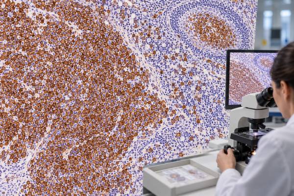
A CD20 test checks whether cells express CD20, a surface protein found on most mature B lymphocytes. In lymphoma diagnosis, CD20 is commonly assessed by immunohistochemistry on tissue and by flow cytometry on blood, bone marrow, or other specimens. Positive staining supports mature B-cell differentiation and is common in many B-cell lymphomas, including follicular lymphoma, mantle cell lymphoma, diffuse large B-cell lymphoma, and other mature B-cell neoplasms. CD20 is also a major treatment target for monoclonal antibodies such as rituximab and for newer CD20-directed bispecific antibodies, so expression can have therapeutic relevance. However, CD20 is not present on every B-cell malignancy and can be weak, heterogeneous, or lost after targeted therapy. A positive result does not identify a specific lymphoma subtype, and a negative result does not automatically exclude B-cell lineage. Pathologists interpret CD20 together with morphology, CD19, PAX5, immunoglobulin light chains, disease-specific markers, genetics, and the patient’s treatment history.
- CD20-positive staining supports mature B-cell lineage, but it does not by itself diagnose a specific B-cell lymphoma.
- CD20 is usually absent from very early B-cell precursors and is often lost as B cells become plasma cells, so some B-lineage cancers are naturally CD20-negative.
- Flow cytometry and immunohistochemistry measure CD20 differently, and reports may describe intensity, percentage, or distribution rather than a universal normal range.
- CD20 expression matters for CD20-targeted treatments, but eligibility and expected benefit depend on the specific disease, drug, and overall clinical setting.
- CD20 can decrease or disappear after anti-CD20 therapy, so repeat biopsy or immunophenotyping can be important when lymphoma relapses.
Table of Contents
- What CD20 is and where it is found
- Why CD20 is tested in B-cell lymphoma
- How CD20 testing is performed
- What positive CD20 staining means
- What negative, dim, or heterogeneous CD20 means
- CD20 as a treatment target and resistance marker
- How CD20 fits with other B-cell markers
What CD20 is and where it is found
CD20 is a membrane protein encoded by the MS4A1 gene. It appears as B cells mature beyond their earliest precursor stages and remains on most mature B lymphocytes. Expression generally falls as B cells terminally differentiate into plasma cells. This developmental pattern makes CD20 a useful marker of mature B-cell identity.
Normal circulating B cells are CD20-positive. Therefore, simply detecting CD20 does not mean cancer is present. The diagnostic question is whether an abnormal population of cells expresses CD20 in a pattern that fits a clonal B-cell neoplasm. Flow cytometry and tissue pathology provide the context needed to make that distinction.
Many mature B-cell lymphomas are strongly CD20-positive. These include most follicular lymphomas, mantle cell lymphomas, marginal zone lymphomas, and diffuse large B-cell lymphomas. CLL/SLL is usually CD20-positive as well, although staining is often relatively dim compared with normal B cells or other lymphomas.
Some B-lineage malignancies are naturally weak or negative for CD20. Precursor B-ALL may lack CD20 or express it only on a subset of blasts because the cells are developmentally immature. Plasma-cell myeloma usually shows loss of conventional mature B-cell markers, including CD20 in many cases, although a subset can retain it. Certain aggressive B-cell lymphomas with plasmablastic differentiation are characteristically CD20-negative or weak.
This is why CD20 should be viewed as a highly useful mature B-cell marker rather than a universal B-cell marker. CD19 testing covers an earlier and broader part of B-cell development, while PAX5 and other markers can help establish B-lineage when CD20 is absent.
Why CD20 is tested in B-cell lymphoma
CD20 is tested for two major reasons: classification and treatment relevance. In a lymph-node biopsy or other tissue, strong membranous CD20 staining can quickly show that a population is composed of mature B cells. Pathologists then add markers that distinguish among lymphoma subtypes.
For example, follicular lymphoma often combines CD20 with CD10 and BCL6 in a germinal-center pattern. Mantle cell lymphoma typically combines CD20 with CD5 and cyclin D1 or a CCND1 rearrangement. CLL/SLL often shows CD20 with CD5 and CD23, although the CD20 intensity is usually dim. Diffuse large B-cell lymphoma is frequently strongly CD20-positive but requires morphology and a broader immunohistochemical panel for classification.
In blood and bone marrow, CD20 contributes to a lymphoma flow cytometry panel. The test identifies an abnormal B-cell population through a combination of CD19, CD20, CD5, CD10, CD23, CD38, CD45, kappa and lambda light chains, and other markers. Clonality and the pattern across markers are far more informative than CD20 alone.
The second reason is therapeutic targeting. Anti-CD20 monoclonal antibodies have become foundational treatments for many B-cell lymphomas. Rituximab, obinutuzumab, and related agents bind CD20 on the malignant B-cell surface and recruit immune mechanisms that kill the targeted cells. CD20 is also the B-cell target for several bispecific antibodies that engage T cells through CD3.
Because the antigen can be changed by prior treatment, a current CD20 result can be more useful than an old report when a patient has relapsed after one or more CD20-directed regimens. Rebiopsy may reveal that the lymphoma remains strongly positive, has become heterogeneous, or has lost expression.
How CD20 testing is performed
Immunohistochemistry, or IHC, is the standard method on formalin-fixed tissue. The pathologist applies an antibody to a tissue section and looks for membranous staining in the cells of interest. The advantage is that staining can be interpreted in the context of tissue architecture: the pathologist can see whether CD20 highlights follicles, sheets of large cells, a diffuse small-cell infiltrate, or only scattered normal B cells.
Flow cytometry is used when viable cells are available from blood, bone marrow, lymph node, body fluid, or another specimen. Fluorescent antibodies measure CD20 and many other surface proteins simultaneously. The report may call expression dim, moderate, bright, partial, or heterogeneous and may give the percentage of cells in a defined gate that are positive.
These methods are related but not numerically interchangeable. A tissue IHC report saying “diffuse strong CD20 expression” cannot be converted into a flow-cytometry percentage. Antibody clones, tissue fixation, gating strategy, and disease distribution all affect the result.
A further difference is that IHC preserves location. A pathologist can tell whether the stained cells are the neoplastic cells, residual normal follicles, or a background population. Flow cytometry loses tissue architecture but can distinguish cell populations with many markers at once and detect small clones with high analytical sensitivity. When the two methods appear discordant, reviewing which cells were assessed often resolves the discrepancy.
There is also no single normal tumor cutoff for CD20. Laboratories validate their own staining and analytical thresholds. In diagnostic pathology, the important question is usually whether the abnormal cells show convincing membranous expression and how that expression compares with internal controls.
Internal control cells can be useful in tissue. Residual normal B lymphocytes may stain strongly even when the lymphoma is CD20-negative, showing that the antibody reaction worked. Conversely, a technically poor specimen can produce weak staining across all cells, which should not be mistaken for biologic antigen loss.
Prior therapy is one of the most important pieces of information to provide to the laboratory. Anti-CD20 antibodies can interfere with antigen detection or select for cells that have reduced expression. A pathologist who knows the treatment history can choose complementary markers and avoid overinterpreting a negative stain.
What positive CD20 staining means
Positive CD20 staining means that the tested cells have detectable CD20 protein. In an abnormal lymphoid population, that finding supports mature B-cell differentiation. It does not determine the lymphoma subtype by itself because many different B-cell neoplasms share CD20 expression.
The pattern and intensity can still add useful information. Strong, uniform staining is common in many mature B-cell lymphomas. Relatively dim CD20 on a CD5-positive clonal population can support CLL/SLL when the rest of the phenotype fits. Heterogeneous staining may reflect biologic variation within the tumor, prior treatment, or technical factors.
A high percentage of CD20-positive cells is not a direct measure of tumor aggressiveness. A lymphoma with nearly all cells positive is not necessarily more advanced or more dangerous than one with partial expression. Stage, histologic subtype, proliferation rate, genetic abnormalities, tumor burden, symptoms, and treatment response determine clinical risk.
Positive CD20 can support use of a CD20-targeted therapy, but the laboratory result is only one part of treatment selection. Some drug approvals and guideline recommendations are based on a disease category in which CD20 expression is expected, not on a single universal percentage threshold. The prior treatment history and the intensity of current antigen expression can become more important in selected relapsed settings.
The presence of CD20 on normal B cells also means that positive staining outside the tumor is expected. In a reactive lymph node, normal follicles can be rich in CD20-positive cells without any lymphoma. Pathologists decide whether those B cells are normally organized and polyclonal or form an abnormal clonal population.
The same principle applies in marrow. A small number of normal CD20-positive B cells can persist during or after treatment and should not automatically be called residual lymphoma. Flow cytometry compares light-chain expression and the complete antigen pattern, while tissue examination assesses distribution and morphology. This distinction is especially important when a report gives a very small percentage of positive cells.
In other words, “CD20 positive” answers a lineage and target-expression question. It does not answer the complete diagnostic, stage, or prognosis question.
What negative, dim, or heterogeneous CD20 means
A CD20-negative result can be biologically real. Some B-cell neoplasms lose mature B-cell markers as they differentiate toward a plasma-cell phenotype. Plasmablastic lymphoma, primary effusion lymphoma, ALK-positive large B-cell lymphoma, and some other uncommon aggressive B-cell neoplasms can be CD20-negative. These diagnoses rely on morphology, plasma-cell markers, viral studies when relevant, and other lineage evidence.
CD20 can also be weak in CLL/SLL or variable in precursor B-ALL. Therefore, negative or dim CD20 does not mean a malignant cell is not B-lineage. CD19, PAX5, CD79a, immunoglobulin gene studies, and other markers can establish B-cell differentiation when CD20 is absent.
After anti-CD20 treatment, loss of expression has a different significance. Therapy can select pre-existing CD20-low clones, drive changes in MS4A1 expression, produce truncating or other mutations, or cause surface-antigen modulation. The relapse may remain the same B-cell lymphoma genetically even though the therapeutic target is no longer detectable.
Heterogeneous expression is particularly important because an average positive percentage can hide a meaningful CD20-negative subpopulation. Pathologists may describe whether staining is strong in most cells, patchy, weak, or absent in specific tumor areas. In a relapsed tumor, that detail may be more useful than a simple positive/negative label.
A negative stain can also be technical. Fixation, decalcification of bone marrow, low viability, antibody clone, recent therapeutic antibody exposure, and very small tumor samples can all affect detection. Internal controls and complementary B-cell markers help distinguish technical failure from true antigen loss.
When CD20 negativity is unexpected, the appropriate response is not to force the original diagnosis to fit. The pathologist reviews the morphology, prior specimen, other B-lineage markers, treatment history, and sometimes molecular findings. In selected cases, a repeat or better-quality specimen is needed.
CD20 as a treatment target and resistance marker
CD20 became one of the most successful targets in lymphoma therapy because it is abundant on many mature malignant B cells and is not shed extensively into the circulation. Rituximab and later anti-CD20 antibodies transformed treatment of numerous B-cell lymphomas. More recently, CD20 × CD3 bispecific antibodies have used the same target to recruit T cells directly against the lymphoma.
Targeted therapy creates selection pressure. If a tumor contains a small population of cells with little or no CD20, those cells may survive while strongly positive cells are eliminated. The surviving population can then dominate at relapse. Tumor cells can also acquire genetic or transcriptional changes that reduce antigen expression.
A 2024 analysis of patients treated with the CD20 × CD3 bispecific antibody mosunetuzumab illustrates the issue. Most pretreatment biopsies had high CD20 expression, but in paired samples taken at progression, CD20 loss was observed in a substantial subset. Genetic changes and reduced MS4A1 transcription explained many of the cases. The study also showed why repeat IHC can be clinically useful at progression.
The exact frequency of CD20 loss varies by disease, therapy, specimen timing, and assay. It should not be assumed that every relapse after rituximab or a bispecific antibody is CD20-negative. Many remain positive and can still be considered for CD20-directed strategies depending on the clinical situation.
Target loss is only one resistance mechanism. Lymphoma can progress despite preserved CD20 because of immune escape, inadequate effector-cell function, tumor microenvironment effects, downstream survival pathways, or other genetic changes. Therefore, a positive CD20 result after progression does not prove that the previous therapy should work again.
When treatment selection depends on target availability, a recent biopsy can answer a more relevant question than the original diagnostic specimen from years earlier.
How CD20 fits with other B-cell markers
B-cell lymphoma diagnosis is built from patterns. CD20 is highly informative when combined with markers that define lineage, maturation stage, and subtype. CD19 and PAX5 support B-cell identity; CD5 and CD23 help distinguish CLL/SLL from other small B-cell neoplasms; CD10 and BCL6 identify germinal-center differentiation; cyclin D1 and SOX11 support mantle cell lymphoma; and plasma-cell markers such as CD138 become important when CD20 is lost.
Clonality is another key layer. In flow cytometry, an abnormal mature B-cell population often shows restriction to kappa or lambda light chain. A normal reactive B-cell population contains a mixture. Light-chain restriction is not interpreted alone, but it helps show that CD20-positive cells belong to a clone rather than normal immune tissue.
Tissue architecture can be equally important. Follicular lymphoma forms abnormal follicles, while diffuse large B-cell lymphoma grows in sheets of large cells. Flow cytometry cannot show architecture, and IHC cannot always provide the same multidimensional surface-marker detail as flow. The methods complement each other.
Treatment history should always be part of the interpretation. A patient whose original lymphoma was strongly CD20-positive may have a relapsed sample that is negative after years of anti-CD20 exposure. The loss can be real and clinically relevant. Conversely, a negative flow result in a low-cellularity specimen should not overturn a convincing tissue IHC pattern without reviewing sample quality.
Useful questions for a pathology report include: Are the abnormal cells uniformly or only partially CD20-positive? Was the test performed by IHC, flow cytometry, or both? Which markers confirm B-cell lineage if CD20 is weak or absent? Has expression changed since the prior biopsy? Is a current CD20-directed therapy being considered?
These questions place the marker in its proper role. CD20 is one of the most useful proteins in B-cell lymphoma diagnosis and treatment, but its meaning comes from the company it keeps—other markers, morphology, genetics, and clinical history.
References
– CD20-Negative Large B-Cell Lymphomas: The Diagnostic Challenge of Tumors with Downregulation of Mature B-Cell Marker Expression. 2025 – Loss of CD20 expression as a mechanism of resistance to mosunetuzumab in relapsed/refractory B-cell lymphomas 2024 – Application of flow cytometry immunophenotypic analysis for the diagnosis of mature B-cell lymphomas/leukemias. 2025 – Mature B- and plasma-cell flow cytometric analysis: A review of the impact of targeted therapy. 2022 – Flow Cytometry for B-Cell Non-Hodgkin and Hodgkin Lymphomas 2025
Disclaimer
This article is for general education and does not replace interpretation by a hematopathologist or treatment advice from a lymphoma specialist. CD20 staining can be affected by disease subtype, assay method, specimen quality, and prior CD20-directed therapy, and a positive or negative result should never be interpreted alone. Treatment decisions require the complete pathology and clinical picture.





