
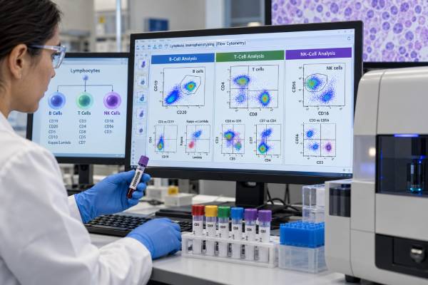
A lymphoma flow cytometry panel looks for abnormal lymphoid cells and characterizes them as B cells, T cells, NK cells, plasma cells, or precursor cells based on their antigen pattern. It is particularly useful for detecting clonal mature B-cell populations, abnormal T-cell populations, and certain leukemic or fluid-based presentations of lymphoma. The test can rapidly narrow the diagnosis, but it usually cannot classify lymphoma by itself because modern lymphoma diagnosis integrates morphology, tissue architecture, immunohistochemistry, cytogenetics, molecular findings, and clinical features.
Flow cytometry works best when fresh viable cells are available from a lymph node aspirate, core-biopsy rinse, blood, bone marrow, cerebrospinal fluid, pleural fluid, or another specimen. The laboratory measures markers such as CD19, CD20, CD5, CD10, CD23, CD38, CD45, surface immunoglobulin light chains, CD2, CD3, CD4, CD7, CD8, CD16, CD56, and newer clonality markers such as TRBC1. The resulting immunophenotype can distinguish many common patterns, flag important differential diagnoses, and guide the next tissue or molecular tests.
- What it measures: The panel measures combinations of lymphoid markers on individual cells to identify lineage, maturation, clonality, and abnormal antigen patterns.
- What a clonal B-cell result means: Marked kappa or lambda light-chain restriction supports a clonal B-cell population, but the final lymphoma type depends on the full phenotype and clinical-pathologic context.
- What an abnormal T-cell result means: Loss, dimming, or unusual combinations of T-cell markers can support a T-cell neoplasm, but reactive T cells can also look atypical, so clonality and tissue findings may be needed.
- NK-cell interpretation: NK cells usually lack surface CD3 and may express CD16 and/or CD56; separating reactive NK-cell expansion from NK-cell neoplasia requires clinical, viral, morphologic, and molecular correlation.
- Preparation: Fasting is not usually needed. Fresh specimen handling and adequate viable-cell recovery are more important than patient preparation.
Table of Contents
- What lymphoma flow cytometry can detect
- B-cell markers and clonality
- T-cell markers and abnormal patterns
- NK-cell markers and interpretation
- Common lymphoma immunophenotype patterns
- Specimens, procedure, and reporting
- Limitations and next tests
What lymphoma flow cytometry can detect
Flow cytometry is designed to answer whether a specimen contains an abnormal lymphoid population and, if so, what biological features that population shows. It does this by measuring multiple fluorescent antibody signals on thousands or millions of cells. Cells with similar size, complexity, and antigen expression form clusters that can be compared with expected normal B-, T-, and NK-cell populations.
The technique is especially strong for mature B-cell neoplasms because B cells normally express immunoglobulin light chains. A population in which nearly all B cells use only kappa or only lambda can indicate clonality. Flow cytometry also recognizes characteristic combinations such as CD5-positive or CD10-positive clonal B cells, which can sharply narrow the differential diagnosis.
For T-cell and NK-cell disorders, the task is more complex because there is no simple equivalent of kappa/lambda restriction for routine screening. Laboratories look for aberrant loss, dimming, overexpression, or unusual combinations of T-cell markers and may use T-cell receptor–based methods such as TRBC1 or TCR-Vβ analysis to support clonality.
The test can help with:
- initial diagnosis of a suspected lymphoproliferative disorder;
- classification of a mature B-cell clone;
- detection of blood or marrow involvement by lymphoma;
- evaluation of lymph node aspirates and tissue suspensions;
- analysis of cerebrospinal, pleural, peritoneal, or other body fluids; and
- targeted follow-up when the prior immunophenotype is known.
A broader hematologic cancer biomarker panel may incorporate genetic or molecular findings that flow cytometry cannot provide. When a lymphoid neoplasm has circulating cells, flow can also overlap conceptually with a leukemia flow cytometry panel, because the same instruments and many of the same markers are used.
B-cell markers and clonality
For mature B-cell lymphoma, the key flow questions are whether the B cells are clonal and which marker pattern best fits the clone. Common B-cell markers include CD19, CD20, CD22, CD79b, and surface immunoglobulin. Laboratories then add markers such as CD5, CD10, CD23, CD25, CD38, CD43, CD103, CD200, FMC7, and others depending on the specimen and differential diagnosis.
Kappa and lambda light-chain restriction
Normal mature B cells contain a mixture of kappa- and lambda-expressing cells. A strongly skewed population, especially when it also has an abnormal antigen pattern, supports clonality. This is called light-chain restriction.
Light-chain interpretation is not a stand-alone cancer diagnosis. Small populations may be difficult to assess, some neoplasms express weak or no surface immunoglobulin, and technical factors can interfere. Monoclonal B-cell lymphocytosis can also produce a clonal blood population without meeting criteria for lymphoma or leukemia requiring treatment.
CD5-positive B-cell populations
A clonal B-cell population that coexpresses CD5 raises a differential that includes chronic lymphocytic leukemia/small lymphocytic lymphoma (CLL/SLL) and mantle cell lymphoma, among other less common possibilities. CLL/SLL often shows CD23 and CD200 with dim surface immunoglobulin and relatively dim CD20, whereas mantle cell lymphoma more often has brighter B-cell markers and usually requires confirmation with cyclin D1 and/or CCND1 rearrangement studies.
A cyclin D1 test for mantle cell lymphoma can therefore resolve a question that flow raises but cannot settle by itself. The same principle applies to CCND1/IGH fusion testing when molecular confirmation is needed.
CD10-positive B-cell populations
CD10 expression can point toward germinal-center-derived B-cell lymphomas, including follicular lymphoma and Burkitt lymphoma, but diffuse large B-cell lymphoma and other entities may also express CD10. Tissue architecture, cell size, BCL2/BCL6 expression, MYC status, and genetic findings become important in this setting.
T-cell markers and abnormal patterns
T-cell flow cytometry relies on pattern recognition. Normal T cells express surface CD3 and usually show orderly distributions of CD2, CD5, CD7, CD4, and CD8. A neoplastic population may lose one or more expected markers, express a marker at abnormal intensity, show an unusual CD4/CD8 pattern, or form a population that is too immunophenotypically uniform for the clinical setting.
Examples of suspicious findings include:
- loss of CD5 or CD7 in a discrete T-cell population;
- unusually bright or dim CD3;
- a markedly expanded CD4-positive or CD8-positive subset with additional aberrancies;
- abnormal coexpression or absence of CD4 and CD8;
- a dominant TCR subtype supported by TRBC1 or TCR-Vβ analysis; and
- expression patterns inconsistent with normal maturation.
The major caution is that reactive T cells can also look unusual. Viral infections, autoimmune disease, drug reactions, immune recovery, and aging can produce skewed or phenotypically atypical T-cell subsets. Loss of CD7 alone, for example, is not enough to diagnose lymphoma.
TRBC1 is increasingly useful because normal αβ T cells contain a mixture of TRBC1-positive and TRBC1-negative cells, whereas a clonal αβ T-cell population may show restriction. The assay has limitations: it does not solve every T-cell clonality question, and interpretation changes in γδ T-cell populations or certain unusual phenotypes.
Tissue-based markers remain essential for many T-cell lymphomas. A CD3 immunohistochemistry test evaluates lineage in tissue architecture, while a CD30 test may be relevant in anaplastic large cell lymphoma and other CD30-positive lymphoid disorders.
NK-cell markers and interpretation
NK cells are lymphocytes of the innate immune system. They typically lack surface CD3 and may express CD16, CD56, CD7, CD2, CD94, and killer-cell receptors. Normal NK cells are heterogeneous, so a suspicious population usually shows both numerical expansion and an abnormal, relatively uniform phenotype.
The distinction between T-cell and NK-cell disease can be challenging because some neoplastic T cells lose surface CD3 and acquire NK-associated markers. Conversely, NK-cell neoplasms can express markers shared with T cells. Cytoplasmic CD3-related staining, surface T-cell receptor expression, TRBC1, TCR gene rearrangement studies, and killer-cell receptor patterns can help establish lineage.
Clinical context is particularly important for NK-cell neoplasms. Some aggressive NK/T-cell disorders are strongly associated with Epstein-Barr virus (EBV), and the diagnosis may require EBV testing in addition to tissue morphology and immunophenotyping. Flow cytometry alone cannot determine whether an NK-cell expansion represents a transient reactive process, an indolent lymphoproliferation, or an aggressive lymphoma.
A result described as “abnormal NK-cell population” therefore needs a next-step plan rather than immediate subtype labeling. The hematopathologist may recommend tissue biopsy, EBV-encoded RNA testing, molecular studies, or correlation with organ involvement and symptoms.
Common lymphoma immunophenotype patterns
Flow cytometry often works as a diagnostic sorting tool. It places a specimen into a smaller set of possibilities and helps select confirmatory tests.
| Flow pattern | Possible interpretation | Common confirmatory context |
|---|---|---|
| CD5+, CD23+, dim CD20, dim surface light chain | CLL/SLL-like phenotype | Blood count, morphology, clinical criteria, and prognostic testing |
| CD5+, CD23 variable, brighter B-cell markers | Mantle cell lymphoma in differential | Cyclin D1/SOX11 and CCND1 rearrangement studies |
| CD10+ clonal B cells | Germinal-center B-cell lymphoma in differential | Tissue architecture, BCL2/BCL6, MYC studies as indicated |
| CD103+, CD25+, CD11c+, CD123+ mature B cells | Hairy cell leukemia pattern | Morphology and BRAF testing |
| Aberrant mature T cells with marker loss | T-cell lymphoproliferative disorder possible | T-cell clonality, tissue pathology, clinical correlation |
| Surface CD3−, CD56+/CD16+ abnormal lymphocytes | NK-lineage process possible | EBV studies, molecular workup, tissue evaluation |
A germinal-center phenotype may lead to FISH testing when high-grade B-cell lymphoma is suspected. A double-hit lymphoma FISH panel evaluates rearrangements that cannot be inferred reliably from flow alone. Likewise, a MYC rearrangement test may be necessary when morphology and phenotype raise concern for Burkitt lymphoma or another aggressive B-cell lymphoma.
Importantly, Hodgkin lymphoma is often not diagnosed by routine flow cytometry because Hodgkin/Reed-Sternberg cells can be rare, fragile, and embedded in a rich reactive background. Specialized flow approaches exist in some laboratories, but excisional or core tissue pathology remains central to diagnosis.
Specimens, procedure, and reporting
The quality of lymphoma flow cytometry depends heavily on the specimen. Fresh tissue is mechanically dispersed into a cell suspension; blood and marrow can be analyzed directly after preparation; body fluids are concentrated when necessary. Delays, low cellularity, necrosis, or prior fixation can reduce the number of viable cells and limit sensitivity.
The laboratory usually starts with a screening strategy that identifies B-, T-, and NK-cell populations. If an abnormal population appears, additional tubes or antibody combinations may be added to refine the phenotype. Some laboratories use broad standardized panels; others tailor testing to the clinical question and the initial findings.
The report may state:
- specimen type and adequacy;
- percentage of B cells, T cells, NK cells, and other populations;
- whether B cells show kappa/lambda restriction;
- whether T cells have antigen loss or an abnormal subset distribution;
- the phenotype of the abnormal population; and
- a differential diagnosis or recommended ancillary testing.
Percentages need context. A clone that is 5% of all analyzed cells may be important in one specimen and incidental in another. Flow also does not preserve tissue architecture, so it cannot determine whether cells grow in follicles, diffuse sheets, sinuses, or another pattern. Those architectural features are often defining in lymphoma classification.
Limitations and next tests
A negative flow result does not rule out lymphoma. Sampling error, necrosis, treatment effects, low tumor burden, fragile cells, or a disease that is poorly represented in suspension can all produce a negative study. Large-cell lymphomas can be especially challenging because large abnormal cells may be lost during processing.
A positive result also has limits. Clonality does not automatically equal clinically significant lymphoma. Small clonal B-cell populations may occur in monoclonal B-cell lymphocytosis, and reactive immune conditions can produce striking T-cell skewing.
The next tests depend on the pattern. They may include:
- histology and immunohistochemistry on core or excisional tissue;
- FISH for CCND1, BCL2, BCL6, MYC, or other rearrangements;
- chromosome analysis in selected settings;
- targeted mutation testing or next-generation sequencing;
- immunoglobulin or T-cell receptor gene rearrangement studies;
- EBV testing for appropriate T/NK-cell entities; and
- imaging and bone marrow evaluation for staging.
The modern WHO and International Consensus classifications define lymphoma entities by integrated evidence. A flow phenotype may be highly characteristic, but the most accurate diagnosis comes from matching the cell phenotype to morphology, genetics, anatomic site, and clinical presentation.
When reviewing your report, ask three questions first: Was an abnormal population found? What lineage and phenotype does it have? What test is required to confirm the subtype? Those questions turn a long list of CD markers into a practical diagnostic plan.
Why tissue type changes the value of flow cytometry
Flow cytometry works best when the specimen contains enough viable cells that can be separated into suspension. Fresh lymph-node tissue, blood, marrow, pleural fluid, and cerebrospinal fluid can all be tested, but they answer different questions. Blood is excellent for a circulating clone yet may be normal in a lymphoma confined to a lymph node. A needle sample can show a convincing clonal population but still provide too little tissue to assess architecture. An excisional or well-targeted core biopsy may therefore be needed even after an abnormal flow result.
The handling interval also matters. Cells can die during transport, and some large lymphoma cells are especially fragile. A report that mentions low viability, low cellularity, or hemodilution is signaling a real sensitivity limitation rather than a minor technical note. When lymphoma remains strongly suspected, a negative low-quality sample should not end the workup.
Patterns that are informative but not disease-specific
Certain phenotypes narrow the differential diagnosis without proving a single entity. CD5-positive clonal B cells can occur in chronic lymphocytic leukemia/small lymphocytic lymphoma, mantle cell lymphoma, and less common B-cell neoplasms. CD10-positive clonal B cells can fit follicular lymphoma, Burkitt lymphoma, or other germinal-center-derived lymphomas. Loss of one or more pan-T-cell markers can support an abnormal T-cell population, but reactive T-cell expansions can also look skewed.
This is why pathologists interpret combinations rather than isolated markers. Light-chain restriction, marker intensity, cell size, aberrant antigen loss, and the relationship between several populations all contribute. The final classification may then require immunohistochemistry, FISH, molecular testing, or tissue architecture to distinguish entities that overlap by flow.
Flow cytometry and immunohistochemistry answer different questions
Flow cytometry measures many markers on individual viable cells and is excellent for identifying small abnormal populations or light-chain restriction. Immunohistochemistry examines markers in intact tissue and preserves where the cells sit relative to follicles, sinuses, vessels, and supporting structures. In lymphoma, that architecture can be diagnostic. A common workflow therefore uses flow for rapid lineage and clonality information while histology and immunohistochemistry establish the tissue-based classification. Neither method should be considered a universal replacement for the other.
A second practical point is that “no monotypic B-cell population” and “no abnormal T-cell population” are assay conclusions, not universal exclusions. Hodgkin lymphoma and some T-cell lymphomas may contain relatively few malignant cells within a large reactive background. If imaging, symptoms, or tissue morphology remain concerning, pathology review continues even when flow is unrevealing.
References
- Application of flow cytometry immunophenotypic analysis for the diagnosis of mature B-cell lymphomas/leukemias 2024 (Review)
- TRBC1 in flow cytometry: Assay development, validation, and reporting considerations 2024 (Review)
- Fifth Edition of the World Health Organization Classification of Tumors of the Hematopoietic and Lymphoid Tissues: Mature T-Cell, NK-Cell, and Stroma-Derived Neoplasms of Lymphoid Tissues 2024 (Review)
- The 5th edition of the World Health Organization Classification of Haematolymphoid Tumours: Lymphoid Neoplasms 2022 (Review)
- The International Consensus Classification of Mature Lymphoid Neoplasms: a report from the Clinical Advisory Committee 2022 (Review)
- Mature B, T and NK-cell, plasma cell and histiocytic/dendritic cell neoplasms: classification according to the World Health Organization and International Consensus Classification 2024 (Review)
Disclaimer
This article provides general educational information and cannot diagnose lymphoma or interpret an individual flow cytometry result. Lymphoma classification requires review by qualified clinicians and pathologists using the complete specimen, clinical history, morphology, and relevant genetic studies. Seek prompt medical care for rapidly enlarging nodes, severe shortness of breath, significant bleeding, high fever, fainting, or other rapidly worsening symptoms.





