
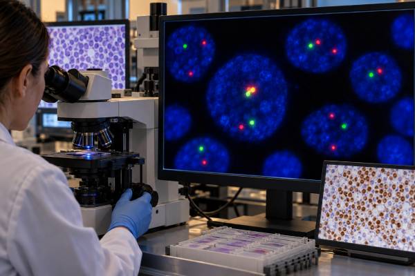
A CCND1/IGH fusion test looks for the genetic rearrangement created by t(11;14)(q13;q32), the hallmark chromosome abnormality in most mantle cell lymphomas (MCL). The translocation places CCND1, the gene encoding cyclin D1, next to regulatory elements of the immunoglobulin heavy-chain gene, IGH. This drives abnormal cyclin D1 expression and helps the lymphoma cells progress through the cell cycle. Fluorescence in situ hybridization (FISH) is a common way to detect the rearrangement in blood, bone marrow, lymph node, or other involved tissue. A positive result strongly supports MCL when the cell morphology and immunophenotype fit, but it is not completely specific: t(11;14) can also occur in plasma-cell neoplasms. Likewise, a negative CCND1/IGH test does not exclude every case of MCL because rare cyclin D1-negative mantle cell lymphomas use alternative cyclin D2 or D3 abnormalities. The final diagnosis therefore combines FISH with pathology, cyclin D1 and SOX11 staining, flow cytometry, and the overall clinical picture.
- A positive CCND1/IGH fusion usually means t(11;14)(q13;q32) is present and strongly supports mantle cell lymphoma in the right pathologic setting.
- The fusion causes cyclin D1 overexpression, which is why cyclin D1 immunostaining and CCND1/IGH FISH often reinforce each other.
- FISH results are usually reported as positive or negative with a percentage of abnormal nuclei and a laboratory-specific cutoff.
- A negative result does not completely exclude MCL because rare cyclin D1-negative cases can have CCND2 or CCND3 rearrangements.
- t(11;14) is not unique to MCL and is also found in a subset of plasma-cell myelomas, so lineage and morphology must be established before interpreting the fusion.
Table of Contents
- What the CCND1/IGH fusion is
- Why the test is used in mantle cell lymphoma
- How CCND1/IGH FISH testing works
- What a positive FISH result means
- What a negative or atypical result means
- How cyclin D1, SOX11, and flow cytometry fit
- How the result affects classification and care
What the CCND1/IGH fusion is
CCND1/IGH is a gene fusion produced by a reciprocal translocation between chromosome 11 and chromosome 14. The cytogenetic notation is t(11;14)(q13;q32). CCND1 sits at 11q13, while the IGH locus is at 14q32. When the rearrangement brings CCND1 under the influence of strong immunoglobulin regulatory elements, cyclin D1 is produced at abnormally high levels.
Cyclin D1 helps normal cells move from the G1 phase into the S phase of the cell cycle. Persistent overexpression removes an important brake on proliferation. In MCL, this abnormal cell-cycle drive is a foundational event, although additional genetic changes are required for the full lymphoma phenotype and influence how aggressive the disease becomes.
Most conventional mantle cell lymphomas carry this rearrangement. That is why t(11;14) and cyclin D1 have become signature findings in the diagnostic workup. The terms “CCND1/IGH fusion,” “IGH::CCND1 rearrangement,” “CCND1-IGH,” and “t(11;14)” may all appear on reports and generally refer to the same core event.
The abnormality is acquired in the B-cell clone; it is not ordinarily an inherited genetic change. A positive result does not imply that family members carry the translocation or need testing.
The fusion also explains why a cyclin D1 IHC test is so useful in tissue. FISH demonstrates the DNA rearrangement, while immunohistochemistry demonstrates the resulting protein overexpression. Concordant findings provide strong, biologically linked evidence.
Why the test is used in mantle cell lymphoma
Mantle cell lymphoma is a mature B-cell neoplasm that can resemble several other small B-cell lymphomas. Typical cells are small to medium sized with irregular nuclear contours, and the immunophenotype usually includes B-cell markers, CD5, and cyclin D1. However, morphology and marker patterns overlap enough that a defining genetic test can be extremely helpful.
CCND1/IGH FISH is ordered when MCL is suspected from a lymph node biopsy, bone marrow specimen, peripheral blood, or another involved site. It may be especially useful when cyclin D1 staining is weak or technically difficult, the tissue sample is small, or the immunophenotype is not classic.
The main differential diagnosis often includes chronic lymphocytic leukemia/small lymphocytic lymphoma, other CD5-positive B-cell lymphomas, and occasionally other mature B-cell neoplasms. MCL commonly has brighter surface immunoglobulin and a different marker profile from CLL/SLL, but no one flow marker is sufficient in every case. A targeted genetic finding can resolve uncertainty.
FISH may also be performed as part of a broader lymphoma panel. This is useful because MCL can acquire secondary chromosome changes that affect disease biology, including alterations involving TP53 and other loci. Still, the primary diagnostic question is usually whether the CCND1 rearrangement is present.
Testing is not used to screen healthy people. Nor is the fusion test a general marker of lymphoma burden. Its primary role is classification. Once MCL is diagnosed, response is usually assessed through clinical findings, imaging, blood or marrow evaluation when appropriate, and increasingly sensitive molecular methods in selected settings rather than by repeatedly measuring a simple CCND1/IGH FISH percentage.
How CCND1/IGH FISH testing works
FISH uses fluorescently labeled DNA probes that bind to specific chromosome regions. A dual-fusion CCND1/IGH assay places one probe around CCND1 and another around IGH. In a normal cell, the fluorescent signals are separate. When the loci are brought together by t(11;14), overlapping or paired fusion signals appear in a characteristic pattern.
The laboratory examines a defined number of nuclei and counts how many show the abnormal signal pattern. The result is compared with a validated cutoff derived from normal control specimens. Because occasional signal overlap can happen by chance, the cutoff is not zero. A result above the laboratory’s threshold is called positive.
Some laboratories use break-apart probes rather than a dual-fusion design. A CCND1 break-apart probe detects disruption of the CCND1 locus regardless of its partner, while an IGH break-apart assay detects rearrangement of IGH without identifying the partner gene. These designs answer slightly different questions. The report should state which probe set was used.
FISH can be performed on fresh cells or formalin-fixed paraffin-embedded tissue, depending on the laboratory. In tissue, a pathologist may mark tumor-rich areas so the assay evaluates the relevant cells. In bone marrow or blood, enrichment is sometimes used for other diseases, but MCL can often be evaluated directly when enough lymphoma cells are present.
| Test | What it shows | Main diagnostic value |
|---|---|---|
| CCND1/IGH FISH | t(11;14) or related targeted rearrangement | Genetic confirmation of the hallmark MCL event |
| Cyclin D1 IHC | Cyclin D1 protein expression in tissue | Rapidly supports classic MCL in morphologic context |
| SOX11 IHC | Nuclear SOX11 expression | Helpful in cyclin D1-negative cases and selected diagnostic dilemmas |
| Flow cytometry | B-cell lineage and surface-marker pattern | Identifies a clonal B-cell population and helps distinguish mimics |
FISH does not show cell morphology and cannot determine whether the positive cells are plasma cells, lymphoma cells, or another lineage unless the specimen and accompanying studies establish that identity. This limitation is central to interpreting t(11;14).
What a positive FISH result means
A positive CCND1/IGH FISH result means that the laboratory detected the targeted fusion pattern above its validated cutoff. In a biopsy showing a mature CD5-positive B-cell lymphoma with cyclin D1 expression, this is powerful confirmation of mantle cell lymphoma.
The reported percentage of positive nuclei is not a universal severity scale. A result of 70% does not necessarily mean a lymphoma is more aggressive than one with 40%. The number can be influenced by tumor purity, specimen type, sectioning artifacts, and how cells were selected. The fusion’s diagnostic presence matters more than treating the percentage as a prognostic score.
If the report includes a laboratory cutoff, that cutoff is analytical rather than prognostic. For example, a lab may require more than a small background percentage of abnormal signal patterns before calling the assay positive. Readers should compare the measured percentage with that specific cutoff, not with a cutoff from another laboratory or publication.
A positive result also does not establish stage. MCL stage depends on the distribution of disease in lymph nodes, spleen, marrow, blood, gastrointestinal tract, and other sites. Nor does t(11;14) by itself determine whether the course will be indolent or aggressive. Proliferation rate, morphology, TP53 status, clinical risk scores, disease distribution, and other features contribute to prognosis.
The most important caveat is that t(11;14) also occurs in plasma-cell neoplasms. In multiple myeloma, the same CCND1/IGH event is a recognized recurrent cytogenetic subtype. Therefore, finding t(11;14) in a marrow containing abnormal plasma cells should not be interpreted as mantle cell lymphoma without evidence of a clonal B-cell lymphoma.
Context usually resolves this quickly. Plasma-cell myeloma has plasma-cell morphology and markers such as CD138, often with monoclonal light-chain restriction and an appropriate clinical protein profile. MCL has a mature B-cell phenotype and characteristic tissue architecture. A lymphoma flow cytometry panel can help establish the lineage of a circulating or marrow population.
Positive CCND1 rearrangement is therefore best understood as a highly characteristic genetic clue whose meaning depends on which abnormal cells carry it.
What a negative or atypical result means
A negative CCND1/IGH FISH result means the assay did not detect the targeted rearrangement above its cutoff. In many diagnostic settings, that makes conventional cyclin D1-positive MCL less likely. It does not, however, eliminate every form of mantle cell lymphoma.
Rare MCL cases lack cyclin D1 overexpression and do not carry the typical CCND1 rearrangement. These cyclin D1-negative tumors can have rearrangements involving CCND2 or CCND3 instead. They can still share the broader molecular and immunophenotypic features of MCL. SOX11 staining is particularly useful in this setting because many cyclin D1-negative MCLs retain strong nuclear SOX11 expression.
A negative result can also be technical. The specimen may contain too few tumor cells, the tissue may be degraded, or the breakpoint may not produce the expected signal configuration for a particular probe design. Laboratories usually report whether the study was technically adequate and how many nuclei were evaluable.
An “atypical” FISH pattern requires specialist interpretation. Extra fusion signals can reflect additional copies of chromosomes or complex rearrangements. Loss of a signal may reflect a deletion. These findings can coexist with the primary translocation and may point to a more complex genome, but they are not interpreted by simply counting colors.
If MCL remains strongly suspected despite negative CCND1/IGH testing, pathologists typically revisit morphology, cyclin D1 and SOX11 staining, the full immunophenotype, and broader molecular or FISH studies. The goal is to avoid both errors: dismissing a genuine cyclin D1-negative MCL and forcing an MCL diagnosis onto a different CD5-positive B-cell neoplasm.
A negative test in a well-characterized non-MCL lymphoma can be reassuring, but no single negative marker substitutes for an integrated classification.
How cyclin D1, SOX11, and flow cytometry fit
Cyclin D1 immunohistochemistry is one of the fastest ways to recognize classic MCL. Most cases show nuclear cyclin D1 positivity in the lymphoma cells. Because the CCND1/IGH translocation directly drives this protein, a positive FISH result and positive cyclin D1 stain are usually biologically concordant.
SOX11 is a transcription factor that is expressed in most conventional MCLs, including many cyclin D1-negative cases. A SOX11 test for mantle cell lymphoma can therefore support the diagnosis when cyclin D1 or CCND1 findings are unusual. However, SOX11 is not perfectly specific and should not be interpreted without the rest of the pathology.
Flow cytometry typically identifies a monoclonal B-cell population expressing CD19, CD20, surface light chain, and often CD5. CD23 is often absent or dim compared with typical CLL/SLL, but marker expression varies. Immunophenotyping is particularly useful in blood and marrow because it shows which lineage carries the abnormal population, something FISH alone cannot demonstrate.
Pathologists also consider morphology. Classic MCL is composed of small to medium lymphoid cells with irregular nuclei, but blastoid and pleomorphic variants have larger, more aggressive-appearing cells and generally worse clinical behavior. Ki-67 proliferation index helps quantify how actively the lymphoma cells are dividing.
The combined pattern is much stronger than any single test. For example, a CD5-positive clonal B-cell population plus cyclin D1 positivity plus CCND1/IGH fusion is a highly coherent MCL profile. By contrast, a t(11;14)-positive plasma-cell population with CD138 expression and an M protein points toward plasma-cell myeloma, not MCL.
This integrated approach also protects against technical pitfalls. Weak cyclin D1 staining can be clarified by FISH; an unusual FISH pattern can be checked against IHC; and a small sample can be supported by flow cytometry if viable cells are available.
How the result affects classification and care
The major clinical value of CCND1/IGH testing is diagnostic certainty. Correctly classifying MCL matters because its expected course, staging workup, and treatment options differ from those of CLL/SLL, follicular lymphoma, marginal zone lymphoma, and plasma-cell neoplasms.
After diagnosis, clinicians determine disease extent and risk. Many patients have stage IV disease because marrow or blood involvement is common, but advanced anatomic stage does not carry exactly the same meaning in lymphoma as it does in many solid tumors. Clinical behavior ranges from indolent forms that may initially be observed to aggressive blastoid disease requiring prompt treatment.
The Mantle Cell Lymphoma International Prognostic Index, performance status, age, lactate dehydrogenase, white blood cell count, proliferation index, TP53 abnormalities, and disease morphology can all influence risk assessment. The presence of the hallmark t(11;14) is essential for classification in most cases but is not, by itself, the variable that separates low-risk from high-risk MCL.
Treatment has evolved to include anti-CD20 antibodies, chemotherapy in selected patients, BTK inhibitors, cellular therapies, and other targeted approaches. The exact strategy depends on age, fitness, symptoms, disease tempo, prior therapy, TP53 status, and whether the disease is newly diagnosed or relapsed. A positive fusion test identifies the lymphoma type rather than directly choosing one drug.
If a report is difficult to understand, the most useful questions are: Was the assay a dual-fusion or break-apart FISH test? What percentage of nuclei was above the lab cutoff? Did the same cells show cyclin D1 and a B-cell phenotype? Was SOX11 tested? Is there any concern for a plasma-cell neoplasm or a cyclin D1-negative MCL variant? These questions connect the molecular result to the actual pathologic diagnosis.
Once the diagnosis is secure, repeating CCND1/IGH FISH is not usually the primary method for routine response monitoring. The fusion can remain the genetic identity of residual lymphoma cells, but surveillance is based on the clinical situation and more appropriate disease-assessment tools.
References
– Mantle cell lymphoma: from pathogenesis to treatment for 2024 and beyond. 2025 – Comprehensive FISH testing using FFPE tissue microarray of primary lymph node tissue identifies secondary cytogenetic abnormalities in Mantle Cell Lymphoma. 2023 – The 5th edition of the World Health Organization Classification of Haematolymphoid Tumours: Lymphoid Neoplasms 2022 – The International Consensus Classification of Mature Lymphoid Neoplasms: a report from the Clinical Advisory Committee 2022 – Mature B, T and NK-cell, plasma cell and histiocytic/dendritic cell neoplasms: classification according to the World Health Organization and International Consensus Classification 2024
Disclaimer
This article is for general education and is not a substitute for diagnosis by a hematopathologist or treatment advice from a lymphoma specialist. CCND1/IGH FISH must be interpreted with morphology, immunophenotyping, cyclin D1 and SOX11 results, and the type of abnormal cells in the specimen. A positive t(11;14) result alone should not be used to distinguish mantle cell lymphoma from a plasma-cell neoplasm.





