
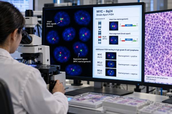
A MYC rearrangement test looks for a structural chromosome change that places the MYC gene under abnormal regulatory control, driving rapid cell growth. In lymphoma, the test is most important when Burkitt lymphoma or an aggressive large/high-grade B-cell lymphoma is suspected. It is usually performed with fluorescence in situ hybridization (FISH), often using a MYC break-apart probe, and may be paired with BCL2 and BCL6 FISH to identify clinically important combinations of rearrangements.
A positive MYC result does not have one universal meaning. Burkitt lymphoma typically has a MYC rearrangement, commonly involving an immunoglobulin partner, but diagnosis still requires the characteristic morphology, germinal-center B-cell phenotype, and overall genetic context. In large B-cell lymphomas, MYC plus BCL2 rearrangements define a particularly important high-grade genetic category in current classifications. MYC protein overexpression by immunohistochemistry is a different finding and must not be confused with a MYC gene rearrangement.
- What it measures: MYC FISH detects structural rearrangement of the MYC locus, usually at chromosome 8q24.
- Burkitt lymphoma: MYC rearrangement is a hallmark, but a positive result alone does not establish Burkitt lymphoma.
- Double-hit meaning: Concurrent MYC and BCL2 rearrangements identify a high-risk genetic lymphoma category; classification of MYC/BCL6 cases differs between WHO and ICC systems.
- Double-expressor is different: High MYC and BCL2 protein expression by immunohistochemistry does not prove gene rearrangements.
- Specimen: Testing is usually performed on lymphoma tissue or another sample containing enough tumor cells; no patient fasting is required.
Table of Contents
- What a MYC rearrangement means
- How MYC FISH is performed
- MYC in Burkitt lymphoma
- Double-hit and high-grade lymphoma
- MYC protein versus MYC gene rearrangement
- How to read a MYC test result
- Limitations and next tests
What a MYC rearrangement means
MYC is a transcription factor that regulates cell growth, metabolism, proliferation, and other fundamental programs. Normal cells tightly control MYC activity. A rearrangement can place MYC next to a powerful enhancer, causing inappropriate expression and accelerating proliferation.
In Burkitt lymphoma, MYC is most often rearranged with an immunoglobulin gene. The classic translocation is t(8;14), involving IGH on chromosome 14, but MYC can also partner with immunoglobulin light-chain loci.
In other B-cell lymphomas, MYC may rearrange with immunoglobulin or non-immunoglobulin partners. The biological and prognostic effect depends on the lymphoma type and accompanying genetic abnormalities.
This is why “MYC rearrangement positive” is not a complete diagnosis. The same basic molecular event can occur in different disease entities.
A lymphoma flow cytometry panel may identify a clonal germinal-center B-cell population, but flow does not show the chromosome architecture that FISH detects. A BCL6 rearrangement test may also be ordered when the morphology and phenotype suggest an aggressive B-cell lymphoma.
How MYC FISH is performed
FISH uses fluorescent DNA probes that bind specific chromosome regions inside tumor-cell nuclei. A common MYC break-apart assay places differently colored probes on either side of the MYC locus. In a normal chromosome, the signals remain together or nearly together. If a rearrangement splits the locus, the colors separate beyond the laboratory’s validated distance threshold.
The laboratory counts a defined number of tumor-cell nuclei and compares the proportion with its assay cutoff. The report may say “MYC rearrangement detected” and give the percentage of abnormal nuclei.
Break-apart FISH answers whether MYC is rearranged but usually does not identify the partner gene. If knowing the partner is important, laboratories may use dual-fusion probes, targeted sequencing, or other molecular methods.
What tissue can be tested?
FISH can often be performed on formalin-fixed paraffin-embedded biopsy tissue. Fresh tissue, bone marrow, or cytology preparations may also be suitable if they contain sufficient tumor. The pathologist selects a tumor-rich area when possible because low tumor content can reduce sensitivity.
No fasting or medication change is generally needed for the FISH assay itself. The main practical issue is obtaining adequate diagnostic tissue without exhausting the sample on other studies.
MYC in Burkitt lymphoma
Burkitt lymphoma is a very aggressive mature B-cell lymphoma with a distinctive combination of morphology, phenotype, proliferation, and genetics. Typical tumor cells are medium-sized, the tissue often shows a “starry-sky” appearance, and the immunophenotype reflects germinal-center differentiation. Ki-67 proliferation is usually extremely high.
MYC rearrangement is a central genetic feature, usually with an immunoglobulin partner. However, MYC positivity does not automatically equal Burkitt lymphoma. Other aggressive B-cell lymphomas can also carry MYC rearrangements.
The pathologist therefore asks whether the entire case fits:
- morphology typical of Burkitt lymphoma;
- mature B-cell and germinal-center phenotype;
- very high proliferation;
- MYC rearrangement, preferably in an appropriate genetic context; and
- absence of findings that better define another lymphoma.
A general MYC rearrangement FISH interpretation focuses on the molecular result, while the final Burkitt diagnosis requires integrated pathology.
Current classifications also recognize lymphomas that can resemble Burkitt but lack the classic MYC pattern, such as high-grade B-cell lymphoma with 11q aberrations. This is another reason morphology plus one FISH result is not enough.
Double-hit and high-grade lymphoma
The term double-hit lymphoma historically described aggressive B-cell lymphomas with MYC rearrangement plus BCL2 and/or BCL6 rearrangement. Modern classifications are more precise and do not group all of these combinations identically.
Under the fifth edition WHO framework, diffuse large B-cell lymphoma/high-grade B-cell lymphoma with MYC and BCL2 rearrangements, with or without an additional BCL6 rearrangement, forms a distinct entity. Cases with MYC and BCL6 rearrangements but no BCL2 rearrangement are classified according to their morphology rather than included in the same MYC/BCL2 entity.
The International Consensus Classification is similar for MYC/BCL2 disease but recognizes MYC/BCL6-rearranged high-grade B-cell lymphoma as a provisional category. This difference can make wording in pathology reports look inconsistent even when the underlying FISH results are the same.
A MYC, BCL2, and BCL6 double-hit lymphoma FISH panel is designed to evaluate these combinations directly.
| Genetic pattern | General significance | Key caution |
|---|---|---|
| MYC rearrangement alone | Can occur in Burkitt lymphoma or other B-cell lymphomas | Not enough to define a disease entity alone |
| MYC + BCL2 rearrangements | Defines an important aggressive large/high-grade B-cell lymphoma category | Must integrate morphology and phenotype |
| MYC + BCL2 + BCL6 rearrangements | Often called triple-hit; generally falls within the MYC/BCL2-rearranged category | Exact report terminology may vary |
| MYC + BCL6, no BCL2 | Aggressive genetic pattern with classification differences between WHO and ICC | Not equivalent to MYC/BCL2 disease |
These rearrangements matter because MYC/BCL2-rearranged lymphomas generally behave more aggressively than morphologically similar lymphomas without the defining cytogenetic combination. That can influence treatment planning and risk discussions.
MYC protein versus MYC gene rearrangement
A common source of confusion is the difference between MYC immunohistochemistry (IHC) and MYC FISH.
IHC measures how much MYC protein is present in tumor-cell nuclei. FISH looks for a structural rearrangement of the MYC gene. A tumor can overexpress MYC protein without a detectable rearrangement, and a rearranged tumor may show variable protein levels.
The same distinction applies to BCL2. “Double-expressor lymphoma” usually refers to a large B-cell lymphoma with high MYC and BCL2 protein expression by IHC. “Double-hit lymphoma” refers to specific gene rearrangements detected by FISH or another genetic method.
These labels should not be used interchangeably.
A BCL2 IHC test measures protein expression, while a BCL2 rearrangement test evaluates the gene itself.
Why IHC is still useful
IHC is fast, preserves tissue architecture, and helps characterize the lymphoma. It can identify a double-expressor phenotype and guide the pathologist toward FISH testing. However, screening only by IHC can miss some rearranged cases, which is why many centers use broader FISH strategies in newly diagnosed large B-cell lymphomas according to local protocols.
How to read a MYC test result
A FISH report may include the specimen, probe type, percentage of abnormal nuclei, laboratory cutoff, and interpretation.
Common phrases include:
- “MYC rearrangement detected” — enough nuclei meet criteria for a rearrangement.
- “MYC rearrangement not detected” — no rearrangement was found above the assay cutoff.
- “Abnormal MYC signal pattern” — the locus shows copy-number or complex signal changes that may need further clarification.
- “Indeterminate” or “insufficient” — tissue quality, tumor content, signal quality, or cell number prevents a reliable conclusion.
A positive MYC result should immediately be read alongside the BCL2 and BCL6 results. If BCL2 is also rearranged, that combination has a different classification and prognostic meaning from isolated MYC rearrangement.
The pathology diagnosis should also state the morphology and immunophenotype. Ask whether the case is being classified as Burkitt lymphoma, DLBCL/HGBL with MYC and BCL2 rearrangements, DLBCL not otherwise specified, HGBL not otherwise specified, or another entity.
A high Ki-67 percentage supports rapid proliferation but is not a substitute for MYC testing. Similarly, LDH may be elevated in aggressive lymphoma but cannot identify a rearrangement.
Limitations and next tests
FISH is targeted. A break-apart probe can show that MYC is rearranged but usually cannot tell which gene is on the other side. A negative result also does not eliminate every mechanism of MYC activation. Copy-number gain, enhancer changes, cryptic rearrangements, and other molecular events may not be captured by a standard assay.
Technical limitations include poor fixation, decalcified bone marrow tissue, low tumor percentage, signal overlap, and borderline percentages near the cutoff.
When the diagnosis remains uncertain, next steps may include:
- BCL2 and BCL6 FISH;
- immunoglobulin-partner MYC probes;
- broader cytogenetic or sequencing studies;
- Ki-67, MYC, BCL2, BCL6, CD10, and other IHC markers;
- flow cytometry for lineage and clonality;
- EBV testing when clinically relevant; and
- expert hematopathology review of the entire biopsy.
The most useful interpretation is not “MYC positive equals bad.” Instead, ask: Is MYC rearranged? What is the partner if known? Are BCL2 or BCL6 also rearranged? What lymphoma entity does the complete pathology support? Those questions determine the actual clinical meaning.
Burkitt lymphoma requires more than a positive MYC FISH result
MYC rearrangement is a defining biologic feature of most Burkitt lymphomas, commonly involving an immunoglobulin partner, but MYC positivity alone does not diagnose Burkitt lymphoma. The pathologist also evaluates the highly characteristic morphology, a germinal-center B-cell immunophenotype, very high proliferative activity, and the broader genetic setting. Other aggressive B-cell lymphomas can carry MYC rearrangements and can look similar in a limited biopsy.
This distinction is clinically important because Burkitt lymphoma is treated with intensive regimens designed for its exceptionally rapid growth. A diagnosis based only on a MYC break-apart signal risks putting biologically different lymphomas into the wrong category. When morphology and FISH appear discordant, expert hematopathology review and additional genetic testing can be valuable.
What “double hit” means in current classification
Historically, the phrase double-hit lymphoma was used broadly for high-grade B-cell lymphomas with MYC plus BCL2 and/or BCL6 rearrangements. Modern WHO and International Consensus classifications have refined that concept. The most clearly recognized molecular category centers on mature B-cell lymphomas with rearrangements involving both MYC and BCL2. Cases with MYC and BCL6 rearrangements are not automatically placed in the same entity and require classification from morphology and the complete phenotype/genetic profile.
This is one reason older medical records and newer pathology reports may use different wording for apparently similar FISH combinations. The raw FISH results remain important, but the final diagnostic line should use the current classification framework applied by the laboratory.
MYC protein expression is not the same as MYC rearrangement
Immunohistochemistry can show high MYC protein expression, and many laboratories also assess BCL2 protein. A lymphoma expressing both proteins may be called a double-expressor phenotype. Double expression can carry prognostic information in some settings, but it is not synonymous with double-hit genetics.
FISH asks whether the gene locus is rearranged. Immunohistochemistry asks whether protein is expressed in the sampled tumor cells. A lymphoma can express MYC strongly without a MYC rearrangement, and a rearranged case may not fit a simple protein-expression threshold. These tests are complementary, not interchangeable.
Why partner genes and specimen quality can matter
A standard break-apart probe detects separation of sequences around MYC and is useful for many rearrangements, but it usually does not identify the partner gene. Additional dual-fusion probes or sequencing can clarify whether an immunoglobulin partner is involved when that information is diagnostically relevant. Conversely, some cryptic or complex rearrangements may be difficult for a single FISH design to detect.
Tumor percentage also affects interpretation. If only a small fraction of the tissue contains lymphoma, an otherwise adequate assay can miss the abnormal clone. Decalcification of bone specimens can damage nucleic acids and FISH signals. Borderline results near the laboratory cutoff may warrant review of the counted cells, alternative tissue, or another method rather than an overconfident positive or negative conclusion.
When pathologists order MYC, BCL2, and BCL6 FISH
Testing strategies differ among laboratories. Some perform FISH broadly on newly diagnosed large B-cell lymphomas, while others use morphology and immunophenotype to select cases most likely to benefit from testing. The reason for testing is not simply prognostic: identifying MYC with BCL2 rearrangement can change the formal disease classification and may affect treatment planning or clinical-trial eligibility.
Because aggressive B-cell lymphomas can progress quickly, tissue triage matters. A small biopsy may need to support histology, immunohistochemistry, flow cytometry, FISH, and molecular studies from limited material. Communicating the concern for Burkitt lymphoma or a high-grade B-cell lymphoma early can help the pathology laboratory preserve enough viable or fixed tissue for the essential studies.
A preliminary diagnosis may sometimes be issued before all FISH probes are complete. In that situation, clinicians should check the final amended pathology report because the MYC/BCL2 result can refine the final classification after treatment planning has already begun.
What a negative MYC rearrangement does not exclude
A negative FISH result rules out the rearrangements detectable by that assay in the tested specimen; it does not make an aggressive lymphoma biologically low risk. High-grade morphology, TP53 abnormalities, strong MYC protein expression, adverse clinical stage, or other molecular features can still matter. Likewise, some tumors activate MYC through mechanisms other than a conventional rearrangement.
The practical interpretation is therefore asymmetric: a positive rearrangement can be highly classification-relevant, while a negative rearrangement should be integrated with the rest of the pathology rather than treated as a reassurance test.
Clinical urgency should not be confused with diagnostic shortcuts. Burkitt lymphoma and other high-grade B-cell lymphomas can have very short doubling times, so staging and treatment planning often move quickly. Even so, the safest rapid pathway is coordinated testing—morphology, immunophenotype, MYC/BCL2 testing, and other necessary studies in parallel—rather than assuming that one marker supplies the complete diagnosis. If the patient is clinically unstable, treatment decisions may begin before every ancillary result is final, with the diagnosis refined as validated results return.
For patients, the key report fields are the final lymphoma name, whether MYC is rearranged, whether BCL2 is rearranged, whether BCL6 was tested, and whether any result is pending. Those details are more actionable than trying to interpret individual FISH signal diagrams without the pathology context.
References
- Burkitt lymphoma 2024 (Review)
- The high-grade B-cell lymphomas: double hit and more 2024 (Review)
- High-grade B-cell lymphomas: Double hit and non-double hit 2024 (Review)
- How I diagnose high-grade B-cell lymphoma 2024 (Review)
- The 5th edition of the World Health Organization Classification of Haematolymphoid Tumours: Lymphoid Neoplasms 2022 (Review)
- The International Consensus Classification of Mature Lymphoid Neoplasms: a report from the Clinical Advisory Committee 2022 (Review)
Disclaimer
This article is for general education and does not replace interpretation by a hematopathologist or lymphoma specialist. MYC rearrangement results must be integrated with morphology, immunophenotype, BCL2/BCL6 testing, and the clinical presentation. Treatment decisions should be based on the final integrated lymphoma diagnosis, not on a single FISH result.





