
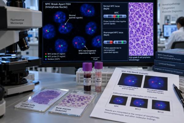
A MYC rearrangement test looks for a structural change involving the MYC gene, usually with fluorescence in situ hybridization (FISH). MYC is a powerful regulator of cell growth and metabolism. When a chromosome rearrangement places MYC next to an active regulatory region, the gene can become abnormally switched on and help drive an aggressive blood cancer. The result is especially important in Burkitt lymphoma and in large or high-grade B-cell lymphomas, where MYC is often tested together with BCL2 and BCL6. A MYC rearrangement can also occur in rare acute lymphoblastic leukemia/lymphoma cases and other hematologic neoplasms. A positive FISH result does not identify a diagnosis by itself, and it is not the same as high MYC protein expression. Pathologists interpret the finding alongside tumor appearance, immunophenotype, other chromosome changes, and clinical features. The combination of rearrangements—not MYC alone—often determines the final disease category and its treatment implications.
- A positive MYC FISH result means the laboratory detected a structural rearrangement involving MYC, usually at chromosome 8q24.
- MYC rearrangement alone does not automatically mean “double-hit lymphoma”; a second defining rearrangement, particularly BCL2, must also be demonstrated.
- High MYC protein on immunohistochemistry is not the same as a MYC gene rearrangement and cannot replace FISH when rearrangement status is required.
- FISH is commonly performed on lymphoma tissue or bone marrow, and blood may be usable when enough abnormal circulating cells are present.
- A negative result does not always exclude a cryptic or uncommon MYC rearrangement; additional molecular testing may be appropriate when morphology and other findings remain strongly suspicious.
Table of Contents
- What the MYC Rearrangement Test Detects
- When MYC Testing Is Used
- How MYC FISH Testing Works
- How to Read a MYC FISH Result
- MYC in High-Grade B-Cell Lymphoma
- MYC in Burkitt Lymphoma and Leukemia
- Next Steps After MYC Testing
What the MYC Rearrangement Test Detects
The test looks for a chromosomal rearrangement involving the MYC gene, located at 8q24. A rearrangement occurs when pieces of chromosomes break and rejoin in a new configuration. In many MYC-driven lymphomas, MYC is moved next to regulatory DNA from an immunoglobulin gene, such as IGH, IGK, or IGL. Those regulatory elements can push MYC activity to unusually high levels.
MYC is a transcription factor, meaning it helps control the expression of many genes. Normal cells use it for tightly regulated growth, metabolism, and proliferation. A rearrangement can uncouple MYC from normal control and give a malignant cell a strong growth advantage.
The test is therefore not measuring a blood concentration or a “MYC level.” It is detecting a genetic architecture change in cancer cells.
Rearrangement is different from mutation, amplification, and protein expression
Several MYC abnormalities can appear on a pathology report, and they are not interchangeable:
- MYC rearrangement: A structural chromosome change involving MYC. FISH is a standard method for detecting it.
- MYC copy-number gain or amplification: Extra copies of the gene are present. This is biologically different from a rearrangement.
- MYC mutation: A change within the DNA sequence of MYC. Sequence variants are not what a standard rearrangement FISH assay is designed to detect.
- MYC protein expression: Immunohistochemistry measures how many tumor cells show MYC protein staining. High expression can occur with or without a rearrangement.
For aggressive B-cell lymphoma, MYC testing often appears beside BCL2 rearrangement testing and BCL6 rearrangement testing. The combined pattern can be much more informative than any one result.
When MYC Testing Is Used
MYC rearrangement testing is most often ordered when a pathologist is evaluating a mature B-cell lymphoma with aggressive morphology or immunophenotype. It can also be used in selected leukemias and in cases where the distinction between Burkitt lymphoma, diffuse large B-cell lymphoma (DLBCL), and other high-grade B-cell lymphomas is difficult.
Common reasons for testing include:
- Newly diagnosed DLBCL or another large B-cell lymphoma in which current classification requires assessment for MYC and BCL2 rearrangements
- A tumor with “high-grade,” blastoid, or Burkitt-like microscopic features
- Strong MYC and BCL2 protein expression on immunohistochemistry, which may trigger genetic testing but does not itself prove rearrangement
- Suspected Burkitt lymphoma
- A lymphoma with unusually rapid growth, high proliferation, or extensive extranodal disease
- A B-cell acute lymphoblastic leukemia/lymphoma with features raising concern for a MYC-rearranged subtype
- A case needing clarification after an initial chromosome study, genomic panel, or RNA-sequencing result
Current hematopathology classifications place greater emphasis on genetic findings than older systems did. A broad hematologic cancer biomarker panel may combine morphology, flow cytometry, immunohistochemistry, FISH, karyotyping, and molecular sequencing because no single method answers every diagnostic question.
Why testing is often done broadly in large B-cell lymphoma
A MYC-rearranged tumor may not look dramatically different from a MYC-negative tumor under the microscope. Likewise, immunohistochemistry cannot reliably identify all rearranged cases. For that reason, many laboratories use a testing strategy that applies FISH to a broad group of newly diagnosed large B-cell lymphomas rather than selecting only those with a particular appearance.
The exact testing algorithm varies among institutions. Some laboratories begin with MYC and reflex to BCL2 or BCL6; others test all three at the same time. The goal is to avoid missing a genetically defined high-risk category that could change classification and treatment planning.
How MYC FISH Testing Works
FISH uses fluorescently labeled DNA probes that bind to defined chromosome regions. The signals are viewed under a fluorescence microscope or analyzed with validated digital systems. For MYC, the most common approach is a break-apart probe.
A break-apart probe places one fluorescent signal on one side of MYC and a differently colored signal on the other side. In a normal chromosome, the signals sit close together and appear fused or adjacent. When a rearrangement breaks the MYC region, the signals separate beyond the laboratory’s validated distance threshold.
Break-apart versus fusion probes
A break-apart assay answers, “Is MYC rearranged?” It generally does not identify the partner gene. A dual-fusion assay can test a specific partner, such as MYC::IGH. Partner identification may add biologic information, but it is not required in every case.
FISH can be performed on several specimen types:
- Formalin-fixed, paraffin-embedded lymph node or tumor tissue
- Bone marrow aspirate or clot material
- Fresh tissue cell suspensions
- Peripheral blood when circulating tumor cells are sufficiently abundant
- Cytology or body-fluid preparations in selected laboratories
The pathologist usually selects an area with adequate tumor content. If the specimen has very few malignant cells, extensive necrosis, poor fixation, or crush artifact, the test may fail or become harder to interpret.
What a laboratory cutoff means
FISH is not interpreted from one abnormal-looking cell. The laboratory counts a validated number of nuclei and compares the percentage with a predefined cutoff established during assay validation. A report may state, for example, that a certain percentage of analyzed nuclei showed a rearranged signal pattern and that this exceeds the laboratory’s positive threshold.
Cutoffs differ by probe design and laboratory. There is therefore no universal percentage that should be applied to every MYC FISH report.
FISH remains a major standard for rearrangement detection, although integrated DNA/RNA genomic profiling can detect many structural variants and may provide additional information. RNA-based fusion testing can be particularly helpful when a cryptic rearrangement is suspected.
Turnaround time and specimen limitations
MYC FISH is often completed within several working days after the laboratory has a suitable specimen, but timing varies with tissue processing, batching, referral to an outside laboratory, and whether reflex BCL2/BCL6 studies are added. A report may be delayed if the pathologist first needs to confirm that enough viable tumor is present.
A technically unsuccessful assay is different from a negative assay. “Insufficient nuclei,” “hybridization failure,” or similar wording means the laboratory could not make a reliable call. In that situation, a different tissue block, bone marrow sample, or another testing method may be used. Decalcified bone specimens can be particularly challenging because some decalcification methods damage DNA and reduce FISH quality.
The percentage of abnormal nuclei also should not be treated as a direct measure of tumor burden. It depends on tumor purity, the area selected for scoring, nuclear truncation in tissue sections, and the specific probe pattern. A higher FISH percentage does not automatically mean a more aggressive lymphoma.
How to Read a MYC FISH Result
A MYC report is best read in layers: result, assay type, percentage of abnormal cells, companion rearrangements, and final pathology diagnosis.
| Report wording | What it usually means | What it does not prove |
|---|---|---|
| MYC rearrangement detected | A structural alteration involving MYC is present above the assay cutoff | It does not by itself define Burkitt lymphoma or double-hit lymphoma |
| MYC rearrangement not detected | No rearrangement pattern was identified by that assay | It cannot exclude every cryptic breakpoint or abnormality outside probe coverage |
| MYC copy-number gain/amplification | Extra MYC signals are present | It is not equivalent to a MYC rearrangement |
| MYC rearranged; BCL2 rearranged | A genetically important double-rearranged pattern is present | The final entity still requires compatible morphology and lineage |
| MYC protein high by IHC; FISH negative | MYC is highly expressed without a detectable MYC rearrangement | Protein expression does not create a FISH-positive result |
Positive does not mean inherited
MYC rearrangements in lymphoma and leukemia are generally somatic, meaning they developed in the cancer cells. A positive tumor FISH result usually does not imply that a person was born with the rearrangement or that relatives should receive predictive genetic testing for the same change.
Why a negative result can need follow-up
Break-apart FISH is highly useful but not perfect. Some rearrangements have unusual breakpoints, complex chromosome structures, or cryptic insertions that are difficult for a particular probe set to detect. If the pathologist sees a strong mismatch between the FISH result and the rest of the case, additional testing may include an alternative FISH probe, conventional cytogenetics, DNA-based structural-variant analysis, or RNA sequencing.
An “equivocal” or borderline result should be interpreted according to the laboratory’s criteria rather than treated as automatically positive.
MYC in High-Grade B-Cell Lymphoma
MYC status is especially important in aggressive large B-cell lymphomas because modern classifications changed how so-called double-hit cases are grouped.
Under the fifth edition of the World Health Organization classification, the key genetically defined entity is DLBCL/high-grade B-cell lymphoma with MYC and BCL2 rearrangements. The 2022 International Consensus Classification likewise separates MYC/BCL2 double-rearranged disease from other high-grade B-cell lymphomas.
Historically, MYC with either BCL2 or BCL6 rearrangement could be grouped together as “double-hit lymphoma.” That terminology is now too broad for precise classification. MYC/BCL6 double-rearranged cases are not simply placed in the same MYC/BCL2 category; they are classified according to their morphology and other features, often within DLBCL, not otherwise specified, or high-grade B-cell lymphoma, not otherwise specified.
A double-hit lymphoma FISH panel therefore provides more useful information than MYC alone when a large B-cell lymphoma is being classified.
Double-hit is not double-expressor
These terms sound similar but refer to different tests:
- Double-hit: Rearrangements involving MYC and BCL2 are demonstrated genetically; older usage sometimes also included MYC/BCL6.
- Triple-hit: MYC, BCL2, and BCL6 rearrangements are present. In current frameworks, cases with MYC and BCL2 remain within the MYC/BCL2-rearranged category even when BCL6 is also rearranged.
- Double-expressor: Immunohistochemistry shows high MYC and BCL2 protein expression. There may be no MYC or BCL2 rearrangement.
This distinction matters because protein-expression cutoffs and genetic rearrangement status capture different biology. A report that says “MYC positive” on immunohistochemistry should not be interpreted as “MYC rearranged” unless FISH or another validated structural-variant assay demonstrates the rearrangement.
MYC rearrangement can correlate with aggressive behavior, but prognosis depends heavily on the lymphoma subtype, the partner abnormalities, stage, patient factors, and treatment. A positive result should therefore be interpreted within the full diagnosis rather than used as a stand-alone prognosis score.
MYC in Burkitt Lymphoma and Leukemia
Burkitt lymphoma is the classic MYC-driven lymphoma. Most cases carry a MYC rearrangement, commonly involving an immunoglobulin partner. The genetic finding supports the diagnosis, but Burkitt lymphoma still requires a matching clinicopathologic pattern: characteristic cell morphology, mature B-cell phenotype, very high proliferation, and exclusion of important mimics.
A MYC rearrangement by itself is therefore not enough to call a tumor Burkitt lymphoma. MYC can also be rearranged in DLBCL and other aggressive B-cell neoplasms.
B-acute lymphoblastic leukemia/lymphoma with MYC rearrangement
Modern classifications and reviews recognize B-ALL/LBL with MYC rearrangement as a rare molecularly defined subtype. These tumors involve precursor B cells rather than mature B cells, so flow cytometry and immunophenotyping are essential. The distinction can be difficult when a high-grade mature B-cell lymphoma looks blastoid or when a leukemic presentation produces many circulating cells.
A leukemia flow cytometry panel helps establish whether the abnormal cells have a precursor phenotype, while FISH or sequencing identifies the rearrangement. Age, immunoglobulin expression, TdT status, morphology, and broader genetic findings may all contribute to classification.
MYC rearrangements are not a routine defining feature of most myeloid leukemias. If a MYC rearrangement appears in an unusual leukemia, the pathology team generally reassesses lineage and the complete cytogenetic context rather than assigning meaning from MYC alone.
Next Steps After MYC Testing
The most important next step is to place the MYC result into the final hematopathology diagnosis. The same positive FISH finding can mean different things in Burkitt lymphoma, DLBCL, high-grade B-cell lymphoma, or B-ALL.
After a positive result, clinicians and pathologists usually review:
- BCL2 and BCL6 rearrangement status. This determines whether the tumor fits a double- or triple-rearranged pattern and helps apply current classification.
- Morphology and immunophenotype. Cell size, growth pattern, CD10, BCL6, MUM1, MYC, BCL2, TdT, surface immunoglobulin, and other markers help establish lineage and subtype.
- Proliferation and disease extent. Ki-67, imaging, bone marrow findings, blood counts, lactate dehydrogenase, and clinical stage provide additional context.
- Other molecular results. Cytogenetics, sequencing, or RNA fusion analysis may refine the diagnosis or resolve a discordant FISH result.
- Treatment implications. The oncology team selects therapy based on the final disease entity, patient fitness, stage, and evidence supporting specific regimens—not simply on one FISH signal pattern.
If MYC FISH is negative but the specimen quality is poor or the tumor strongly resembles a MYC-driven neoplasm, the report may recommend repeat or complementary testing. Conversely, if FISH is positive but the morphology does not fit the expected entity, the positive result should not override the rest of the case.
For patients reading their report, three questions usually clarify the result quickly: Was MYC rearranged? Were BCL2 or BCL6 also rearranged? What final lymphoma or leukemia diagnosis did the pathologist assign? Those answers are more informative than the word “positive” alone.
References
- The 5th edition of the World Health Organization Classification of Haematolymphoid Tumours: Lymphoid Neoplasms 2022 (Position Statement)
- The International Consensus Classification of Mature Lymphoid Neoplasms: a report from the Clinical Advisory Committee 2022 (Position Statement)
- Diagnostic approaches and future directions in Burkitt lymphoma and high-grade B-cell lymphoma 2022 (Review)
- Integrated Genomic DNA/RNA Profiling vs Fluorescence in Situ Hybridization in the Detection of MYC and BCL2 (and BCL6) Rearrangements in Large B-Cell Lymphomas: Updates Amid the New WHO Classification of Lymphoid Neoplasms 2023
- Emerging molecular subtypes and therapies in acute lymphoblastic leukemia 2023 (Review)
Disclaimer
This article provides general information about MYC rearrangement testing and does not replace interpretation by a hematopathologist or oncology team. The significance of a MYC result depends on the specimen, assay, lymphoma or leukemia subtype, companion BCL2/BCL6 findings, and the complete clinical picture. Treatment decisions should be based on the final integrated diagnosis rather than a MYC FISH result alone.





