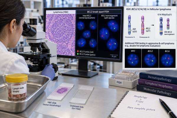
A BCL2 rearrangement test looks for a structural chromosome change involving the BCL2 gene, most often by fluorescence in situ hybridization (FISH). In conventional follicular lymphoma, the classic finding is t(14;18)(q32;q21), which creates an IGH::BCL2 rearrangement and drives excess BCL2 protein, helping abnormal B cells avoid programmed cell death. BCL2 testing is also important in aggressive large B-cell lymphomas because a BCL2 rearrangement combined with a MYC rearrangement can define a biologically high-risk “double-hit” lymphoma category. A positive result does not diagnose lymphoma by itself and should not be confused with BCL2 protein positivity on immunohistochemistry. The pathologist integrates FISH with tissue morphology, immunophenotype, MYC testing, BCL6 testing, and the clinical picture. A negative BCL2 FISH result also does not exclude follicular lymphoma, because a minority of follicular lymphomas lack the classic BCL2 rearrangement and may have different molecular features.
- A positive BCL2 rearrangement result means FISH or another validated molecular method detected a structural change involving BCL2.
- In conventional follicular lymphoma, the typical abnormality is IGH::BCL2 from t(14;18), but about 10%–15% of follicular lymphomas can lack BCL2 rearrangement.
- BCL2 rearrangement plus MYC rearrangement can define a double-hit large B-cell lymphoma; BCL2 protein overexpression alone does not.
- BCL2 FISH and BCL2 IHC measure different things: FISH detects a gene rearrangement, while IHC detects protein expression.
- No special patient preparation is needed because testing is usually performed on an existing lymph node, marrow, or other tissue specimen.
Table of Contents
- What BCL2 Rearrangement Means
- When BCL2 Rearrangement Testing Is Used
- How BCL2 FISH Testing Works
- How to Interpret a BCL2 FISH Result
- BCL2 in Follicular Lymphoma
- BCL2, MYC, and Double-Hit Lymphoma
- What Happens After BCL2 Testing
What BCL2 Rearrangement Means
A BCL2 rearrangement is a chromosome-level change that places BCL2 next to another genetic control region, causing abnormal survival signaling in B cells. BCL2 encodes an anti-apoptotic protein. Apoptosis is the cell’s built-in process for self-destruction when a cell is damaged or no longer needed. Too much BCL2 protein can allow abnormal lymphocytes to survive longer than they should.
The best-known BCL2 rearrangement is t(14;18)(q32;q21). This translocation joins BCL2 on chromosome 18 to the immunoglobulin heavy-chain locus, IGH, on chromosome 14. The resulting IGH::BCL2 rearrangement places BCL2 under strong immunoglobulin regulatory control.
This abnormality is a hallmark of conventional follicular lymphoma, but it is not completely specific. Rare B cells carrying t(14;18) can be found at low levels in some healthy adults, and BCL2 rearrangement can occur in other germinal-center B-cell lymphomas. Therefore, the diagnosis still depends on tissue architecture, cell appearance, immunophenotype, and clinical findings.
A BCL2 IHC test is related but not equivalent. IHC shows whether lymphoma cells express the BCL2 protein. FISH shows whether the BCL2 gene is structurally rearranged. Protein can be overexpressed without a rearrangement, and some rearranged tumors can show variable staining because of technical or biological factors.
When BCL2 Rearrangement Testing Is Used
BCL2 FISH is most commonly used to support classification of follicular lymphoma and to evaluate aggressive B-cell lymphomas for double-hit genetics. The test is usually ordered by a hematopathologist after reviewing the biopsy.
Common situations include:
- A lymph node biopsy with a follicular growth pattern and germinal-center markers such as CD10 or BCL6.
- A suspected follicular lymphoma with unusual morphology or unexpectedly weak BCL2 protein staining.
- Diffuse large B-cell lymphoma or high-grade B-cell lymphoma in which MYC and BCL2 rearrangements could affect classification and risk.
- A transformed lymphoma arising from a known or suspected follicular lymphoma.
- A case with discordant morphology and immunohistochemistry that needs molecular clarification.
In follicular lymphoma, the test is not always required when morphology and immunophenotype are classic, but it can be valuable when the diagnosis is difficult or when distinguishing conventional BCL2-rearranged follicular lymphoma from newer BCL2-rearrangement-negative entities.
In aggressive B-cell lymphoma, BCL2 testing is usually paired with MYC rearrangement testing. A broader double-hit lymphoma FISH panel may assess MYC, BCL2, and BCL6 together.
How BCL2 FISH Testing Works
FISH uses fluorescent DNA probes to detect abnormal chromosome structure directly inside tumor-cell nuclei. It works on formalin-fixed paraffin-embedded tissue, making it well suited to routine lymphoma biopsies.
Two broad probe strategies may be used.
BCL2 break-apart FISH
Break-apart probes bind on opposite sides of the BCL2 locus. In a normal chromosome, the colored signals remain close together and appear fused or adjacent. When BCL2 is rearranged, the signals separate beyond the laboratory’s validated cutoff.
A break-apart assay answers the question “Is BCL2 rearranged?” but does not necessarily identify the partner gene.
IGH::BCL2 dual-fusion FISH
A dual-fusion assay uses probes for both IGH and BCL2. A qualifying fusion pattern supports the classic t(14;18) rearrangement. This approach can be particularly useful in follicular lymphoma because IGH is the common partner.
Each laboratory validates its own scoring criteria, minimum number of nuclei, and positivity cutoff. The report may provide a percentage of abnormal nuclei, but that percentage is not a tumor-marker concentration and should not be interpreted as a direct measure of tumor burden.
Poor tissue quality, crush artifact, decalcification, low tumor content, and overlapping nuclei can make FISH difficult to interpret. Bone marrow specimens exposed to strong acid decalcification can be especially problematic. A failed or technically inadequate test should not be treated as a true negative.
FISH also has a narrower purpose than next-generation sequencing. NGS can provide a larger mutational profile, but standard DNA panels may not detect all lymphoma rearrangements equally well. For many hematopathology questions, targeted FISH remains fast and clinically practical.
Some laboratories use both a BCL2 break-apart probe and an IGH::BCL2 dual-fusion probe when the first study is negative but suspicion remains high. This can help with uncommon signal patterns, cryptic events, or cases in which the rearrangement partner is important. Rare BCL2 rearrangements involve non-IGH immunoglobulin loci or other partners, so a negative IGH::BCL2 fusion study is not always identical to a negative BCL2 break-apart study. The report should make the probe design clear.
Sample selection also matters. The highest-yield material is usually the tissue block containing the greatest concentration of viable lymphoma cells. If a lymph node biopsy contains only a small suspicious focus surrounded by reactive tissue, the abnormal clone may be diluted below the assay cutoff. In transformed disease, testing the aggressive component is preferable when possible because the genetic profile may differ from an older indolent specimen.
FISH is generally performed once for classification rather than repeated serially to measure response. Imaging, clinical examination, blood counts, and other disease-specific tools are better suited to routine monitoring. A falling percentage of abnormal nuclei on separate biopsies is not a validated substitute for standard response assessment.
How to Interpret a BCL2 FISH Result
A BCL2 FISH result is interpreted as positive, negative, equivocal, or technically unsuccessful according to the laboratory’s validated criteria. The clinical meaning then depends on the lymphoma type.
| Result | What it means | What it does not mean |
|---|---|---|
| BCL2 rearrangement positive | A qualifying structural BCL2 abnormality was detected | It does not diagnose lymphoma by itself |
| IGH::BCL2 fusion positive | The classic t(14;18)-type fusion pattern was detected | It does not determine stage or treatment alone |
| BCL2 rearrangement negative | No qualifying rearrangement was found by that assay | It does not exclude follicular lymphoma |
| Equivocal or borderline | Signals are near the cutoff or technically difficult | It should not be forced into a positive or negative category without review |
The percentage of abnormal nuclei should be interpreted against the assay cutoff. A report showing 65% abnormal nuclei does not mean “65% severity” or predict survival by itself. It simply reflects how many scored nuclei met the abnormal signal pattern.
A negative result can have several explanations. The lymphoma may truly lack BCL2 rearrangement, the tumor fraction may be too low, the breakpoint may not be captured by the probe design, or tissue quality may be poor. If the morphology strongly suggests follicular lymphoma but BCL2 FISH is negative, the pathologist may review BCL2 IHC, CD10, BCL6, other genetic findings, and whether the case fits a BCL2-rearrangement-negative follicular lymphoma subtype.
BCL2 in Follicular Lymphoma
IGH::BCL2 is the defining molecular hallmark of most conventional follicular lymphomas, but it is neither necessary nor sufficient for every diagnosis. Current classification recognizes biologic diversity among follicular lymphomas.
Conventional follicular lymphoma is a germinal-center B-cell neoplasm with a follicular architecture and expression of markers such as CD10 and BCL6. Most cases also show strong BCL2 protein expression because of t(14;18). A BCL6 IHC test helps establish germinal-center differentiation but does not substitute for a BCL2 rearrangement study.
Modern reviews estimate that roughly 10%–15% of follicular lymphomas lack BCL2 rearrangement. This group is heterogeneous. Some cases have alternative genetic alterations and clinical features that differ from classic t(14;18)-positive disease. The 2022 International Consensus Classification and WHO fifth-edition framework recognize several related follicular lymphoma entities rather than forcing all follicular-pattern lymphomas into one molecular category.
BCL2 rearrangement also is not sufficient by itself to prove overt follicular lymphoma. The translocation can arise early in B-cell development and can be detected in rare circulating B-cell clones or in situ follicular neoplasia. Overt lymphoma requires the corresponding pathologic and clinical context.
For prognosis, the presence of t(14;18) alone is not a simple good-versus-bad marker in established conventional follicular lymphoma. Clinical stage, tumor burden, symptoms, grade, transformation, response to therapy, and other molecular changes are more useful in day-to-day risk assessment.
Follicular lymphoma grading and transformation should also be kept separate from BCL2 status. Grades 1–2 and many grade 3A cases usually retain follicular lymphoma biology, while a diffuse large-cell component can indicate transformation to diffuse large B-cell lymphoma. Transformation changes clinical urgency and treatment far more than the mere presence of t(14;18). A transformed tumor may retain the original IGH::BCL2 rearrangement because it descended from the same follicular lymphoma clone, while acquiring additional changes that drive aggressive behavior.
BCL2-rearranged follicular lymphoma can also be found at very early stages. In situ follicular neoplasia describes small collections of BCL2-positive, t(14;18)-bearing germinal-center B cells within otherwise reactive follicles. It is not managed like overt disseminated follicular lymphoma. This is another example of why the same molecular abnormality can carry very different implications depending on tissue architecture and extent of disease.
BCL2, MYC, and Double-Hit Lymphoma
The most important high-grade use of BCL2 FISH is determining whether a tumor has both MYC and BCL2 rearrangements. This combination creates a distinct aggressive biology in many large B-cell lymphomas.
MYC drives proliferation. BCL2 blocks apoptosis. When both pathways are activated by rearrangements, the tumor gains a powerful combination of rapid growth and resistance to cell death. These tumors have historically been called double-hit lymphomas.
Current classification has become more precise. The WHO fifth edition groups large B-cell/high-grade B-cell lymphomas with MYC and BCL2 rearrangements as a distinct entity. Cases with MYC and BCL6 rearrangements but no BCL2 rearrangement are no longer automatically placed in the same double-hit category because their biology is more heterogeneous.
This is why a BCL6 rearrangement test can still be useful, but the interpretation of MYC/BCL6 is not identical to MYC/BCL2.
Double hit is not the same as double expressor
A common source of confusion is the term double-expressor lymphoma. Double expression refers to high MYC and BCL2 protein levels by IHC. Double hit refers to actual gene rearrangements, typically demonstrated by FISH or another molecular method.
A tumor can be double expressor without being double hit, and not every double-hit tumor has the same IHC percentages. The two findings carry different classification and prognostic implications, so an IHC report should never be substituted for cytogenetic testing when double-hit status matters.
Double-hit lymphomas often present with aggressive clinical features, but prognosis is not determined by rearrangement status alone. Age, stage, performance status, LDH, extranodal disease, central nervous system involvement, treatment regimen, and response all contribute. Treatment decisions should be made by a lymphoma specialist using the current disease classification rather than relying on an older label from a previous WHO edition.
The rearrangement result can influence how urgently staging and treatment planning proceed. Many centers consider intensified chemoimmunotherapy approaches for fit patients with MYC/BCL2-rearranged disease, although the optimal regimen remains an area of active study and practice variation. Relapsed or refractory disease may be treated with cellular therapies, bispecific antibodies, antibody-drug conjugates, or other lymphoma-directed strategies depending on prior therapy and eligibility. The FISH result helps define the disease; it does not prescribe a single regimen on its own.
Central nervous system risk is also assessed separately. Some patients with MYC/BCL2-rearranged lymphoma have clinical features associated with higher CNS relapse risk, but prophylaxis decisions use the full disease pattern rather than the BCL2 result alone. Similarly, a positive BCL2 rearrangement is not itself a reason to perform stem-cell transplantation.
What Happens After BCL2 Testing
The next step is to integrate the BCL2 result with the biopsy diagnosis and the rest of the lymphoma workup. A positive result can support a diagnosis, refine classification, or trigger additional FISH testing.
For suspected follicular lymphoma, the team may review:
- Tissue architecture and grade.
- CD20, CD10, BCL6, BCL2, and Ki-67 staining.
- Whether IGH::BCL2 or another BCL2 rearrangement was found.
- Stage based on imaging and, when indicated, bone marrow evaluation.
- Symptoms and tumor burden to decide whether immediate treatment is needed.
For DLBCL or high-grade B-cell lymphoma, a positive BCL2 rearrangement should usually be interpreted together with MYC rearrangement status. If MYC is also rearranged, the diagnostic label and treatment discussion may change substantially.
Useful questions for the hematopathologist or oncologist include:
- Was BCL2 tested by break-apart FISH, IGH::BCL2 dual-fusion FISH, or another method?
- Does the result fit conventional follicular lymphoma?
- If BCL2 is negative, does the biopsy fit a recognized BCL2-rearrangement-negative follicular lymphoma subtype?
- Was MYC also tested?
- Was BCL6 tested, and how does it affect the current classification?
- Is this a true double-hit lymphoma or only a MYC/BCL2 double-expressor pattern by IHC?
- Did specimen quality limit interpretation?
A BCL2 rearrangement result is most useful when it answers a specific pathology question. In follicular lymphoma, it often confirms the classic molecular pathway. In aggressive B-cell lymphoma, its greatest impact comes from determining whether MYC and BCL2 are rearranged together.
When terminology on an older report is unclear, re-review by a hematopathologist can align the diagnosis with current WHO and ICC categories before treatment decisions.
When old and new biopsies are available, testing the current clinically important specimen is often best. A longstanding indolent follicular lymphoma can acquire additional abnormalities during transformation, so an archival BCL2-positive result may establish clonal continuity without fully describing the aggressive tumor now being treated. Reviewing both specimens can clarify which alterations were present from the beginning and which appeared during progression.
References
- The 5th edition of the World Health Organization Classification of Haematolymphoid Tumours: Lymphoid Neoplasms 2022 (Guideline)
- Follicular Lymphoma: a Focus on Current and Emerging Therapies 2022 (Review)
- Large B-Cell Lymphomas in the 5th Edition of the WHO-Classification of Haematolymphoid Neoplasms-Updated Classification and New Concepts 2023 (Review)
- The pathobiology of follicular lymphoma 2023 (Review)
- The high-grade B-cell lymphomas: double hit and more 2024 (Review)
- Diagnosis and Classification of Follicular Lymphoma and Related Entities 2025 (Review)
Disclaimer
This article is for general education and does not replace interpretation by a hematopathologist or lymphoma specialist. BCL2 FISH must be assessed with biopsy morphology, immunohistochemistry, MYC/BCL6 studies, specimen quality, and the current lymphoma classification. Do not make treatment decisions from a BCL2 rearrangement result alone.