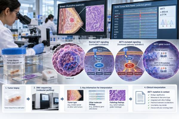
An AKT1 mutation test looks for DNA changes in the AKT1 gene, a key part of the PI3K-AKT-mTOR growth-signaling pathway. In cancer, the best-known AKT1 change is p.E17K, an activating mutation that can keep growth signals switched on even when a cell should slow down. AKT1 testing is most often performed on tumor tissue or, in some settings, circulating tumor DNA from blood. It may be ordered as part of a broad next-generation sequencing panel rather than as a single-gene test. A positive result can help define the molecular profile of a tumor and, for certain advanced breast cancers, can identify patients whose tumors meet biomarker criteria for AKT-targeted treatment. The result does not by itself diagnose cancer, determine stage, or predict the entire course of disease. Its meaning depends on the exact variant, cancer type, other biomarkers, prior treatment, and whether the alteration is somatic or inherited.
- A positive AKT1 result usually means an activating tumor mutation was detected; AKT1 p.E17K is the most common clinically recognized hotspot.
- In HR-positive, HER2-negative advanced breast cancer, qualifying AKT1 alterations can help identify patients eligible for capivasertib plus fulvestrant after specified prior endocrine treatment.
- Most cancer-related AKT1 findings are somatic, meaning they arose in the tumor and are not automatically inherited.
- A negative result means no reportable AKT1 alteration was found by that test; it does not rule out cancer or all pathway abnormalities.
- No fasting or special preparation is usually needed for blood-based testing, while tissue testing depends on an existing biopsy or surgical specimen.
Table of Contents
- What the AKT1 Mutation Test Detects
- When AKT1 Testing Is Used
- How AKT1 Testing Is Performed
- How to Read an AKT1 Test Result
- AKT1 in Breast Cancer and Other Solid Tumors
- Treatment and Follow-Up After AKT1 Testing
- Common Questions About AKT1 Mutation Testing
What the AKT1 Mutation Test Detects
The test detects sequence changes in AKT1 that may alter how tumor cells receive and transmit growth signals. AKT1 encodes a serine-threonine kinase, an enzyme that helps control cell growth, survival, metabolism, and division. It sits downstream of PI3K in the PI3K-AKT-mTOR pathway, one of the signaling networks most often altered in human cancer.
The best-known AKT1 cancer mutation is c.49G>A, which produces the protein change p.Glu17Lys, usually written p.E17K. This change affects the pleckstrin homology domain of the AKT1 protein and can cause abnormal membrane localization and persistent pathway activation. In practical terms, the mutation can give a tumor cell a growth advantage.
AKT1 p.E17K is not the only AKT1 alteration a laboratory may detect. Broad sequencing can identify other substitutions, insertions, deletions, or occasionally copy-number changes. However, not every detected change has the same biological or treatment significance. A molecular report should therefore state the exact DNA and protein change, its classification, and any known clinical interpretation.
AKT1 is closely connected with other pathway genes. For example, a PIK3CA mutation test assesses an upstream driver that can activate the same pathway, while PTEN loss removes an important brake on PI3K-AKT signaling. This matters because modern treatment decisions may use a group of pathway alterations rather than AKT1 alone.
An AKT1 tumor test is not a blood tumor-marker level like CEA or CA 15-3. It does not produce a high or low concentration. Instead, the result is usually qualitative—detected or not detected—with additional details such as variant allele fraction, pathogenicity, and technical quality metrics.
When AKT1 Testing Is Used
AKT1 testing is most useful when a tumor’s molecular profile could affect treatment selection, clinical-trial eligibility, or diagnostic interpretation. It is commonly included in multigene next-generation sequencing rather than ordered alone.
In advanced breast cancer, testing can be especially relevant for people with hormone receptor-positive, HER2-negative disease that has progressed after endocrine therapy. The treatment landscape now includes an AKT inhibitor for tumors with qualifying PIK3CA, AKT1, or PTEN alterations, so identifying an AKT1 mutation can have direct therapeutic importance.
A broader hormone receptor-positive breast cancer biomarker panel may also assess ER, PR, HER2, ESR1, PIK3CA, and other markers that answer different treatment questions. AKT1 should be interpreted alongside—not instead of—these markers.
Doctors may also order AKT1 testing in other solid tumors when comprehensive genomic profiling is appropriate. AKT1 mutations have been reported in several cancer types, including breast, endometrial, cervical, colorectal, lung, ovarian, and prostate cancers, although frequency and clinical actionability differ widely. In many non-breast tumors, an AKT1 finding is more likely to support trial matching or broader molecular characterization than to point to a standard approved treatment.
Testing may be considered at several points:
- At diagnosis of an advanced or metastatic cancer when broad genomic profiling is recommended.
- At recurrence or progression, especially if new targeted treatment options are being considered.
- When earlier limited testing did not include AKT1 or other relevant pathway genes.
- When tissue is insufficient and a validated plasma circulating-tumor-DNA assay could provide useful molecular information.
- When enrollment in a clinical trial requires a specific AKT-pathway alteration.
AKT1 testing is generally not a screening test for healthy people and is not used to estimate population cancer risk in the way germline BRCA1, BRCA2, ATM, or APC testing can. If the clinical question is inherited susceptibility, the laboratory must use a germline-testing strategy designed for that purpose.
How AKT1 Testing Is Performed
Most AKT1 mutation testing uses DNA sequencing from tumor tissue, although some assays can analyze circulating tumor DNA in blood. The method chosen affects what the result can detect and how a negative result should be interpreted.
Tumor tissue testing
A pathology laboratory may use tissue from a biopsy, surgery, or another archived tumor specimen. The pathologist first confirms that enough tumor is present. DNA is then extracted and analyzed, most often through a multigene next-generation sequencing panel. A solid tumor NGS panel can assess many genes in one run and may also detect selected copy-number changes, fusions, or other genomic features depending on the assay.
Testing an older specimen can be appropriate when the sample is adequate, but a newer metastatic biopsy may sometimes better reflect the current cancer after treatment-related evolution. The best specimen depends on tumor type, accessibility, prior testing, and the clinical question.
Blood-based testing
A liquid biopsy analyzes DNA fragments shed by tumor cells into the bloodstream. It avoids a tissue procedure and can be useful when tissue is unavailable or obtaining another biopsy is difficult. However, the amount of circulating tumor DNA varies. A low-shedding tumor or a small amount of disease may produce a false-negative plasma result even when an AKT1 mutation is present in tumor tissue.
For that reason, a negative liquid-biopsy result may need follow-up with tissue testing when knowing the mutation status would change treatment and suitable tissue can be obtained.
Common laboratory methods
AKT1 can be assessed by:
- Next-generation sequencing: preferred when many actionable genes need evaluation at once.
- Targeted PCR or hotspot assays: designed to detect specific known mutations such as p.E17K.
- Digital PCR: highly sensitive for selected variants and sometimes used in research or focused monitoring settings.
Preparation is usually simple. A blood draw typically requires no fasting. Tissue testing generally uses material already collected, so the genetic test itself does not add another procedure unless a new biopsy is clinically needed. Turnaround commonly ranges from several days to a few weeks, depending on the laboratory, specimen quality, and breadth of testing.
How to Read an AKT1 Test Result
The most important parts of the report are the exact variant, its classification, the specimen tested, and whether the result is clinically actionable for that cancer. “Positive” and “negative” are useful shorthand, but the details determine meaning.
| Report finding | What it usually means | What to check next |
|---|---|---|
| AKT1 pathogenic or likely pathogenic variant detected | A biologically important AKT1 alteration was found in the tested sample | Exact variant, cancer type, treatment relevance, other biomarkers |
| AKT1 p.E17K detected | A recognized activating hotspot mutation is present | Whether it qualifies for an approved therapy or trial in this setting |
| No reportable AKT1 alteration detected | The assay did not find an AKT1 change meeting its reporting criteria | Assay coverage, sample quality, ctDNA shedding, other pathway genes |
| Variant of uncertain significance | A change was found, but evidence is insufficient to classify it as disease-causing or actionable | Do not treat it as a positive predictive biomarker unless evidence changes |
A variant allele fraction, often abbreviated VAF, may also appear. It is the percentage of sequencing reads that carry the variant. A VAF of 20%, for example, means about one in five DNA reads at that position contained the change. VAF can be influenced by tumor purity, copy number, tumor heterogeneity, and whether the specimen is tissue or plasma. It is not a simple measure of how aggressive the cancer is.
A negative result also needs context. Every assay has a limit of detection. Some panels examine the entire coding region of AKT1, while others test only selected hotspots. Plasma assays can miss mutations when little tumor DNA is circulating. The phrase “not detected” therefore means not found by this test in this sample, not that the tumor definitely lacks every AKT1 abnormality.
A variant of uncertain significance, or VUS, should not be treated like a proven driver. Laboratories periodically reclassify variants as new evidence accumulates. If a report contains a VUS, the useful question is whether there is a separate pathogenic alteration that has established treatment significance.
AKT1 in Breast Cancer and Other Solid Tumors
AKT1 alterations are most clinically established in a subset of advanced breast cancers, but they occur across several solid-tumor types. The p.E17K hotspot accounts for a large share of AKT1 mutations seen in breast tumors.
Large genomic datasets suggest AKT1 alterations occur in only a minority of breast cancers overall, with prevalence around a few percent rather than tens of percent. They are enriched in hormone receptor-positive disease compared with some other breast-cancer subtypes. This lower prevalence is one reason broad panel testing can be more efficient than ordering AKT1 alone.
AKT1 is part of the same signaling network as PIK3CA and PTEN, but these alterations are not interchangeable. A tumor may have one, several, or none of them. In hormone receptor-positive breast cancer, the pathway can also interact with endocrine-resistance mechanisms. An ESR1 mutation test, for example, answers a different question about estrogen-receptor pathway resistance and may be clinically relevant at the same point in advanced disease.
AKT1 mutations also appear in endometrial, cervical, colorectal, ovarian, lung, and prostate tumors, among others. Yet a mutation that is actionable in one cancer is not automatically actionable in another. Regulatory approvals, evidence from clinical trials, tumor biology, and the exact variant all matter.
For lung cancer, a broad lung cancer NGS panel usually prioritizes established drivers such as EGFR, ALK, ROS1, BRAF, MET, RET, KRAS, ERBB2, and NTRK. An incidental AKT1 alteration may provide biological information, but it generally does not replace those standard predictive biomarkers.
It is also important to separate somatic AKT1 mutations from rare constitutional disorders. Most AKT1 mutations detected during tumor profiling arose in cancer cells and are not present in every cell of the body. A somatic tumor result therefore does not automatically imply that children, siblings, or parents carry the same change. If the laboratory or clinical history raises concern about an inherited or mosaic condition, confirmatory testing should use a genetics laboratory and an appropriate non-tumor specimen.
Treatment and Follow-Up After AKT1 Testing
For eligible HR-positive, HER2-negative advanced breast cancer, a qualifying AKT1 alteration can directly influence treatment selection. In 2023, the U.S. Food and Drug Administration approved capivasertib with fulvestrant for adults with locally advanced or metastatic HR-positive, HER2-negative breast cancer harboring one or more qualifying PIK3CA, AKT1, or PTEN alterations after progression on specified endocrine therapy or qualifying early recurrence.
The phase 3 CAPItello-291 trial evaluated capivasertib plus fulvestrant against placebo plus fulvestrant. The trial included an AKT-pathway-altered population defined by PIK3CA, AKT1, or PTEN changes. This is why a positive AKT1 result can be more than descriptive in the correct breast-cancer setting: it can function as a predictive biomarker linked to an approved treatment strategy.
The result should still be integrated with the full breast-cancer profile. ER, PR, HER2, disease location, symptoms, prior endocrine therapy, prior CDK4/6 inhibitor exposure, comorbidities, and other mutations all influence the treatment plan. A breast cancer biomarker panel provides context that no single mutation can supply.
Capivasertib also has important toxicity considerations. Diarrhea, skin reactions, and hyperglycemia are among the clinically significant adverse effects reported with treatment, so eligibility for a targeted drug is not the same as saying the drug is automatically the best option. Clinicians weigh expected benefit against prior treatments, other available therapies, diabetes risk, performance status, and patient preferences.
Outside an approved indication, an AKT1 mutation may support consideration of a clinical trial. Trial eligibility can be highly specific: some studies require p.E17K, others accept broader AKT-pathway alterations, and some restrict enrollment by tumor type or previous treatment.
After receiving a result, useful questions for the oncology team include:
- What exact AKT1 variant was detected?
- Is it classified as pathogenic or likely pathogenic, or is it a VUS?
- Was the test performed on tumor tissue or plasma?
- Does this alteration qualify for an approved treatment in my cancer type and stage?
- Were PIK3CA, PTEN, ESR1, HER2, and other relevant biomarkers also tested?
- If the result was negative in plasma, would tissue testing add useful information?
- Is there any reason to consider germline genetic counseling or testing?
Common Questions About AKT1 Mutation Testing
Does an AKT1 mutation mean the cancer is hereditary?
Usually not. Most AKT1 mutations found on cancer profiling are somatic, meaning they developed in the tumor. A tumor-only sequencing report cannot always prove whether a variant is somatic or germline, but AKT1 is not typically approached like BRCA1 or APC in routine hereditary cancer evaluation. If the finding or personal history is unusual, a genetics professional can determine whether testing normal tissue is appropriate.
Is AKT1 p.E17K the same as PIK3CA mutation?
No. They are changes in different genes, although both can activate the PI3K-AKT pathway. A tumor may carry an AKT1 mutation without a PIK3CA mutation, or vice versa. Treatment labels and clinical trials may group them together when evidence supports a shared pathway-targeting strategy.
Can a positive AKT1 result diagnose breast cancer?
No. Diagnosis still depends on pathology from a biopsy or surgical specimen, together with imaging and clinical findings. AKT1 testing adds molecular information after cancer is suspected or confirmed.
Can an AKT1 mutation disappear after treatment?
Tumor populations can change over time. A clone carrying an AKT1 mutation may become more or less prominent under treatment pressure, and plasma detectability can vary with disease burden. However, a later “not detected” plasma result does not necessarily prove that every cancer cell lacks the mutation. Serial molecular testing should be interpreted with disease status and assay sensitivity.
Should everyone with metastatic cancer get AKT1 testing?
Not necessarily as a stand-alone test. Many patients with advanced solid tumors benefit from broad molecular profiling because it can evaluate several actionable genes at once. The ideal panel depends on the cancer type and current treatment guidelines. In breast cancer, AKT1 is especially relevant when the result can affect use of AKT-pathway-directed therapy.
What if the report says AKT1 VUS?
A VUS is not the same as a clinically actionable AKT1 mutation. It means the laboratory found a change but current evidence is not strong enough to classify it as pathogenic or benign. Treatment decisions should not assume a VUS predicts benefit from an AKT inhibitor unless future evidence and a qualified molecular interpretation support that conclusion.
References
- Panel Sequencing for Targeted Therapy Selection in Solid Tumors 2022 (Review)
- Capivasertib in Hormone Receptor-Positive Advanced Breast Cancer 2023 (RCT)
- FDA approves capivasertib with fulvestrant for breast cancer 2023
- US Food and Drug Administration Approval Summary: Capivasertib With Fulvestrant for Hormone Receptor-Positive, Human Epidermal Growth Factor Receptor 2-Negative Locally Advanced or Metastatic Breast Cancer With PIK3CA/ AKT1/ PTEN Alterations 2024 (Review)
- Clinicogenomic Landscape and Function of PIK3CA, AKT1, and PTEN Mutations in Breast Cancer 2026
Disclaimer
This article is for general education and does not replace medical advice, pathology review, or molecular interpretation by your oncology team. AKT1 results must be interpreted with the cancer type, exact variant, specimen, other biomarkers, and current treatment approvals. Do not start, stop, or change cancer treatment based on a genetic result without discussing it with a qualified clinician.