
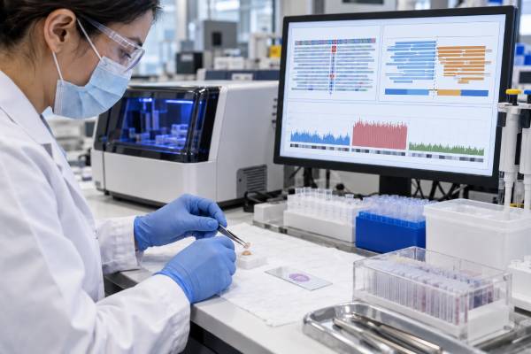
A solid tumor next-generation sequencing (NGS) panel analyzes many cancer-related genes at once to identify mutations, gene fusions, copy-number changes, and sometimes genomic biomarkers such as microsatellite instability (MSI) or tumor mutational burden (TMB). The goal is not simply to label a tumor “positive” or “negative.” A useful report connects each alteration to diagnosis, prognosis, an approved treatment, a resistance mechanism, or a clinical trial—and explains what the assay could not reliably measure. Results depend on tumor type, sample quality, tumor percentage, sequencing design, whether RNA was included, and whether a matched normal sample was tested. A “pathogenic” tumor variant can be biologically important without being inherited, while a finding in a hereditary cancer gene may still need separate germline confirmation. NGS is most valuable when the pathology diagnosis and clinical question are clear before testing and when the report is reviewed in context by the oncology and molecular pathology team. A broad panel can reveal highly actionable drivers, but it can also produce uncertain findings that should not be overinterpreted.
- A solid tumor NGS panel can test dozens to hundreds of genes in one assay for mutations, fusions, and copy-number changes.
- A detected alteration is clinically useful only when its evidence level matches the patient’s cancer type, stage, and treatment setting.
- RNA sequencing often improves fusion detection because some rearrangements are difficult to capture with DNA-only panels.
- Tumor-only sequencing cannot reliably determine whether every hereditary-cancer-gene variant is germline; confirmatory testing may be needed.
- A negative panel does not mean “no cancer mutation”; it means no reportable alteration was found within that assay’s validated scope and sample limits.
Table of Contents
- What a Solid Tumor NGS Panel Measures
- When NGS Testing Is Used
- How the Sample and Assay Affect Results
- How to Read a Molecular Report
- How Different Alteration Types Are Interpreted
- Treatment Actionability and Tumor-Agnostic Markers
- Limitations, Germline Findings, and Next Steps
What a Solid Tumor NGS Panel Measures
NGS uses massively parallel sequencing to read many DNA or RNA regions at the same time. Instead of ordering a separate test for EGFR, BRAF, KRAS, RET, NTRK, and other genes, a broad panel can evaluate them together from one tumor specimen.
Panel size varies. A small focused assay may analyze a few dozen genes with established relevance to one cancer. A comprehensive genomic profile may analyze hundreds of genes and calculate additional biomarkers. Bigger is not automatically better: the best test is the one that reliably covers the alterations that matter for the clinical question while preserving tissue and producing an interpretable report.
Common alteration classes include:
- Single-nucleotide variants (SNVs): one DNA base is changed, such as BRAF V600E.
- Small insertions and deletions (indels): a few DNA bases are added or removed, sometimes causing a frameshift.
- Copy-number changes: genes may be amplified, gained, or deleted.
- Gene fusions and rearrangements: parts of two genes join, potentially creating an oncogenic driver such as RET, ALK, ROS1, or NTRK fusion.
- Splice alterations: variants may cause abnormal exon usage, such as MET exon 14 skipping.
- Genomic signatures: some assays estimate MSI, TMB, homologous-recombination-related metrics, or other composite markers.
The report should state which categories were validated. A DNA panel that detects point mutations well may have weaker fusion coverage. An RNA panel may be excellent for expressed fusions but cannot replace DNA testing for all mutation and copy-number questions.
When NGS Testing Is Used
NGS is increasingly used when multiple biomarkers can affect treatment, especially in advanced or metastatic cancers. Current ESMO recommendations support routine tumor NGS in several advanced cancers and have expanded its use as precision-medicine evidence has grown. ASCO likewise recommends genomic testing when a biomarker-linked therapy is available or when the result can meaningfully guide management.
The strongest use cases occur when the cancer has several established targetable drivers. Advanced non-squamous NSCLC is a classic example because EGFR, ALK, ROS1, BRAF, MET, RET, NTRK, KRAS, and ERBB2 can each affect treatment selection. Similar logic applies to many advanced colorectal, prostate, ovarian, cholangiocarcinoma, breast, thyroid, sarcoma, and rare-tumor settings.
NGS may also help classify a difficult tumor. Certain fusions are strongly associated with specific sarcomas, salivary tumors, renal cancers, and other entities. In that setting, the molecular result supports diagnosis rather than simply finding a drug target.
Testing can be ordered on the original resection specimen, a metastatic biopsy, or a new sample obtained at progression. A more recent specimen may better reflect treatment-selected resistance, but the oldest high-quality tissue can still be informative when the key driver is expected to persist.
When tissue is unavailable or unsafe to obtain, circulating tumor DNA can sometimes provide a blood-based alternative. A ctDNA mutation panel may detect actionable alterations in advanced disease, but a negative plasma result can occur simply because the tumor sheds little DNA.
How the Sample and Assay Affect Results
The most important hidden variable in tumor sequencing is tumor content. A pathology section contains a mixture of cancer cells, stromal cells, inflammatory cells, blood vessels, and normal tissue. If only 10% of nucleated cells are tumor, a heterozygous mutation present in every tumor cell may appear at roughly 5% variant allele fraction before copy-number and purity effects are considered.
Laboratories often mark a tumor-rich area for macrodissection to increase the cancer fraction. Very low tumor percentage can make low-frequency variants, copy-number changes, and some fusions harder to detect.
Fixation matters too. Formalin can damage nucleic acids, and old blocks may contain fragmented DNA and RNA. Decalcified bone specimens are particularly problematic because some decalcification methods degrade nucleic acids. A failed quality-control result is therefore a specimen problem, not evidence that the tumor lacks molecular abnormalities.
The assay’s gene list is only the first page to read. Coverage details matter:
- Which exons and introns are captured?
- Are full coding regions sequenced or only hotspots?
- Is deletion/duplication analysis validated?
- Can the assay detect large structural variants?
- Does it include RNA fusion testing?
- What is the minimum variant allele fraction reported?
- Are MSI and TMB analytically validated on this platform?
These questions explain why two panels with similar gene counts can have different clinical sensitivity.
How to Read a Molecular Report
A good NGS report separates what was found from what it means. The findings section usually lists gene, variant notation, alteration type, VAF or copy-number estimate, and classification. The interpretation section then assigns clinical evidence.
Many laboratories use a tiered system derived from professional consensus. Tier I variants have strong clinical significance, such as an alteration linked to an approved therapy or major guideline in the relevant cancer. Tier II findings have potential clinical significance, which may include evidence from another tumor type or an active investigational strategy. Tier III findings are variants of uncertain significance. Benign or likely benign variants are usually omitted.
A tumor VUS should not be treated as a proven driver. Large panels generate many rare changes simply because so much DNA is being examined. A VUS may later be reclassified, but treatment should not be based on speculation when stronger biomarkers are available.
Variant allele fraction (VAF) is useful but easy to misuse. It is the proportion of sequencing reads carrying the variant, not the percentage of cancer cells. Tumor purity, local copy number, loss of heterozygosity, subclones, and technical factors all influence VAF.
The report may also list “negative” biomarkers. These can be as important as positive findings. For example, absence of RAS mutations may be required before certain anti-EGFR strategies are considered in colorectal cancer. The meaning of a negative call still depends on assay sensitivity and sample quality.
Finally, check the date and knowledge base. Actionability changes as drugs receive approvals, resistance evidence grows, and variant classifications are updated. A molecular report is a time-stamped interpretation, not a permanent treatment menu.
How Different Alteration Types Are Interpreted
Point mutations and indels
Activating mutations in oncogenes can function like a stuck accelerator. Examples include BRAF V600E, PIK3CA hotspot mutations, or KRAS G12C. Loss-of-function mutations in tumor suppressors such as TP53, PTEN, or RB1 are different: the cancer has lost a brake, which is often harder to target directly.
The same gene can contain both actionable and non-actionable variants. “BRAF mutated” is less informative than the exact variant because V600E and non-V600 alterations can have different biology and treatment evidence.
Gene fusions
A fusion may be highly actionable when it creates an intact kinase domain and is known to drive cancer. The exact partner, reading frame, transcript evidence, and cancer type matter. An NTRK fusion, for example, can have tumor-agnostic treatment significance, while many incidental rearrangements do not.
RNA-based sequencing can improve fusion sensitivity and can show that the rearrangement is actually transcribed. DNA-only panels may miss fusions when breakpoints fall in long or repetitive introns outside captured regions.
Copy-number changes
Amplification means extra copies of a gene, but the threshold varies by assay. A low-level gain is not equivalent to high-level focal amplification. Tumor purity and whole-chromosome changes can complicate estimates. The clinical meaning also depends on the gene: ERBB2 amplification can be a validated target in several settings, whereas amplification of another gene may be only exploratory.
Deletions should likewise be distinguished as single-copy loss versus biallelic/homozygous loss when possible. Complete loss of both tumor-suppressor copies is usually stronger evidence of functional inactivation.
Treatment Actionability and Tumor-Agnostic Markers
The most valuable NGS finding is one that changes a real clinical decision. Actionability generally falls into several levels: an approved therapy for that exact cancer and biomarker; a guideline-supported use; an approval in another tumor type with biological rationale; an investigational clinical trial; or preclinical evidence only.
The report should not collapse these levels into a single word such as “targetable.” A therapy supported by a randomized trial in the patient’s cancer is not equivalent to a drug that inhibited a cell line.
Some biomarkers are tumor agnostic, meaning the treatment indication is based primarily on the molecular feature rather than the organ of origin. Examples include certain NTRK fusions and, in defined settings, MSI-high/mismatch-repair-deficient status or high TMB. Even then, the indication has exact regulatory and clinical criteria.
A tumor mutational burden test may be calculated from the same panel, but TMB is assay dependent. Mutations per megabase, panel size, germline filtering, variant types counted, and bioinformatic pipeline can all change the score. A numeric cutoff should therefore be interpreted using the assay and treatment context that validated it.
NGS also helps avoid ineffective therapy. Identifying one dominant driver may make another treatment less attractive, and resistance mutations can explain why a previously effective drug no longer works.
Clinical trials are another major use. Rare genomic subsets may be too uncommon for traditional organ-specific trials, so basket trials enroll patients based on a shared molecular alteration. The report’s trial list is a starting point, not proof of eligibility; location, stage, prior therapy, organ function, and enrollment status still matter.
Limitations, Germline Findings, and Next Steps
Tumor-only NGS has several important limits. First, it does not directly compare tumor DNA with the person’s normal DNA. A pathogenic variant in BRCA1, BRCA2, PALB2, mismatch repair genes, TP53, or another hereditary cancer gene may therefore be somatic or germline. VAF can raise suspicion but cannot settle the question.
When a tumor finding could represent inherited risk, confirmatory germline testing from a non-tumor sample is usually required. The reverse is also true: a negative tumor panel cannot replace germline testing in someone who meets hereditary-testing criteria. Tumor-only sequencing can miss germline copy-number variants, intronic variants, repetitive insertions, or regions that were not adequately covered.
Second, a negative NGS report does not mean the tumor is genetically normal. The driver may be outside the panel, in a noncoding region, below the detection threshold, epigenetic, or present as an alteration type the assay does not detect.
Third, tumor heterogeneity means one biopsy may not represent every cancer site. Treatment can select resistant subclones over time. A new biopsy or liquid biopsy at progression may reveal clinically meaningful evolution.
A practical way to review any solid tumor NGS report is to ask:
- Was the sample adequate, and what was the estimated tumor percentage?
- Did the assay include both DNA and RNA when fusion detection matters?
- Which findings are Tier I/strongly actionable for this exact cancer?
- Are any results only investigational or VUS?
- Do copy-number calls represent high-level focal changes or broad gains/losses?
- Are MSI, TMB, or other signatures validated on this platform?
- Could any pathogenic variant be germline and require genetics referral?
- Would repeat testing at progression have a realistic chance of changing treatment?
NGS works best as a decision tool, not a data dump. The report becomes clinically meaningful only after the findings are ranked by evidence, technical reliability, cancer context, and the patient’s treatment goals.
References
- Recommendations for the use of next-generation sequencing (NGS) for patients with advanced cancer in 2024: a report from the ESMO Precision Medicine Working Group 2024 (Guideline)
- Somatic Genomic Testing in Patients With Metastatic or Advanced Cancer: ASCO Provisional Clinical Opinion 2022 (Guideline)
- Diagnostic impact of RNA-based next-generation sequencing fusion panel for solid tumors: A single-institution experience 2024
- Comprehensive assessment of germline pathogenic variant detection in tumor-only sequencing 2022
- Standards and Guidelines for the Interpretation and Reporting of Sequence Variants in Cancer: A Joint Consensus Recommendation of the Association for Molecular Pathology, American Society of Clinical Oncology, and College of American Pathologists 2017 (Guideline)
Disclaimer
Solid tumor NGS results are highly dependent on specimen quality, assay design, cancer type, and current evidence for each biomarker. Treatment and germline-testing decisions should be made with qualified oncology, pathology, and genetics professionals who can review the complete report. This article is educational and does not replace medical care.





