
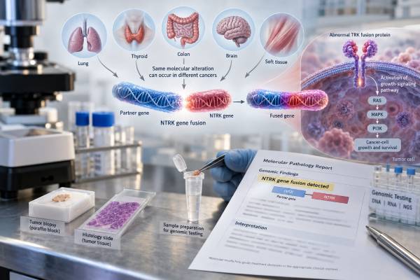
An NTRK fusion test looks for abnormal joining of an NTRK gene to another gene in a cancer cell. These fusions can create a continuously active TRK signaling protein that drives tumor growth, and they matter because some cancers with a confirmed NTRK1, NTRK2, or NTRK3 fusion can be treated with TRK inhibitors regardless of where the cancer started. That is why NTRK is called a tumor-agnostic biomarker. The clinically meaningful finding is usually a functional NTRK gene fusion, not simply an NTRK mutation or increased TRK staining. Testing may use RNA or DNA next-generation sequencing, fluorescence in situ hybridization, reverse-transcription PCR, or pan-TRK immunohistochemistry as a screening method. Because NTRK fusions are rare in most common cancers but frequent in a few uncommon tumor types, the best testing strategy depends on the diagnosis, available tissue, and assay design. A positive or negative report should always be interpreted with those technical details in mind.
- A positive NTRK fusion result means an oncogenic fusion involving NTRK1, NTRK2, or NTRK3 was detected and may create eligibility for TRK-targeted therapy.
- NTRK fusion-positive is not the same as having any NTRK mutation; most isolated NTRK sequence variants are not tumor-agnostic treatment markers.
- RNA-based NGS is often preferred for confirming expressed fusion transcripts, especially when the fusion partner is unusual.
- Pan-TRK immunohistochemistry can screen for TRK protein expression, but positive staining usually requires molecular confirmation because staining is not specific for a fusion.
- A negative result is only as reliable as the assay’s fusion coverage, specimen quality, tumor content, and ability to detect NTRK1, NTRK2, and NTRK3 events.
Table of Contents
- What an NTRK Fusion Is and Why It Matters
- Which Tumors Can Have NTRK Fusions
- How NTRK Fusions Are Tested
- Positive, Negative, and Uncertain Result Meaning
- NTRK as a Tumor-Agnostic Treatment Marker
- Resistance and Repeat Molecular Testing
- Questions to Ask About an NTRK Report
What an NTRK Fusion Is and Why It Matters
NTRK1, NTRK2, and NTRK3 are genes that encode the TRKA, TRKB, and TRKC receptor tyrosine kinases. In normal cells, these receptors respond to neurotrophins, which are signaling molecules involved in development and nervous-system function. A cancer-driving NTRK fusion occurs when part of an NTRK gene becomes joined to part of another gene. The partner gene often provides a structure that keeps the TRK kinase switched on without the normal external signal.
The resulting fusion protein can activate growth pathways such as RAS-MAPK and PI3K-AKT. In a tumor that depends on this signal, blocking TRK can produce substantial tumor shrinkage. This biology is the basis for tumor-agnostic therapy: treatment is selected by the molecular driver rather than solely by the organ in which the cancer arose.
A fusion is different from a point mutation. A report may list an NTRK1 missense variant, for example, without showing that the gene is fused to a partner. Unless that variant is known to be oncogenic and clinically actionable in the specific setting, it should not be interpreted as equivalent to an NTRK fusion. Likewise, NTRK gene amplification and TRK protein expression do not automatically establish a fusion.
Fusion reports often name both partners, such as ETV6-NTRK3, TPM3-NTRK1, or LMNA-NTRK1. ETV6-NTRK3 is a classic driver in secretory carcinoma of the breast and salivary gland and in infantile fibrosarcoma. Many other partners have been described. A well-designed molecular assay should determine whether the rearrangement preserves the NTRK kinase domain and is expected to create a functional transcript.
NTRK testing is commonly included in a broader solid-tumor NGS panel, particularly for advanced cancers in which comprehensive molecular profiling is already standard. This approach can conserve tissue because multiple biomarkers are assessed at once.
Which Tumors Can Have NTRK Fusions
NTRK fusions have been reported across many adult and pediatric solid tumors, but their frequency varies dramatically. Across unselected common solid tumors, the overall prevalence is low—often around a few tenths of one percent. By contrast, some rare tumor types are strongly enriched for specific NTRK fusions.
Examples of tumors with a high or meaningful fusion frequency include infantile fibrosarcoma, secretory breast carcinoma, mammary analogue secretory carcinoma/secretory carcinoma of salivary glands, and selected pediatric thyroid cancers. NTRK fusions also occur at low frequencies in more common cancers such as non-small cell lung cancer, colorectal cancer, melanoma, and common adult thyroid carcinomas.
This uneven distribution affects how testing is organized. In a tumor type known to have a high frequency of NTRK fusions, direct molecular testing can be efficient. In a common cancer where the prevalence is very low, NTRK may be assessed as part of broad NGS or through a screening strategy followed by confirmation.
For colorectal cancer, an NTRK fusion test in colon cancer is particularly relevant in selected advanced tumors without more common drivers. Some NTRK-positive colorectal cancers have distinctive molecular contexts, including an association in certain series with mismatch-repair deficiency and absence of common RAS/BRAF drivers, but the exact testing algorithm depends on current guidelines and local panel practices.
In lung cancer, NTRK fusions are rare but actionable. Comprehensive profiling can identify them while simultaneously evaluating more common drivers. A dedicated NTRK fusion result in lung cancer should be interpreted alongside EGFR, ALK, ROS1, RET, BRAF, MET, KRAS, and other clinically relevant markers.
The low frequency in common cancers is one reason clinicians should not infer fusion status from age, symptoms, imaging, or routine pathology alone. Molecular testing is required to establish the biomarker.
How NTRK Fusions Are Tested
No single method is perfect in every tumor. The main approaches are RNA-based NGS, DNA-based NGS, immunohistochemistry, FISH, and RT-PCR. The choice depends on prevalence, tissue, laboratory resources, and whether the test is intended for screening or definitive confirmation.
| Method | Main strength | Important limitation |
|---|---|---|
| RNA-based NGS | Detects expressed fusion transcripts and can identify many partners | RNA can degrade in older or poorly handled tissue |
| DNA-based NGS | Can profile mutations, copy changes, and some fusions in one assay | Large intronic regions, especially in NTRK2/3, may reduce fusion sensitivity depending on panel design |
| Pan-TRK IHC | Fast, tissue-sparing screening tool | Protein staining is not specific for a gene fusion and can be difficult to interpret in neural or smooth-muscle tissues |
| Break-apart FISH | Can detect rearrangement of a specific NTRK gene | Separate probes may be needed for NTRK1, NTRK2, and NTRK3; partner and transcript function may remain unknown |
| RT-PCR | Highly focused and sensitive for known transcripts | May miss novel or unexpected fusion partners |
Why RNA testing is often valuable
RNA-based sequencing assesses the transcript produced after genes are expressed. That makes it especially useful for showing that a DNA rearrangement actually forms an in-frame fusion transcript containing the NTRK kinase domain. Consensus recommendations emphasize careful RNA quality control, appropriate read thresholds, and confirmation of unusual calls when needed.
Why IHC is usually a screen, not the final answer
Pan-TRK immunohistochemistry uses antibodies that detect TRK proteins. Strong or characteristic staining can flag a tumor for molecular testing, particularly when fusion prevalence is low and comprehensive NGS is not already being done. However, normal tissues and some tumors can express TRK protein without an oncogenic fusion. Conversely, some genuine fusions can produce weak or difficult-to-recognize staining. A treatment decision should therefore rely on a validated fusion result rather than IHC alone when molecular confirmation is feasible.
Sample quality matters for every method. Tumor percentage, fixation, decalcification, nucleic-acid degradation, and the amount of available tissue can all affect sensitivity. A “not detected” result from a small, damaged specimen deserves more caution than a negative result from a high-quality sample tested with a well-validated fusion assay.
Another technical issue is assay architecture. Some DNA panels cover selected introns rather than every possible breakpoint, and NTRK2 and NTRK3 contain large intronic regions that can be challenging to capture. RNA panels avoid some of that genomic complexity because they interrogate the expressed transcript, but RNA quality may be poor in small formalin-fixed samples. Laboratories therefore validate their own sensitivity and specify when a negative result has limitations.
The pathology context can also change the value of pan-TRK staining. Cytoplasmic, membranous, nuclear, or perinuclear patterns may occur depending on the fusion, but staining pattern alone does not reliably identify the partner. Normal neural tissue can express TRK, which creates a background signal in some specimens. These practical details explain why a two-step screening-and-confirmation strategy may be appropriate in one setting while direct sequencing is preferred in another.
Positive, Negative, and Uncertain Result Meaning
A positive NTRK fusion result should identify which NTRK gene is involved and, whenever possible, the partner gene and fusion structure. The report may describe the event as pathogenic, oncogenic, or therapeutically actionable. The key question is whether the alteration produces a functional TRK fusion with an intact kinase domain.
A positive result can have treatment significance even in a cancer where NTRK fusions are extremely rare. This is the essence of a tumor-agnostic marker. However, eligibility for a particular drug still depends on factors such as disease stage, prior treatments, age, regulatory indication, ability to take the drug, and whether there is a satisfactory alternative therapy.
A negative NTRK result means no qualifying fusion was detected within the assay’s capabilities. It does not mean the patient has “no targetable mutations.” Other genes may be actionable. It also does not completely exclude an NTRK fusion if the assay had limited coverage or the specimen was inadequate.
One common issue is a DNA test that reports an NTRK rearrangement of uncertain functional significance. If the breakpoint or partner cannot establish an expressed oncogenic fusion, RNA testing may clarify the finding. Similarly, a positive pan-TRK IHC result may lead to RNA or DNA confirmation rather than being treated as a definitive positive.
A variant of uncertain significance in NTRK1, NTRK2, or NTRK3 is not the same as a fusion-positive result. Patients should not assume that any line containing “NTRK” makes them eligible for tumor-agnostic TRK therapy. The alteration class and evidence level matter.
If a plasma ctDNA panel is negative, tissue testing may still be appropriate. Circulating tumor DNA assays can miss fusions, and some tumors release little DNA into the bloodstream. A non-informative liquid biopsy should not automatically close the question when tissue testing is clinically important and feasible.
NTRK as a Tumor-Agnostic Treatment Marker
NTRK fusions were among the biomarkers that established the modern tumor-agnostic treatment model. Larotrectinib and entrectinib are first-generation TRK inhibitors with regulatory approvals in multiple jurisdictions for appropriately selected patients with NTRK fusion-positive solid tumors. Their approvals and exact age or treatment-history criteria vary by regulator, so clinicians use the current local label and guideline.
The reason this approach works is that a true NTRK fusion can act as the dominant growth driver in cancers from very different organs. Clinical trials pooled patients across numerous tumor types and showed that many fusion-positive tumors can respond substantially and durably to TRK inhibition. Responses have been observed in adults and children and in cancers of the lung, thyroid, salivary gland, soft tissue, colon, and other sites.
Tumor-agnostic does not mean tumor context is irrelevant. A person’s cancer type still affects surgery, radiation, local treatment, prognosis, competing systemic options, response assessment, and timing. It means the molecular fusion can justify a targeted drug across histologies when the treatment indication is otherwise met.
It is also important to confirm that the NTRK fusion is the actionable driver rather than merely an incidental laboratory signal. Highly validated fusion calls are most persuasive when they are mutually exclusive with another strong oncogenic driver, although exceptions and complex tumors can occur.
TRK inhibitors can cause adverse effects and drug interactions, so a positive biomarker is not the same as an automatic prescription. Clinicians weigh expected benefit against neurologic effects, dizziness, weight gain, liver-test abnormalities, gastrointestinal effects, cardiovascular considerations, and the patient’s overall treatment plan. Dose adjustments may be needed for toxicity or interacting medications.
Resistance and Repeat Molecular Testing
Even when a TRK fusion-positive cancer initially responds, resistance can develop. Some resistance mechanisms alter the TRK kinase itself so that the first-generation drug binds less effectively. Examples include solvent-front, gatekeeper, and xDFG-region substitutions. Other cancers bypass TRK signaling by activating alternative pathways such as MAPK signaling through different molecular changes.
When a cancer progresses on a TRK inhibitor, repeat molecular testing can sometimes identify the resistance mechanism and help determine whether a clinical trial or next-generation TRK inhibitor is rational. Testing may use a new tumor biopsy, plasma ctDNA, or both, depending on the disease and the suspected mechanism.
A repeat test is not automatically necessary after every scan. It is most useful when the result could change the next treatment. The oncologist also considers whether progression occurred in one site or many, whether local therapy is possible, whether the patient still benefits from the current drug, and what approved alternatives exist.
Second-generation TRK inhibitors have been developed to address some on-target resistance mutations, and this remains an active area of clinical research. As new agents and approvals emerge, the precise therapeutic meaning of a resistance mutation can change. Reports therefore need current interpretation rather than relying only on an older annotation generated when the original tumor was tested.
Questions to Ask About an NTRK Report
A few details can determine whether an NTRK result is clinically actionable. Useful questions include:
- Does the report show a true NTRK1, NTRK2, or NTRK3 fusion, or only a mutation, amplification, rearrangement, or positive IHC stain?
- What is the fusion partner, and is the fusion predicted to be in-frame with an intact NTRK kinase domain?
- Was the result detected by RNA sequencing, DNA sequencing, FISH, RT-PCR, or immunohistochemistry?
- If IHC was positive, was molecular confirmation performed?
- If DNA testing found an uncertain rearrangement, would RNA testing help confirm expression?
- If the result was negative, did the assay adequately cover all three NTRK genes and was the sample quality acceptable?
- Is the cancer advanced or unresectable in a setting where a TRK inhibitor is clinically appropriate?
- Which TRK inhibitors are currently approved or recommended for this patient’s age, country, and treatment history?
- If the cancer progressed after TRK therapy, could repeat sequencing identify a resistance mutation?
The most important distinction is simple: an actionable NTRK fusion is a specific molecular event, not any abnormality involving an NTRK gene. Once a validated fusion is confirmed, it can become one of the most consequential findings on a tumor profile because it may connect a rare molecular subgroup to a highly specific targeted treatment.
References
- Japanese Society of Medical Oncology/Japan Society of Clinical Oncology/Japanese Society of Pediatric Hematology/Oncology-led clinical recommendations on the diagnosis and use of tropomyosin receptor kinase inhibitors in adult and pediatric patients with neurotrophic receptor tyrosine kinase fusion-positive advanced solid tumors 2023 (Guideline)
- Consensus Recommendations to Optimize the Detection and Reporting of NTRK Gene Fusions by RNA-Based Next-Generation Sequencing 2023 (Consensus)
- NTRK fusions in solid tumours: what every pathologist needs to know. 2023 (Review)
- Evaluating Targeted Next-Generation Sequencing Assays and Reference Materials for NTRK Fusion Detection 2022
- Diagnosis and management of TRK fusion cancer. 2022 (Review)
Disclaimer
NTRK testing and TRK-inhibitor treatment should be interpreted by an oncology and molecular-pathology team using the exact fusion, assay method, cancer type, disease stage, and current drug approvals. A positive screening test may require molecular confirmation, and a negative result may not exclude a fusion when the sample or assay is limited. This article provides general educational information and is not individualized medical advice.





