
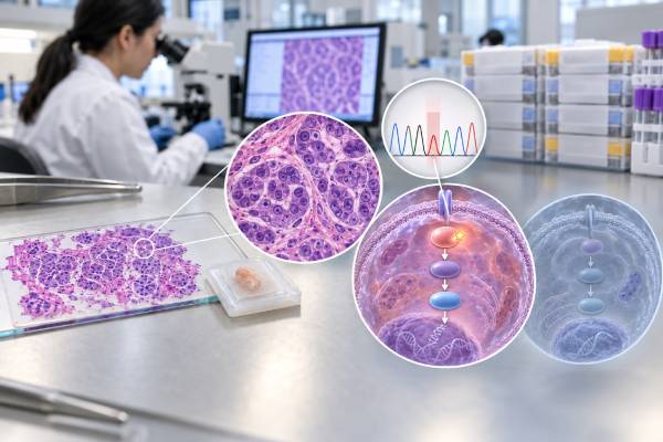
A PIK3CA mutation test looks for activating changes in the gene that encodes the p110α catalytic subunit of PI3K, a central part of the PI3K-AKT-mTOR growth pathway. PIK3CA mutations occur in many solid tumors, but their clearest routine treatment role is in hormone receptor-positive, HER2-negative advanced breast cancer, where a qualifying mutation can help identify patients for PI3K-pathway targeted therapy. Common hotspots include E542K and E545K in exon 9 and H1047R or H1047L in exon 20, although broader sequencing can detect additional pathogenic variants. Testing can use tumor tissue or circulating tumor DNA from plasma. A positive result is not automatically a sign of inherited cancer risk because most PIK3CA mutations found in tumors are somatic. A negative blood result may also need tissue confirmation because some cancers shed too little DNA into plasma. The exact cancer type, variant, assay, disease stage, and treatment history determine how clinically important a PIK3CA result is.
- A positive PIK3CA result means an activating PIK3CA mutation was detected in the tested cancer sample; it usually reflects a somatic tumor alteration.
- In HR-positive/HER2-negative advanced breast cancer, PIK3CA status can determine eligibility for PI3K-pathway targeted regimens such as alpelisib- or inavolisib-based therapy in appropriate settings.
- Common hotspot mutations include E542K, E545K, and H1047R, but clinically relevant variants occur outside the classic hotspots.
- If plasma ctDNA is negative for PIK3CA, tumor-tissue testing may still be needed because liquid biopsy can produce false-negative results when ctDNA levels are low.
- PIK3CA mutation status is not a universal treatment marker across all solid tumors; actionability depends on tumor type and current evidence.
Table of Contents
- What PIK3CA Does in the PI3K Pathway
- PIK3CA Mutations in Breast Cancer
- PIK3CA in Other Solid Tumors
- Positive, Negative, and Variant Result Meaning
- Tissue Testing Versus Liquid Biopsy
- How PIK3CA Results Guide Treatment
- Questions, Safety Issues, and Follow-Up
What PIK3CA Does in the PI3K Pathway
PIK3CA encodes p110α, the catalytic component of class IA phosphatidylinositol 3-kinase alpha. When growth-factor receptors at the cell surface are activated, PI3K helps convert membrane lipids into signaling molecules that activate AKT and downstream targets including mTOR. The pathway regulates cell growth, metabolism, protein synthesis, survival, and proliferation.
Activating PIK3CA mutations increase PI3Kα signaling even when the normal upstream signal is weak or absent. In a cancer cell, this can support uncontrolled growth and can contribute to resistance to other therapies. The gene is among the most frequently mutated oncogenes across human solid tumors.
Most clinically important PIK3CA changes are missense mutations, meaning one amino acid in the protein is replaced by another. The best-known hotspots occur in the helical domain encoded by exon 9 and the kinase domain encoded by exon 20. Examples include:
- E542K and E545K, classic helical-domain activating mutations.
- H1047R and H1047L, classic kinase-domain activating mutations.
- C420R, E726K, Q546K, and other less common variants, some of which also have functional and treatment relevance.
Older PCR companion tests were designed around a fixed list of common mutations. Broader solid-tumor NGS panels can detect a wider spectrum of PIK3CA variants and simultaneously assess other genes. That can matter because a meaningful fraction of pathogenic PIK3CA mutations in breast cancer fall outside the original trial-defined hotspot list.
PIK3CA is different from PTEN and AKT1, although all three can alter the same signaling pathway. A report that describes an “AKT pathway alteration” may therefore include PIK3CA mutation, AKT1 mutation, or PTEN loss depending on the therapy and assay being discussed.
PIK3CA Mutations in Breast Cancer
PIK3CA mutations are particularly common in hormone receptor-positive breast cancer. In advanced HR-positive/HER2-negative disease, roughly one-third to two-fifths of tumors carry a PIK3CA mutation, with estimates varying by cohort and assay. The mutation can be present in the primary tumor, a metastasis, or detectable in circulating tumor DNA.
The major clinical reason to test is predictive, not simply prognostic. PIK3CA status can identify a subgroup that may benefit from drugs designed to inhibit PI3Kα or from regimens selected for PI3K-AKT-pathway alterations. A hormone receptor-positive breast cancer biomarker profile may also include ER, PR, HER2, ESR1, BRCA1/2 or PALB2 status, and other markers depending on disease stage and treatment history.
Alpelisib, a PI3Kα-selective inhibitor, established PIK3CA as a routine therapeutic biomarker in HR-positive/HER2-negative advanced breast cancer. It is used with endocrine therapy in appropriate patients whose tumors carry qualifying PIK3CA mutations. Later clinical data, including the BYLieve study, supported activity after prior CDK4/6 inhibitor therapy, a common modern treatment sequence.
The treatment landscape has expanded further. Inavolisib is a potent PI3Kα inhibitor that also promotes degradation of mutant p110α. In the phase 3 INAVO120 trial, adding inavolisib to palbociclib and fulvestrant in a defined group with endocrine-resistant, PIK3CA-mutated HR-positive/HER2-negative advanced breast cancer significantly prolonged progression-free survival, and later follow-up showed an overall-survival benefit. The U.S. FDA approved that combination in October 2024 for the specified clinical setting.
PIK3CA can also be part of the biomarker definition for capivasertib, an AKT inhibitor. Capivasertib plus fulvestrant has demonstrated benefit in HR-positive advanced breast cancer, with particular regulatory relevance to tumors carrying one or more alterations in PIK3CA, AKT1, or PTEN. This is an example of why the same PIK3CA mutation can qualify a patient for more than one pathway-directed strategy, while the best choice depends on prior treatment, disease timing, comorbidities, availability, and current guidelines.
A dedicated PIK3CA breast cancer result should therefore be interpreted in the context of the whole treatment sequence rather than as a stand-alone prescription.
PIK3CA in Other Solid Tumors
PIK3CA mutations occur in many cancers, including endometrial, cervical, head and neck, colorectal, bladder, and other solid tumors. Frequency and hotspot patterns vary by tissue type. Some squamous carcinomas, for example, have relatively high rates of PI3K-pathway activation.
The important caution is that a mutation can be biologically oncogenic without being clinically actionable in the same way in every cancer. Tumor-agnostic approval does not currently apply to an ordinary PIK3CA mutation simply because it is activating. Evidence for a PI3K inhibitor in one breast cancer setting cannot automatically be transferred to colorectal, lung, head and neck, or another cancer.
Why does tumor type matter? Cancers contain different co-drivers and feedback pathways. In colorectal cancer, for instance, RAS and MAPK signaling can influence the effect of PI3K inhibition. In other tumors, PTEN loss, receptor tyrosine kinase activity, hormone signaling, or parallel pathways may determine whether blocking PI3Kα produces meaningful benefit.
PIK3CA can also be detected in nonmalignant overgrowth disorders caused by mosaic activating variants, collectively called PIK3CA-related overgrowth spectrum. That is a very different clinical context from tumor sequencing. A somatic PIK3CA hotspot in a breast tumor should not be confused with constitutional or mosaic testing for an overgrowth syndrome.
Likewise, rare germline PIK3CA variants are not interpreted using the same framework as common tumor hotspots. Most oncology reports concern somatic changes acquired in the cancer. If a laboratory flags possible germline significance, confirmatory testing using an appropriate normal-tissue sample and genetics consultation may be needed.
For solid tumors outside breast cancer, the result may still be useful for clinical-trial matching, molecular classification, or understanding pathway biology. Its treatment value should be graded according to evidence for that exact tumor and therapy.
Positive, Negative, and Variant Result Meaning
A PIK3CA test does not have a normal numeric range. Results are usually expressed as detected/not detected and then classified by the exact variant.
| Result | Typical meaning | Key follow-up question |
|---|---|---|
| Pathogenic/activating PIK3CA mutation detected | The tumor contains a known or strongly supported activating alteration | Is it actionable in this cancer and treatment setting? |
| No PIK3CA mutation detected | No reportable mutation was found within assay coverage and sensitivity | Was the specimen adequate, and should tissue be tested after a negative plasma result? |
| Variant of uncertain significance | The change has insufficient evidence for oncogenic or therapeutic classification | Do not treat it as a proven qualifying PIK3CA mutation without supporting evidence |
| Indeterminate / insufficient | The test could not provide a reliable result | Can another block, biopsy, or plasma sample be tested? |
A positive result is most useful when the report names the DNA and protein change, classifies pathogenicity, and states whether it matches a companion-diagnostic or guideline-defined alteration. A classic H1047R mutation, for example, has extensive functional and treatment evidence. A rare noncanonical variant may need database review, functional evidence, or molecular tumor-board interpretation.
A negative result has different weight depending on the sample. A high-quality tissue NGS result with complete PIK3CA coding coverage is more comprehensive than a narrow hotspot assay. A negative plasma result is also less definitive if little tumor DNA is circulating.
Some reports list a variant allele fraction (VAF). This is the percentage of sequencing reads containing the variant, not the percentage of the patient’s body affected and not a direct measure of how aggressive the cancer is. In plasma, VAF can fluctuate with tumor burden and DNA shedding; in tissue, it depends on tumor purity, copy number, and clonality.
Multiple PIK3CA mutations can occasionally appear in the same tumor. “Double PIK3CA” mutations may increase pathway activation in some biological studies, but their routine treatment interpretation still depends on the approved assay and clinical context. The presence of more than one variant should not be converted into a simple “more positive” score.
Tissue Testing Versus Liquid Biopsy
PIK3CA can be tested from formalin-fixed tumor tissue or from plasma ctDNA. Both approaches can be clinically valid in advanced breast cancer when performed with an appropriately validated assay.
Tumor tissue
Tissue has the advantage of directly sampling cancer cells and can often provide a broad molecular profile. Archival primary-tumor tissue may be sufficient because PIK3CA mutations often persist through the disease course. However, tissue can be exhausted, old, decalcified, or difficult to obtain from a metastatic site.
Plasma ctDNA
A ctDNA mutation panel requires a blood draw and can return results without a new invasive biopsy. It may also capture DNA from multiple metastatic sites. This makes plasma especially useful when tissue is unavailable or when rapid molecular reassessment is needed.
The main limitation is false-negative plasma testing. Not every tumor sheds enough DNA into blood. Low-volume disease, treatment response, and certain anatomic sites can reduce circulating tumor fraction. ASCO guidance has therefore recommended testing tumor tissue, if available, when a candidate for PIK3CA-targeted treatment has a negative ctDNA result.
Assay design also matters. A PCR test may target a defined set of hotspots, while NGS can cover many more variants. The oncologist should confirm that the assay used is acceptable for the treatment being considered, especially when a drug label requires an FDA-approved or otherwise regulator-approved companion diagnostic.
Timing can influence plasma detection. Drawing ctDNA during a period of very low disease burden can lower sensitivity, while progression may increase the amount of detectable tumor DNA. That does not mean clinicians should delay clinically necessary therapy just to increase test positivity; it means negative results should be interpreted with the biology of ctDNA in mind.
When both tissue and plasma are available, discordant results can occur. A mutation found in plasma but not in an older primary-tumor block may reflect assay sensitivity, tumor evolution, or sampling differences. Conversely, a tissue-positive/plasma-negative result often reflects limited shedding rather than true loss of the mutation. Clinical interpretation should favor the validated result that best fits the testing question and specimen quality.
How PIK3CA Results Guide Treatment
The clearest treatment use is in advanced HR-positive/HER2-negative breast cancer. PIK3CA testing helps identify patients for therapies that target PI3Kα directly or the broader AKT pathway.
For an alpelisib-based regimen, clinicians consider whether the cancer meets the approved disease setting, prior endocrine treatment, mutation result, and whether the patient can safely receive the drug. Alpelisib’s major toxicities include hyperglycemia, rash, diarrhea, stomatitis, and other effects. Baseline glucose and hemoglobin A1c are important because diabetes or poor glycemic control can make treatment more difficult. Severe cutaneous reactions and pneumonitis are less common but clinically important.
Inavolisib-based therapy has a different place in the sequence. The 2024 U.S. approval specifies adults with endocrine-resistant, PIK3CA-mutated HR-positive/HER2-negative locally advanced or metastatic breast cancer following recurrence on or after completion of adjuvant endocrine therapy, used with palbociclib and fulvestrant. Because drug labels can change, the exact current indication should be checked when treatment is being planned.
Capivasertib targets AKT rather than PI3Kα and can be relevant when the tumor has a qualifying PIK3CA, AKT1, or PTEN alteration in the approved clinical setting. Its toxicity profile differs, with diarrhea, rash, hyperglycemia, and other adverse effects requiring monitoring.
A positive PIK3CA mutation does not mean endocrine therapy has stopped mattering. These targeted regimens are commonly paired with endocrine treatment because estrogen-receptor and PI3K-pathway signaling interact. Treatment selection therefore integrates ER/HER2 status, endocrine sensitivity, prior CDK4/6 inhibitor exposure, visceral disease, symptoms, genomic findings, and patient preferences.
In other solid tumors, the treatment consequence may be much less direct. A PIK3CA mutation can support enrollment in a clinical trial but may not justify an approved PI3K inhibitor. The evidence level for the exact cancer should always be checked before extrapolating from breast cancer.
Questions, Safety Issues, and Follow-Up
PIK3CA testing is usually done to make a treatment decision, so a useful report should answer more than whether the gene is “abnormal.” Questions to ask include:
- What exact PIK3CA mutation was detected, and is it classified as pathogenic or activating?
- Was the test performed on tumor tissue or plasma ctDNA?
- If plasma was negative, is tumor tissue available for reflex testing?
- Did the assay test only common hotspots or the full coding region?
- Is this mutation accepted for the specific drug being considered?
- What are my ER, PR, HER2, ESR1, BRCA1/2, AKT1, and PTEN results where relevant?
- Which PIK3CA- or AKT-pathway treatment fits my prior therapies and timing of relapse?
- If alpelisib or inavolisib is being considered, how will blood glucose be monitored and managed?
- Could another medical condition or medication increase the risk of treatment toxicity?
- If the mutation was found on tumor sequencing, is there any reason to suspect a hereditary finding?
A PIK3CA mutation usually does not need to be repeatedly measured as a routine surveillance marker. Imaging, symptoms, examination, and standard disease-specific assessments remain the foundation of response monitoring. Serial ctDNA can provide additional molecular information in selected settings, but changes in PIK3CA VAF are not a universal stand-alone rule for continuing or stopping therapy.
The central interpretation is straightforward: PIK3CA is a common oncogenic driver, but its clinical meaning is context-dependent. In HR-positive/HER2-negative advanced breast cancer, it is an established predictive biomarker with multiple pathway-directed treatment implications. Elsewhere, the same molecular finding may be mainly classificatory or investigational. Reading the variant, specimen, cancer type, and treatment setting together prevents both missed opportunities and overinterpretation.
References
- Biomarkers for Systemic Therapy in Metastatic Breast Cancer: ASCO Guideline Update 2022 (Guideline)
- Integrating PIK3CA Testing into Clinical Practice for Advanced HR+/HER2- Breast Cancer: An Expert Consensus 2026 (Consensus)
- Inavolisib-Based Therapy in PIK3CA-Mutated Advanced Breast Cancer 2024 (RCT)
- Overall Survival with Inavolisib in PIK3CA-Mutated Advanced Breast Cancer 2025 (RCT)
- Capivasertib in Hormone Receptor-Positive Advanced Breast Cancer. 2023 (RCT)
- Biology and Targetability of the Extended Spectrum of PIK3CA Mutations Detected in Breast Carcinoma 2022
Disclaimer
PIK3CA results should be interpreted by an oncology team using the exact mutation, specimen type, breast cancer subtype or other tumor type, prior treatments, comorbidities, and current drug labeling. A negative plasma result may require tissue testing, and a PIK3CA VUS should not be treated as a proven actionable mutation. This article is educational and does not replace personalized medical advice.





