
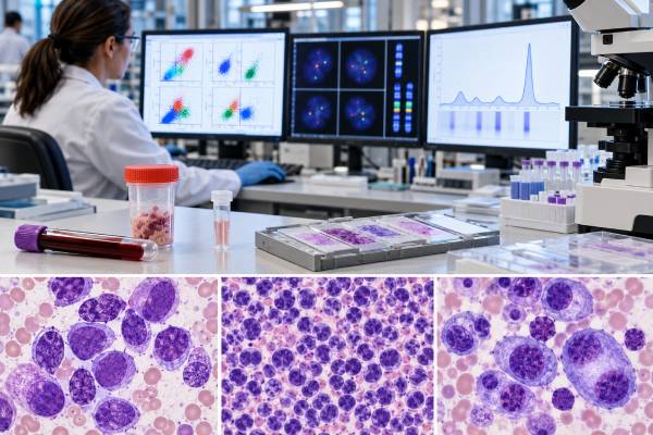
A hematologic cancer biomarker panel is not one universal blood test. It is a coordinated set of laboratory methods used to identify, classify, risk-stratify, and sometimes monitor leukemia, lymphoma, myeloma, and related blood cancers. The exact combination depends on the suspected disease and the specimen. A leukemia workup may combine blood and bone marrow morphology, flow cytometry, chromosome analysis, FISH, PCR, and next-generation sequencing. Lymphoma usually requires a tissue biopsy with immunohistochemistry, often supplemented by flow cytometry and targeted genetic testing. Myeloma adds serum and urine protein studies, free light chains, bone marrow plasma-cell markers, and myeloma-specific FISH. No single marker proves “blood cancer” across all these diseases. Instead, each result contributes a different layer: cell lineage, clonality, defining genetic abnormality, prognosis, treatment target, or disease burden. The most accurate diagnosis comes from integrating those layers with symptoms, blood counts, imaging, and pathology.
- There is no single standard hematologic cancer panel; the markers and methods should match the suspected leukemia, lymphoma, myeloma, or myeloid neoplasm.
- Flow cytometry identifies abnormal cell populations by their surface and intracellular markers, while FISH, PCR, and NGS identify chromosome or gene abnormalities.
- Lymphoma generally requires tissue architecture from a biopsy; a blood biomarker panel alone cannot classify most lymphomas.
- Myeloma testing commonly combines SPEP, immunofixation, serum free light chains, bone marrow plasma-cell assessment, and cytogenetic/FISH risk testing.
- A negative result on one panel does not rule out hematologic cancer if the wrong specimen, wrong marker set, or insufficiently sensitive method was used.
Table of Contents
- What a Hematologic Cancer Biomarker Panel Is
- Major Testing Methods and What They Show
- Common Leukemia Biomarkers
- Common Lymphoma Biomarkers
- Common Myeloma Biomarkers
- How Results Are Interpreted Together
- Limitations, Follow-Up, and Next Steps
What a Hematologic Cancer Biomarker Panel Is
A hematologic cancer biomarker panel is best understood as a diagnostic toolkit rather than a fixed menu of markers. Blood cancers arise from different stages of blood-cell development, so the useful biomarkers for one disease can be irrelevant to another.
Modern classifications of hematologic malignancies integrate several types of evidence. Morphology shows what the cells look like and how they are arranged. Immunophenotyping identifies proteins that reveal cell lineage and maturation. Cytogenetic testing shows whole-chromosome or large structural abnormalities. Molecular tests identify gene mutations, fusions, and rearrangements that can define a disease or guide treatment.
That integration is especially important because one marker can appear in multiple diseases. CD20 identifies mature B cells, for example, but many different B-cell lymphomas express CD20. CD34 can mark immature blasts but does not specify which leukemia is present. A JAK2 mutation strongly supports a clonal myeloproliferative process in the right setting, yet the exact diagnosis still depends on blood counts and marrow morphology.
The specimen matters just as much as the marker. Leukemia often involves blood and bone marrow, so both can be informative. Lymphoma may be confined to a lymph node or extranodal tissue, making a tissue biopsy essential. Myeloma involves plasma cells in bone marrow but also produces measurable monoclonal proteins or free light chains in blood and urine.
A broad molecular panel may be called a hematologic malignancy NGS panel, but NGS is only one part of the workup. It cannot show tissue architecture, reliably quantify all proteins, or replace every FISH or PCR assay. Likewise, flow cytometry cannot identify every gene fusion that determines modern disease classification.
The best panel therefore starts with a clinical question: What disease is suspected, what specimen contains the abnormal cells, and what information is needed for diagnosis, prognosis, treatment selection, or monitoring?
Major Testing Methods and What They Show
Different laboratory methods answer different questions. A useful panel combines complementary methods rather than repeating the same information.
| Method | What it measures | Common use |
|---|---|---|
| Morphology | Cell appearance, blast percentage, tissue architecture | Initial classification and confirmation of marrow or tissue disease |
| Flow cytometry | Multiple cell-surface and intracellular proteins on individual cells | Leukemia lineage, lymphoma clonality, abnormal plasma-cell phenotype, MRD |
| Immunohistochemistry | Protein expression in intact tissue | Lymphoma classification, plasma-cell identification, lineage markers |
| Karyotype | Whole chromosomes and large structural abnormalities | Myeloid neoplasm and leukemia classification and risk |
| FISH | Targeted gains, losses, rearrangements, or fusions | Rapid detection of disease-defining or prognostic chromosome changes |
| PCR | Specific mutations or fusion transcripts | Rapid diagnosis and highly sensitive monitoring |
| NGS | Many gene variants and, on some platforms, fusions or copy changes | Classification, risk, targeted therapy, clonal assessment |
| Serum/urine protein studies | Monoclonal immunoglobulins and free light chains | Myeloma and plasma-cell disorder diagnosis and monitoring |
A leukemia flow cytometry panel can distinguish myeloid, B-lymphoid, T-lymphoid, or mixed-lineage blasts within hours. However, a genetic test may still be required to name the exact WHO or ICC entity.
Turnaround time varies by method. Flow cytometry and targeted PCR or FISH may return quickly, while karyotyping and broad NGS often take longer. In acute leukemia, laboratories frequently prioritize rapid tests for abnormalities that immediately change therapy, such as PML::RARA, BCR::ABL1, FLT3, or selected actionable mutations.
No special preparation is needed for most biomarker assays beyond the requirements of the blood draw, bone marrow biopsy, or tissue biopsy. The main preanalytic concerns are collecting the right specimen, preserving viable cells for flow cytometry, and avoiding tissue processing that damages DNA or antigens.
The report should also be read in light of each assay’s limit of detection and variant scope. A negative targeted PCR result means the specific alteration was not detected above that assay’s threshold; it does not prove the entire gene is normal. Likewise, an NGS panel may sequence dozens or hundreds of genes but still have limited ability to detect certain long insertions, repetitive regions, balanced rearrangements, or low-level copy-number changes. Laboratories should state which variant types and genomic regions are covered.
Another important distinction is somatic versus germline testing. Most cancer panels are designed to detect acquired changes in tumor or blood cells, not inherited cancer-predisposition variants. A variant near a 50% allele fraction can raise the possibility of a germline alteration, but that percentage alone cannot establish inheritance because tumor purity, copy-number changes, and clonal structure can produce similar values. When an inherited predisposition is suspected, confirmatory testing generally uses nonhematopoietic tissue and follows genetic-counseling standards rather than relying on the diagnostic cancer specimen.
Common Leukemia Biomarkers
Leukemia panels usually begin by asking what lineage the abnormal cells belong to and how immature they are. Flow cytometry uses combinations of markers rather than one protein.
In AML, commonly evaluated markers include myeloid-associated proteins such as CD13, CD33, CD117, and myeloperoxidase, plus immaturity markers such as CD34 and HLA-DR. Some AML subtypes have distinctive patterns; acute promyelocytic leukemia, for example, often lacks HLA-DR and has strong myeloid marker expression, but the diagnosis requires rapid demonstration of PML::RARA.
Molecular profiling in AML now carries major diagnostic and treatment weight. Key examples include:
- NPM1 and CEBPA, which can define biologically important AML groups;
- FLT3, which can affect risk and identify patients for FLT3-directed therapy;
- IDH1 and IDH2, which can identify targeted treatment options;
- TP53 and myelodysplasia-related gene patterns, which can indicate high-risk biology;
- PML::RARA, RUNX1::RUNX1T1, CBFB::MYH11, KMT2A, MECOM, and other fusions/rearrangements, which may define the leukemia subtype.
A FLT3 mutation test illustrates why the panel must go beyond diagnosis: FLT3 is not needed to prove AML exists, but its presence can change frontline therapy and relapse management.
For B-cell acute lymphoblastic leukemia, flow markers can include CD19, CD22, CD79a, CD10, TdT, and others. Genetic testing looks for BCR::ABL1, ETV6::RUNX1, KMT2A rearrangements, hyperdiploidy, and newer molecular subgroups. T-ALL uses T-lineage markers such as cytoplasmic or surface CD3, CD7, and related antigens.
Chronic leukemias require different biomarkers. Chronic myeloid leukemia is defined by BCR::ABL1, while CLL uses a characteristic mature B-cell immunophenotype and genetic tests such as FISH and TP53/IGHV assessment for prognosis and treatment planning.
This is why a “leukemia panel” should never be assumed to cover every leukemia equally. The correct panel depends on whether the concern is acute or chronic, myeloid or lymphoid, and diagnostic or follow-up testing.
Common Lymphoma Biomarkers
Lymphoma diagnosis usually starts with tissue architecture, which is why excisional or core biopsy material is often more informative than blood alone. The pathologist examines the pattern of lymph node or tissue involvement and then applies immunohistochemical markers.
B-cell markers include CD20, PAX5, CD19, and CD79a. T-cell markers include CD3 and other T-lineage antigens. Additional markers help separate specific entities. CD5 and cyclin D1 support mantle cell lymphoma in the correct context; CD10, BCL6, and BCL2 help characterize germinal-center-derived lymphomas; CD30 is central in classical Hodgkin lymphoma and anaplastic large cell lymphoma; ALK can define a major subtype of anaplastic large cell lymphoma.
A cyclin D1 test is a good example of an immunohistochemical biomarker that becomes highly informative only when matched to cell morphology and other B-cell markers.
FISH is essential for several lymphoma questions. Follicular lymphoma commonly has a BCL2 rearrangement. Mantle cell lymphoma commonly has an IG::CCND1 rearrangement. Aggressive large B-cell lymphomas may require MYC, BCL2, and BCL6 FISH to identify high-risk rearrangement patterns. A double-hit lymphoma FISH panel distinguishes genetic MYC/BCL2 disease from protein-only “double expression.”
Flow cytometry can identify a clonal B-cell or abnormal T-cell population and is valuable in lymph node aspirates, blood, marrow, and fluids. However, flow loses tissue architecture. Some lymphomas also yield few viable tumor cells, making flow falsely negative even when histology is diagnostic.
Molecular assays increasingly refine lymphoma classification by detecting rearrangements, mutations, and gene-expression patterns. Still, most lymphoma diagnoses remain integrated tissue diagnoses rather than purely sequence-based labels.
Common Myeloma Biomarkers
Myeloma requires a different biomarker strategy because the malignant cell is a plasma cell and the disease often produces a monoclonal immunoglobulin or light chain.
Core laboratory tests commonly include serum protein electrophoresis (SPEP), serum immunofixation, quantitative immunoglobulins, and serum free kappa and lambda light chains. Urine electrophoresis or immunofixation may add information, especially for light-chain excretion. These tests identify and quantify the monoclonal protein but do not by themselves prove symptomatic myeloma.
A multiple myeloma test panel integrates these protein studies with bone marrow and clinical evidence. Bone marrow examination measures plasma-cell percentage and confirms clonality. CD138 highlights plasma cells in tissue, while flow cytometry can identify an abnormal plasma-cell phenotype.
The serum free light chain assay provides kappa, lambda, and a kappa/lambda ratio. A markedly involved/uninvolved free light chain ratio can serve as one myeloma-defining biomarker in the proper clinical context, but kidney function and inflammatory states can complicate interpretation. The serum free light chain test should therefore be read with SPEP, immunofixation, renal function, and marrow findings.
Myeloma FISH is performed on enriched or identified plasma cells when possible and looks for risk-relevant abnormalities such as del(17p), 1q gain/amplification, t(4;14), t(14;16), t(11;14), and other changes. Some abnormalities have treatment implications; others mainly contribute to risk stratification.
Beta-2 microglobulin, albumin, and LDH help stage or risk-stratify myeloma but are not specific tumor markers. Imaging for lytic bone lesions and organ assessment for anemia, kidney injury, hypercalcemia, and bone disease complete the diagnostic picture.
How Results Are Interpreted Together
The most important principle is concordance. A strong diagnosis usually emerges when morphology, immunophenotype, and genetics point to the same entity.
For example, a lymph node showing a CD20-positive, CD5-positive B-cell lymphoma with diffuse nuclear cyclin D1 expression and an IG::CCND1 rearrangement provides mutually reinforcing evidence for mantle cell lymphoma. A marrow with myeloblasts, an AML immunophenotype, and an NPM1 mutation likewise creates an integrated diagnosis.
Discordant results require investigation rather than averaging. A mutation may represent clonal hematopoiesis rather than the tumor. A FISH abnormality may occur as a secondary event in more than one disease. Flow cytometry may miss cells damaged during processing. NGS may detect a variant of uncertain significance that should not be treated as a disease-defining mutation.
Variant allele frequency adds another layer. A mutation at 35% VAF often represents a substantial clone, while one at 2% may represent a small clone, but VAF is not a direct tumor-cell percentage. Copy number, zygosity, sample purity, and lineage all affect the number.
The purpose of the test also changes interpretation. A 2% mutation may be adequate evidence of clonal hematopoiesis in a diagnostic workup but far too high a detection limit for MRD testing. A diagnostic NGS panel and a validated leukemia MRD test may therefore use completely different sensitivity thresholds.
Reports also separate pathogenic variants from variants of uncertain significance (VUS). A VUS is not established as a disease-causing or treatment-predictive change and should not be promoted to a diagnostic marker simply because it appears on a cancer panel. Its classification can change as evidence accumulates, so clinically important reinterpretation may require review of the original report or updated laboratory classification.
Results should also be dated. Hematologic cancers evolve under treatment. A mutation detected at diagnosis may disappear, while a resistance mutation can emerge at relapse. The current specimen often matters more for a new treatment decision than a molecular profile from years earlier.
Limitations, Follow-Up, and Next Steps
A biomarker panel can fail for several reasons even when a hematologic cancer is present. The specimen may not contain enough tumor. The disease may be in tissue while only blood was tested. The panel may not include the relevant gene, fusion, or copy-number change. The assay may detect point mutations well but miss structural rearrangements. Tissue decalcification can reduce the quality of IHC, FISH, or DNA testing.
A negative broad NGS panel therefore does not mean “no cancer.” Some hematologic neoplasms are diagnosed primarily by morphology, immunophenotype, or chromosome structure. Likewise, a positive mutation panel does not automatically mean cancer because age-related clonal hematopoiesis can produce mutations in genes such as DNMT3A, TET2, and ASXL1.
When the initial workup is incomplete, the next step is usually targeted rather than simply ordering a larger panel. Examples include:
- obtaining a bone marrow biopsy when blood testing shows unexplained cytopenias or blasts;
- obtaining an adequate lymph node or tissue biopsy when lymphoma is suspected;
- adding rapid FISH or PCR for a specific defining abnormality;
- performing plasma-cell-enriched FISH when myeloma risk needs clarification; or
- repeating molecular testing at relapse when treatment targets may have changed.
Patients can make a complex report easier to understand by asking what each test was meant to answer. Was it establishing lineage? Confirming clonality? Naming the disease? Assigning risk? Selecting a drug? Measuring response? The same “positive” result can carry very different weight depending on that purpose.
A complete hematologic cancer diagnosis is therefore closer to a multidimensional profile than a single biomarker score. The most useful final report is an integrated interpretation that names the disease, lists the defining findings, identifies prognostic and actionable markers, notes any technical limitations, and recommends additional testing only when it can change classification or management.
References
- The 5th edition of the World Health Organization Classification of Haematolymphoid Tumours: Myeloid and Histiocytic/Dendritic Neoplasms 2022 (Review)
- The 5th edition of the World Health Organization Classification of Haematolymphoid Tumours: Lymphoid Neoplasms 2022 (Review)
- Molecular Diagnostic Testing for Hematopoietic Neoplasms: Linking Pathogenic Drivers to Personalized Diagnosis 2022 (Review)
- Comprehensive biomarker profiles in hematological malignancies: improving diagnosis, prognosis, and treatment 2025 (Review)
- NGS panel enhance precise diagnosis of myeloid neoplasms under WHO-HAEM5 and International Consensus Classification: An observational study 2024
Disclaimer
Hematologic cancer biomarker panels must be selected and interpreted for the specific suspected disease and specimen. No single panel can reliably diagnose or exclude every leukemia, lymphoma, myeloma, or myeloid neoplasm. Review abnormal or conflicting results with a hematologist and, when relevant, a hematopathologist who can integrate morphology, immunophenotype, cytogenetics, and molecular findings.





