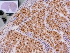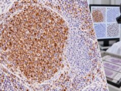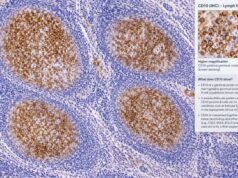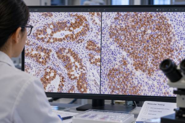
ALK immunohistochemistry (IHC) is a tissue stain that detects abnormal expression of anaplastic lymphoma kinase protein. Its meaning depends heavily on the tumor being tested. In non-small cell lung cancer, strong ALK staining commonly serves as a surrogate for an ALK gene fusion that can make a tumor eligible for ALK-targeted therapy. In anaplastic large cell lymphoma (ALCL), ALK staining helps classify the lymphoma as ALK positive or ALK negative and can show patterns that reflect the underlying fusion protein.
A positive ALK IHC result does not have one universal interpretation across all cancers. The pathologist considers the tumor type, staining location and intensity, assay used, morphology, and companion tests. Discordant or unusual findings may need confirmation with fluorescence in situ hybridization (FISH), RNA-based testing, or next-generation sequencing (NGS). ALK IHC is therefore both a diagnostic marker and, in selected cancers, a practical fusion-screening tool rather than a stand-alone cancer test.
- ALK IHC detects ALK protein in tumor tissue, not the ALK gene alteration itself.
- In lung adenocarcinoma, clearly positive validated ALK IHC strongly supports an ALK fusion and may guide targeted therapy.
- In ALCL, ALK positivity helps define ALK-positive anaplastic large cell lymphoma, usually together with strong CD30 expression and compatible morphology.
- Weak, unusual, or discordant ALK staining may need molecular confirmation with FISH or DNA/RNA-based testing.
- There is no blood “normal range” or universal percent-positive cutoff for ALK IHC; interpretation is assay- and tumor-specific.
Table of Contents
- What the ALK IHC Test Detects
- How ALK IHC Is Performed and Reported
- ALK IHC in Lung Cancer
- ALK IHC in Lymphoma
- Positive, Negative, and Equivocal ALK Results
- ALK IHC Versus FISH and NGS
- What Happens After ALK Testing
What the ALK IHC Test Detects
ALK is a receptor tyrosine kinase. In most normal adult tissues, ALK protein expression is low or absent. Certain cancers acquire rearrangements that fuse the ALK gene to another gene, creating an abnormal signaling protein that can drive tumor growth. Other ALK alterations, including mutations or amplification, occur in selected tumor types but are different from the classic fusion-driven setting.
IHC detects the protein produced in the tissue. It does not directly read the DNA or RNA sequence. This distinction is important because a positive protein stain can act as a highly useful surrogate for an ALK fusion in a validated clinical setting, but it is not technically the same test as FISH or sequencing.
ALK IHC is used most often for two very different clinical questions:
- Does a non-small cell lung cancer express ALK in a pattern that supports an actionable ALK fusion?
- Does a CD30-positive large-cell lymphoma represent ALK-positive ALCL?
It may also be used in the workup of inflammatory myofibroblastic tumor and several rarer ALK-associated neoplasms. Because the differential diagnosis changes by organ and morphology, ALK is generally ordered as part of a larger tumor IHC panel rather than interpreted alone.
In lung cancer, ALK expression is unusual in normal lung epithelium. That creates a favorable signal-to-background relationship for sensitive ALK antibodies. In lymphoma, the location of ALK staining within the cell can also be informative because different fusion partners can direct the abnormal protein to different cellular compartments.
ALK positivity is therefore best understood as a biologic clue whose strength depends on context. In one biopsy it may predict an oncogenic fusion and targeted therapy; in another it may define a lymphoma subtype; in a third it may trigger additional molecular testing before a diagnosis is finalized.
How ALK IHC Is Performed and Reported
ALK IHC is performed on a thin section of formalin-fixed, paraffin-embedded tissue. Lung specimens may come from a resection, core biopsy, endobronchial biopsy, or properly prepared cytology cell block. Lymphoma specimens may come from a lymph node, extranodal mass, skin, or other involved tissue.
The laboratory applies an antibody against ALK, uses a detection system to create visible staining, and checks appropriate controls. The pathologist then asks which cells stain, how strong the reaction is, and where the stain appears within the cells.
In non-small cell lung cancer, commonly used validated assays are designed so that a convincingly positive tumor usually shows strong granular cytoplasmic staining. Some assay systems use a binary interpretation: positive or negative. Background elements and weak nonspecific staining should not be mistaken for true tumor positivity.
In ALCL, the pattern can be more varied. The common NPM::ALK fusion often produces both nuclear and cytoplasmic staining because the fusion partner influences where the protein localizes. Other ALK fusion partners may produce cytoplasmic, membranous, granular, or other patterns. The exact pattern is supportive but usually does not replace molecular identification of the fusion when knowing the partner is clinically necessary.
A report may include:
- The antibody or assay platform used.
- Whether tumor cells are positive or negative.
- The staining intensity and distribution.
- A comment on whether the result supports an ALK rearrangement or ALK-positive ALCL.
- A recommendation for confirmatory testing when the result is weak, atypical, or discordant.
There is no meaningful serum-style reference range such as “0–10 units.” ALK IHC is a tissue-pattern test. The threshold that matters is the validated interpretation rule for the specific laboratory assay and tumor setting.
ALK IHC in Lung Cancer
ALK testing is an important part of biomarker evaluation in non-small cell lung cancer, especially adenocarcinoma and other nonsquamous tumors in which actionable driver alterations are routinely sought. ALK fusions occur in a minority of NSCLC cases, often quoted at roughly 2%–7% depending on the population and testing method. They are enriched in younger patients and people with little or no smoking history, but clinical features should not be used to exclude an otherwise eligible tumor from testing.
The classic lung cancer alteration is an EML4::ALK fusion, although multiple ALK partners and fusion variants exist. The fusion creates constitutive kinase signaling that can be blocked by ALK tyrosine kinase inhibitors. This makes accurate testing clinically important even though the alteration is uncommon.
For lung tumors, ALK IHC can be a highly effective primary test when the assay has been validated. Modern antibodies, including D5F3-based assays, are sensitive enough that clearly positive staining is accepted in many diagnostic algorithms as evidence of ALK protein overexpression caused by a rearrangement. Historic practice relied heavily on break-apart FISH, but IHC is faster, tissue-efficient, and easier to integrate into routine pathology laboratories.
The lung diagnosis itself is usually established before ALK is interpreted. For example, TTF-1 IHC and Napsin A staining can support lung adenocarcinoma differentiation. ALK then answers a different question: whether the tumor has an actionable oncogenic driver.
A positive ALK IHC result in an appropriate NSCLC may be sufficient for treatment selection under a validated testing program. However, molecular testing is increasingly performed with broad NGS panels that can identify ALK fusions alongside EGFR, ROS1, RET, NTRK, MET, KRAS, BRAF, ERBB2, and other relevant alterations. RNA-based NGS can be particularly useful for gene fusions because it detects the expressed fusion transcript.
Discordant results deserve investigation. Examples include strongly positive IHC with negative FISH, an unusual weak stain, or a sequencing result that identifies an ALK rearrangement of uncertain functional significance. In these cases, another method or specimen can help determine whether a true expressed oncogenic fusion is present.
ALK IHC in Lymphoma
ALK IHC has a different role in lymphoma. It is a classification marker, especially for anaplastic large cell lymphoma, rather than simply a predictive screen for a lung cancer drug target.
ALCL is a mature T-cell lymphoma characterized by large anaplastic cells and strong CD30 expression. A CD30 IHC result is therefore central to the diagnosis. Once morphology and immunophenotype raise ALCL, ALK staining separates systemic ALCL into ALK-positive and ALK-negative disease categories.
ALK-positive ALCL is driven by an ALK rearrangement. The most common is t(2;5), which creates NPM::ALK. It occurs frequently in children, adolescents, and young adults, although it can occur at other ages. The tumor cells may lose conventional T-cell antigens despite their T-cell origin, so the diagnosis uses an integrated panel rather than requiring every tumor to express CD3 or other lineage markers.
The ALK staining pattern may provide a clue to the fusion partner:
- Nuclear and cytoplasmic staining strongly suggests NPM::ALK.
- Diffuse cytoplasmic staining may occur with other fusion partners.
- Membranous or granular patterns can occur with selected non-NPM partners.
This pattern recognition can be diagnostically helpful, but it should not be overinterpreted. Molecular testing is more definitive when the exact genetic alteration is important.
ALK-negative ALCL has the same strong CD30 phenotype but no ALK expression and no ALK rearrangement. It is genetically heterogeneous. Selected cases may carry DUSP22, TP63, JAK/STAT pathway, or other abnormalities. Therefore, a negative ALK stain does not mean “not ALCL.” It means that if the lymphoma otherwise meets ALCL criteria, it belongs to the ALK-negative group and may need additional classification work.
A CD3 IHC test and other T-cell markers are often included, but ALCL can lose CD3. Conversely, classic Hodgkin lymphoma is also strongly CD30 positive but is generally ALK negative. PAX5, CD15, EBV studies, T-cell markers, morphology, and clinical setting help distinguish these entities.
ALK expression also occurs in rare ALK-positive large B-cell lymphoma, which is biologically distinct from ALCL. This tumor usually has plasmablastic features and a B-cell/plasma-cell phenotype rather than the classic CD30-rich ALCL phenotype. Recognizing that rare possibility prevents the simple but incorrect assumption that every ALK-positive hematolymphoid tumor is ALCL.
ALK expression also occurs in a small set of tumors outside the two major settings above. Inflammatory myofibroblastic tumors can harbor ALK rearrangements, especially in younger patients, and may show cytoplasmic ALK staining. Rare renal, histiocytic, and other neoplasms also have ALK-driven subsets. The diagnostic value in those settings depends on the morphology and the expected alteration for that tumor family, so a positive stain should trigger a tumor-specific workup rather than be interpreted by lung-cancer rules.
This distinction matters because the same antibody can answer different questions. A strong lung adenocarcinoma stain may function as a predictive fusion screen, whereas an ALCL stain helps name a lymphoma subtype and an inflammatory myofibroblastic tumor stain may support a molecularly defined driver. The report should make clear which clinical question the laboratory is answering.
Positive, Negative, and Equivocal ALK Results
The words “positive” and “negative” mean different things depending on what is being tested. The report should always be read alongside the tumor diagnosis.
| Setting | Positive ALK IHC | Negative ALK IHC |
|---|---|---|
| Non-small cell lung cancer | Strong validated staining supports an ALK fusion and potential ALK-targeted therapy | Usually argues against ALK fusion, but molecular testing may still identify unusual discordant cases |
| Suspected systemic ALCL | Supports ALK-positive ALCL when morphology and CD30 phenotype fit | Does not exclude ALCL; may support ALK-negative ALCL if other criteria are met |
| Other tumor | May suggest an ALK-driven neoplasm but requires tumor-specific interpretation | Meaning depends on the differential diagnosis and expected sensitivity |
A clearly positive lung cancer stain is generally strong and granular in tumor cell cytoplasm. Very weak or focal staining is more difficult and may reflect technical background, true low-level expression, limited tissue, or unusual tumor biology. Laboratories should define how such cases are handled during assay validation.
In lymphoma, intensity and cellular localization matter more than a numeric percentage alone. ALK-positive ALCL usually shows convincing tumor-cell staining, while background elements should not drive the interpretation. The pathologist also checks that the large CD30-positive cells are the same cells expressing ALK.
Equivocal, unexpected, or discordant findings are a reason to resolve the result rather than force a binary conclusion. Repeat IHC on another block, FISH, RNA sequencing, or broader NGS can clarify the biology. This is especially useful when a treatment decision depends on the result.
ALK IHC Versus FISH and NGS
No single ALK method is ideal for every specimen. IHC, FISH, and sequencing examine different aspects of the same problem.
IHC detects protein expression. It is fast, relatively inexpensive, tissue-sparing, and widely available. In validated NSCLC testing, it can perform extremely well. Its limitation is that it infers an underlying genomic event rather than identifying the exact fusion sequence.
FISH uses labeled DNA probes, commonly a break-apart design, to show rearrangement of the ALK locus. It was historically a companion diagnostic standard in lung cancer. It can work on small samples but requires specialized microscopy and interpretation, and unusual genomic configurations may create borderline or discordant patterns.
NGS can test many cancer genes at once. DNA-based NGS may detect rearrangements when the breakpoints are covered, while RNA-based NGS directly identifies expressed fusion transcripts and their partners. RNA quality can be a limitation in older or poorly preserved tissue, but RNA methods are particularly valuable for fusion-rich cancers.
A practical modern approach is molecular redundancy: when a result is unexpected or uncertain, use an orthogonal method that measures the alteration differently. For example, a questionable ALK IHC result may be resolved with RNA NGS rather than simply repeating the same assay. This reduces the risk that one technical limitation determines treatment.
What Happens After ALK Testing
For lung cancer, a positive ALK result usually enters the molecular pathology report with other predictive biomarkers. The oncology team then considers an ALK tyrosine kinase inhibitor according to disease stage, prior treatment, current guidelines, drug availability, central nervous system involvement, and patient factors. The ALK result is predictive because it identifies a tumor that may depend on ALK signaling.
For lymphoma, the result is incorporated into the final subtype. ALK-positive ALCL and ALK-negative ALCL are distinct diagnostic categories with different epidemiology, genetics, and prognostic patterns. Treatment decisions depend on the complete lymphoma classification, stage, patient age and fitness, and other clinical factors rather than ALK staining alone.
If the result is negative but clinical suspicion remains high, broad molecular testing may still be appropriate. A small biopsy can be false-negative because of poor preservation or low tumor content, and some assays have different sensitivity for uncommon fusion variants. Conversely, a positive stain in a tumor that does not fit an ALK-associated entity should prompt verification rather than an automatic diagnosis.
Patients reviewing an ALK report can ask four useful questions: What tumor type was tested? Was ALK IHC clearly positive or only weak/equivocal? Does the result need molecular confirmation? Does it change the diagnosis, treatment options, or both? Those questions connect the stain to the decision it was intended to support.
References
- A narrative review of methods for the identification of ALK fusions in patients with non-small cell lung carcinoma 2023 (Review)
- New update to the guidelines on testing predictive biomarkers in non-small-cell lung cancer: a National Consensus of the Spanish Society of Pathology and the Spanish Society of Medical Oncology 2023 (Guideline)
- Fifth Edition of the World Health Organization Classification of Tumors of the Hematopoietic and Lymphoid Tissues: Mature T-Cell, NK-Cell, and Stroma-Derived Neoplasms of Lymphoid Tissues 2024 (Review)
- Updates in pathobiological aspects of anaplastic large cell lymphoma 2023 (Review)
- Updated Molecular Testing Guideline for the Selection of Lung Cancer Patients for Treatment With Targeted Tyrosine Kinase Inhibitors: Guideline From the College of American Pathologists, the International Association for the Study of Lung Cancer, and the Association for Molecular Pathology 2018 (Guideline)
Disclaimer
ALK IHC must be interpreted by a qualified pathologist with the tumor morphology, clinical setting, assay validation, and other laboratory results. A positive or negative stain alone cannot establish every ALK-associated diagnosis, and equivocal or discordant results may require molecular confirmation. Treatment choices should be made with the oncology team using the complete pathology and molecular report.