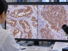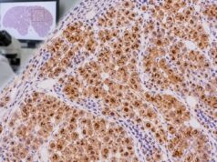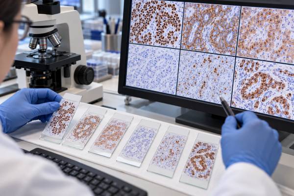
A tumor immunohistochemistry (IHC) panel is a group of tissue stains selected to identify a cancer’s lineage, subtype, and likely site of origin. Instead of asking one marker to provide the whole diagnosis, pathologists combine several positive and negative results with the tumor’s microscopic appearance and the patient’s clinical information. This approach is especially important in poorly differentiated tumors, small biopsies, metastatic cancer, and carcinoma of unknown primary. A panel may begin with broad lineage markers to distinguish carcinoma, lymphoma, melanoma, and sarcoma, then move to organ-associated markers such as TTF-1, PAX8, GATA3, CDX2, SATB2, NKX3.1, or hepatocellular markers. Neuroendocrine markers may be added when morphology suggests that phenotype. The most useful panel is usually focused rather than exhaustive: unnecessary stains can waste tissue and create confusing incidental positivity. IHC narrows probabilities; it does not replace morphology, imaging, molecular testing, or multidisciplinary clinical judgment.
- An IHC panel uses multiple protein stains together to classify tumor lineage and narrow the likely primary site.
- Panels are chosen from the microscopic differential diagnosis, not from a fixed universal list.
- Positive and negative markers are both informative; a coherent pattern is more reliable than one positive stain.
- Small biopsies require tissue conservation so enough material remains for molecular and predictive biomarker testing.
- IHC can strongly suggest tumor origin but may not identify a primary site in every metastatic cancer.
Table of Contents
- What a tumor IHC panel is
- Step one: establish the tumor lineage
- Step two: narrow the primary site in a carcinoma
- Specialized panels for common tumor patterns
- Why IHC panels can be misleading
- IHC versus molecular and genomic testing
- How to read an IHC panel in a pathology report
What a tumor IHC panel is
Immunohistochemistry uses antibodies to detect proteins in tissue sections. Each stain highlights a protein associated with a cell lineage, differentiation program, mutation surrogate, receptor, proliferation state, or other biologic feature. A panel combines several stains because most individual proteins are shared by more than one tumor type.
The pathologist begins with hematoxylin-and-eosin morphology. If the tumor looks gland-forming, squamous, spindle-cell, lymphoid, melanocytic, or neuroendocrine, that appearance determines the first set of stains. A pancytokeratin IHC stain can support epithelial differentiation, while CD45 supports hematolymphoid lineage.
The result is an immunophenotype—a pattern of markers—not a simple positive/negative cancer test.
Step one: establish the tumor lineage
For a very poorly differentiated malignancy, the first goal may be to determine whether it is carcinoma, lymphoma, melanoma, sarcoma, or another neoplasm. Broad epithelial keratins, CD45, SOX10/S100, and selected mesenchymal markers can be used in a small initial panel.
This first step prevents a common diagnostic error: applying organ-specific carcinoma markers before proving that the tumor is actually epithelial. For example, GATA3 can be positive in several non-breast tumors, and SOX10 can occur in melanoma, nerve sheath tumors, breast carcinomas, and salivary neoplasms. Their meaning changes depending on lineage.
Once lineage is established, the panel can become more specific. A carcinoma may be subdivided by CK7 and CK20 pattern; a lymphoma by B- or T-cell markers; a melanoma by melanocytic markers; and a sarcoma by morphology-driven muscle, vascular, neural, or other stains.
Step two: narrow the primary site in a carcinoma
For metastatic carcinoma, CK7 and CK20 are common organizing markers but rarely give the final answer. A CK7 stain and CK20 stain create broad patterns that guide the next antibodies.
| Likely lineage or site | Commonly useful markers | Important caveat |
|---|---|---|
| Lung adenocarcinoma | TTF-1, Napsin A | Some lung cancers are negative; rare non-lung tumors can be positive |
| Colorectal / lower GI | CDX2, SATB2, CK20 | Intestinal markers can occur outside colorectum |
| Müllerian / renal / thyroid | PAX8 plus context-specific markers | PAX8 spans several organ systems |
| Breast / urothelial | GATA3 plus organ-specific companions | GATA3 is not specific to one site |
| Prostate | NKX3.1, PSA, PSAP | Treatment can reduce some marker expression |
| Hepatocellular | Arginase-1, HepPar-1, glypican-3 | Each has sensitivity/specificity limitations |
The site suggested by IHC is then checked against imaging and the pattern of metastasis.
Specialized panels for common tumor patterns
Neuroendocrine morphology may prompt synaptophysin, chromogranin A, and INSM1, followed by Ki-67 and site-specific transcription factors. Melanocytic tumors may use SOX10, S100, Melan-A, HMB-45, and PRAME. Mesothelioma differentials may use calretinin, WT1, D2-40, CK5/6, claudin-4, and other epithelial exclusion markers.
Gynecologic tumors often use PAX8, WT1, ER, p53, and p16 according to morphology. Germ cell tumors may require SALL4, OCT4, SOX2 or SOX17, CD30, PLAP, glypican-3, and AFP. Hematolymphoid lesions use a separate lineage and maturation framework.
The key is that a marker’s value is conditional. PAX8 positivity, for example, means something different in an ovarian mass than in a renal mass or thyroid lesion. Panels are built around the actual competing diagnoses.
Why IHC panels can be misleading
No IHC marker is perfectly sensitive and specific. Tumors can lose expected differentiation as they become high grade, gain unusual proteins, change after treatment, or show intratumoral heterogeneity. Small biopsies may sample only one component of a mixed cancer.
Technical factors also matter. Fixation, decalcification, necrosis, antibody clone, antigen retrieval, and laboratory validation can alter staining. Internal controls help show whether a negative result is biologically credible.
Another danger is “shotgun” testing. Large indiscriminate panels increase the chance of incidental focal positivity, which can create false trails. A morphology-driven sequence is usually more accurate: ask one focused question, interpret the answer, then choose the next stain only if needed.
IHC versus molecular and genomic testing
IHC and molecular testing answer overlapping but different questions. IHC can identify lineage, protein expression, receptor status, proliferation, and surrogate patterns for selected genomic changes. DNA/RNA assays can detect mutations, fusions, copy-number changes, microsatellite instability, tumor mutational burden, and other molecular features.
Modern cancer diagnosis often needs both. A small metastatic lung adenocarcinoma may require TTF-1/Napsin A for lineage and then broad molecular profiling for actionable alterations. A breast carcinoma needs ER, PR, and HER2 assessment. A mismatch-repair IHC panel may trigger or complement molecular MSI testing.
Tissue conservation is therefore part of panel design. The best IHC workup reaches a defensible diagnosis while leaving sufficient material for predictive biomarkers that may directly affect therapy.
How to read an IHC panel in a pathology report
A pathology report may list stains in a table or narrative. Focus first on the final diagnosis and comment, because those sections integrate the full pattern. A long list of positive markers is not meant to be interpreted independently by counting positives.
Ask which diagnosis each stain was intended to support or exclude. A negative marker can be as important as a positive one. For example, TTF-1 positivity plus Napsin A positivity and lack of a competing organ marker is stronger evidence for lung adenocarcinoma than TTF-1 alone.
In cancer of unknown primary, even a well-designed panel may narrow the tumor to a likely family without identifying one site with certainty. Current guidelines support combining histopathology, clinical evaluation, imaging, and molecular testing where appropriate rather than forcing an unsupported primary-site label.
References
- Carcinoma of unknown primary (CUP): an update for histopathologists 2023 (Review)
- Cancer of unknown primary: ESMO Clinical Practice Guideline for diagnosis, treatment and follow-up 2023 (Guideline)
- Detecting undetectable – epidemiology, etiology, and diagnosis of carcinoma of unknown primary – systematic review 2023 (Systematic Review)
- Carcinoma of Unknown Primary Origin: Application of Immunohistochemistry With Emphasis to Different Cytokeratin 7 and 20 Staining Patterns. 2022 (Study)
- Immunohistochemistry for Diagnosis of Metastatic Carcinomas of Unknown Primary Site 2018 (Review)
- Practical Applications in Immunohistochemistry: Carcinomas of Unknown Primary Site 2016 (Review)
Disclaimer
Tumor IHC panels are interpreted by pathologists in the context of morphology, specimen quality, clinical history, imaging, and other laboratory studies. No single panel can identify every tumor or primary site with certainty. Molecular testing and additional sampling may be needed when the immunophenotype is inconclusive.