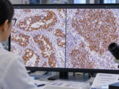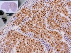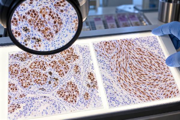
SOX10 immunohistochemistry (IHC) is a nuclear tissue stain that helps identify tumors showing melanocytic, Schwann-cell, or related neural-crest differentiation. It is widely used in the diagnosis of melanoma and peripheral nerve sheath tumors because many of these neoplasms show clear nuclear SOX10 expression. SOX10 can be particularly valuable in spindle-cell and desmoplastic melanoma, where other melanocytic markers such as HMB-45 or Melan-A may be weak or absent. The marker is not limited to melanoma or nerve sheath tumors, however. Normal melanocytes and Schwann cells stain, and subsets of breast, salivary, myoepithelial, and other tumors may also be positive. A positive result therefore indicates a differentiation pattern rather than a single cancer type. Pathologists interpret SOX10 together with microscopic features, anatomic site, clinical history, and a targeted panel that may include S100, Melan-A, HMB-45, PRAME, cytokeratins, and additional lineage-specific stains.
- SOX10-positive staining is nuclear and strongly supports melanocytic or Schwannian differentiation in the appropriate setting.
- Melanoma is commonly SOX10 positive, including many desmoplastic and spindle-cell melanomas.
- Schwannoma and neurofibroma are usually positive, while malignant peripheral nerve sheath tumor is less consistently positive.
- SOX10 is not melanoma-specific: some breast, salivary, myoepithelial, and other neoplasms can express it.
- The result is interpreted in a panel, not by a universal percentage cutoff or blood reference range.
Table of Contents
- What the SOX10 IHC test detects
- SOX10 staining in melanoma
- SOX10 staining in nerve sheath tumors
- Other tumors that can be SOX10 positive
- How positive, negative, and focal SOX10 results are interpreted
- How SOX10 fits into a tumor-origin IHC panel
- What the SOX10 result means for care
What the SOX10 IHC test detects
SOX10 is a transcription factor involved in neural crest development and differentiation. In tissue, a validated antibody produces a nuclear signal in cells that express the protein. Pathologists use this crisp nuclear pattern because it can be easier to localize to tumor cells than the diffuse cytoplasmic staining produced by some older markers.
A SOX10 stain is usually ordered after routine microscopy suggests a melanocytic lesion, a peripheral nerve sheath tumor, or another SOX10-associated neoplasm. It may also be included in an unknown-primary panel when melanoma is a serious possibility. An S100 IHC stain often complements SOX10 because S100 is very sensitive for melanocytic and Schwannian lesions but is less specific.
SOX10 IHC is not a genetic test for a SOX10 mutation and is not a blood test. It measures protein expression in the sampled tissue and does not have a normal serum range.
SOX10 staining in melanoma
Most conventional melanomas show nuclear SOX10 staining. The marker is especially useful in metastatic melanoma and in spindle-cell or desmoplastic melanoma, where melanocytic differentiation can be subtle. Studies have found high sensitivity in these difficult variants, and the nuclear pattern can separate melanoma cells from many background fibroblasts and histiocytes that may complicate S100 interpretation.
SOX10 does not distinguish a benign nevus from melanoma because benign melanocytes and nevi also express the protein. Diagnosis of malignancy therefore still depends on architecture, cytologic atypia, growth pattern, clinical context, and—in selected cases—additional ancillary testing.
Pathologists commonly pair SOX10 with HMB-45 and Melan-A. PRAME may add information in selected melanocytic lesions. Desmoplastic melanoma may retain SOX10 and S100 while showing little or no HMB-45 or Melan-A, which is an important practical pattern.
SOX10 staining in nerve sheath tumors
Schwannomas and neurofibromas generally show strong SOX10 expression because the neoplastic cells have Schwann-cell differentiation. This makes SOX10 a valuable marker when a spindle-cell soft tissue tumor is being assessed for nerve sheath origin.
Malignant peripheral nerve sheath tumor (MPNST) is more difficult. SOX10 sensitivity is lower in MPNST than in benign Schwannian tumors, and high-grade tumors may lose markers of mature Schwann-cell differentiation. A negative SOX10 result therefore does not exclude MPNST. The diagnosis relies heavily on morphology, relationship to a nerve or neurofibroma, clinical features such as neurofibromatosis type 1, and other stains or molecular findings.
In small biopsies, pathologists also have to distinguish nerve sheath tumors from melanoma, clear cell sarcoma, synovial sarcoma, and other spindle or epithelioid neoplasms. SOX10 narrows the field, but the remaining differential determines which stains or molecular tests come next.
Other tumors that can be SOX10 positive
SOX10 positivity outside melanoma and nerve sheath tumors is a major interpretive pitfall. A significant subset of triple-negative and basal-like breast cancers can express SOX10. Some salivary gland tumors and myoepithelial neoplasms are also positive because SOX10 participates in normal salivary and myoepithelial differentiation.
This overlap becomes important in metastases. A SOX10-positive tumor in an axillary lymph node or lung is not automatically melanoma. Cytokeratins, GATA3, TRPS1, mammaglobin, and other breast markers may be needed if breast carcinoma is plausible. Salivary-site tumors require a different panel based on morphology.
SOX10 also labels normal structures, including melanocytes and Schwann cells, which can act as internal controls but can also be mistaken for tumor staining in tiny samples. The pathologist identifies exactly which nuclei are positive before assigning diagnostic weight.
How positive, negative, and focal SOX10 results are interpreted
A strong diffuse nuclear pattern in a morphologically compatible tumor is generally convincing. Focal staining is still potentially meaningful, especially in a heterogeneous or treated tumor, but carries less weight. The percentage of positive cells may be recorded, yet there is no universal diagnostic cutoff that applies to every disease.
A negative result reduces support for a melanocytic or Schwannian lineage but does not eliminate it. Marker loss can occur in malignant tumors, and tissue quality can reduce staining. For melanoma, pathologists may use S100, Melan-A, HMB-45, PRAME, or other melanocytic markers when SOX10 is negative but suspicion remains.
The phrase “SOX10 positive” should therefore be read as one line of evidence. It becomes much stronger when the morphology and other markers point in the same direction.
How SOX10 fits into a tumor-origin IHC panel
In an undifferentiated tumor, SOX10 can be used alongside broad lineage stains. Pancytokeratin supports epithelial differentiation, CD45 supports hematolymphoid lineage, and SOX10/S100 can raise melanocytic or Schwannian possibilities. Once lineage is narrowed, more specific stains are added.
For suspected melanoma, a panel might include SOX10, S100, Melan-A, HMB-45, and PRAME. For a nerve sheath tumor, morphology and S100/SOX10 are combined with markers chosen to exclude mimics. For a possible triple-negative breast carcinoma, epithelial markers and breast-lineage stains are critical because SOX10 alone can mislead.
This stepwise approach preserves tissue and reduces false certainty. It mirrors the principles of a broader tumor-origin IHC panel, where no individual marker is expected to answer every diagnostic question.
What the SOX10 result means for care
SOX10 itself usually does not determine cancer stage or select a specific drug. Its main clinical value is helping the pathology team classify the tumor accurately. That classification then directs staging, molecular testing, surgery, systemic therapy, and surveillance.
If a melanoma diagnosis is confirmed, treatment decisions may depend on stage and molecular findings such as BRAF status rather than the amount of SOX10 staining. If a nerve sheath tumor is diagnosed, grade, resection status, location, and associated syndromes become more important.
When reviewing a pathology report, ask which cells were SOX10 positive, which diagnoses the stain supports, what competing diagnoses were excluded, and which other markers agreed with the interpretation. Those details give the result its practical meaning.
References
- SOX10 2023 (Review)
- Performance of preferentially expressed antigen in melanoma (PRAME) immunohistochemistry for metastatic melanoma in cytology specimens 2024 (Study)
- Immunohistochemical Markers for Distinguishing Metastatic Breast Carcinoma from Other Common Malignancies: Update and Revisit 2022 (Review)
- A newly developed mouse monoclonal SOX10 antibody is a highly sensitive and specific marker for malignant melanoma, including spindle cell and desmoplastic melanomas 2015 (Study)
- Sox10 and S100 in the diagnosis of soft-tissue neoplasms 2012 (Study)
Disclaimer
SOX10 IHC is an ancillary tissue stain and cannot diagnose melanoma, a nerve sheath tumor, or another cancer by itself. Interpretation depends on microscopic findings, the anatomic site, internal controls, and complementary stains. Discuss the final integrated diagnosis with the treating clinician or pathologist.