
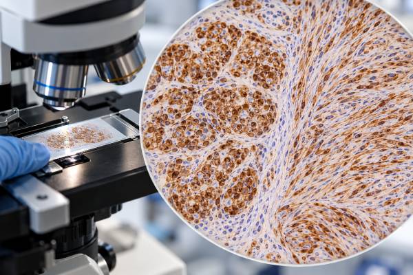
The S100 immunohistochemistry (IHC) test is a sensitive tissue stain used in the diagnosis of melanoma, peripheral nerve sheath tumors, and several other neoplasms. S100 proteins are found in many normal cell types, so positive staining is broad rather than disease-specific. In melanoma, S100 is highly sensitive and can highlight tumor cells that are difficult to see, but it also stains dendritic cells, Schwann cells, adipocytes, cartilage, myoepithelial cells, and numerous other tumors. For that reason, pathologists usually combine it with more specific melanocytic markers such as SOX10, Melan-A, or HMB-45. In nerve sheath pathology, diffuse strong S100 staining is characteristic of many schwannomas, while malignant peripheral nerve sheath tumors may show only focal staining or lose S100 altogether. The expected reaction often includes both nuclear and cytoplasmic staining. A positive S100 result therefore supports a lineage or differentiation pattern; it does not prove melanoma, prove a tumor is benign, determine its origin, or establish cancer stage by itself.
- What S100 detects: a family of calcium-binding proteins expressed in melanocytic, Schwannian, dendritic, adipocytic, cartilaginous, and other cell types.
- What positive staining can support: melanoma or nerve sheath differentiation when the morphology and companion marker panel agree.
- Why S100 is not specific: many benign cells and nonmelanoma tumors are S100 positive, so a positive stain cannot establish melanoma alone.
- In nerve sheath tumors: schwannomas are often strongly and diffusely positive, while high-grade malignant peripheral nerve sheath tumors can be focal or negative.
- How results are confirmed: SOX10, Melan-A, HMB-45, keratins, and other markers are selected according to the suspected diagnosis.
Table of Contents
- What the S100 IHC Test Detects
- S100 Staining in Melanoma
- S100 in Nerve Sheath Tumors
- What Positive and Negative S100 Results Mean
- How S100 Fits Into IHC Marker Panels
- Limitations and Diagnostic Pitfalls
- What Happens After an S100 Result
What the S100 IHC Test Detects
S100 refers to a family of small calcium-binding proteins involved in intracellular and extracellular signaling. Several S100 family members are present in normal melanocytes, Schwann cells, dendritic cells, chondrocytes, adipocytes, myoepithelial cells, and other tissues. A commonly used S100 IHC antibody detects proteins shared across this family and therefore labels a wide range of cell types.
The test is performed on a thin section of biopsy or surgical tissue. When the antibody binds, positive cells typically show nuclear and cytoplasmic staining. The exact intensity and distribution vary by tissue and tumor. The pathologist identifies which cells are staining and compares that pattern with the routine microscopic appearance.
S100 is best thought of as a sensitive lineage-associated marker rather than a specific cancer marker. In melanoma, its sensitivity is one of its main advantages. In peripheral nerve sheath tumors, the pattern can show Schwannian differentiation. However, the same broad sensitivity means many unrelated tissues are also positive.
The test is not a serum S100 assay and does not produce a blood concentration. Tissue IHC answers a spatial question: which cells in this specimen express S100? That is different from research or clinical blood assays of individual S100 proteins.
S100 is also not a genetic test. It does not identify BRAF, NF1, NF2, PRKAR1A, or other mutations that may be relevant to melanoma or nerve sheath tumors. Molecular tests and IHC answer different questions and may both be needed in difficult cases.
Because S100 can highlight normal dendritic cells or nerves in a tumor, a pathologist must always correlate staining with the cells that look neoplastic. Strong staining in the wrong cell population does not support the suspected diagnosis.
S100 Staining in Melanoma
S100 has long been used as a highly sensitive marker for melanocytic differentiation. Most conventional melanomas show S100 expression, making it useful for detecting tumor cells in primary and metastatic lesions. In difficult specimens, the stain can make subtle infiltrating cells easier to recognize.
High sensitivity is not the same as high specificity. S100 labels Langerhans cells, other dendritic cells, Schwann cells, and many mesenchymal or myoepithelial tumors. In skin, this background can make a heavily inflamed or scarred specimen harder to interpret. A brown S100-positive cell is not automatically a melanoma cell.
SOX10 has become an important companion and, in many settings, a preferred melanocytic nuclear marker because it has similar high sensitivity with a cleaner pattern in certain tissues. The SOX10 IHC test is still not absolutely melanoma-specific, because Schwannian and some myoepithelial tumors also express SOX10.
Melan-A and HMB-45 provide additional melanocytic evidence. The HMB-45 IHC test recognizes a melanocytic differentiation antigen and is generally more specific but less uniformly sensitive than S100. Melan-A has a different sensitivity/specificity balance and may be very helpful in a panel.
Some melanoma subtypes can lose conventional melanocytic markers, particularly when they are dedifferentiated, spindle-cell, or treated. Desmoplastic melanoma is a classic example in which S100 and SOX10 can remain strong while HMB-45 and Melan-A are often absent. This is why the marker panel must fit the morphology rather than follow one fixed recipe.
S100 positivity does not measure melanoma depth, ulceration, lymph-node status, or metastatic stage. Those features are determined by the pathology architecture and clinical staging workup.
S100 in Nerve Sheath Tumors
S100 is also central to the evaluation of peripheral nerve sheath tumors because Schwann cells normally express S100 proteins. The pattern can help distinguish different benign and malignant nerve sheath lesions, although no result should be interpreted alone.
Schwannoma usually shows strong, diffuse S100 staining in the neoplastic Schwann cells. SOX10 is also commonly diffuse. This pattern fits the relatively pure Schwann-cell composition of the tumor. Histologic features such as Antoni A and Antoni B areas, Verocay bodies, capsule, and degenerative change provide the structural context.
Neurofibroma can also be S100 positive, but the tumor contains a mixed population of Schwann cells, fibroblastic cells, mast cells, and other elements. The staining can therefore be less uniformly intense than in schwannoma. Architecture, clinical setting, and other markers help distinguish the two.
Malignant peripheral nerve sheath tumor (MPNST) is more challenging. High-grade MPNST may show only focal S100 or SOX10 staining, and some cases are completely negative. Loss of H3K27me3 can support MPNST in selected clinical and morphologic settings, particularly certain sporadic and radiation-associated tumors, but that marker is not universally lost and is not specific enough to stand alone.
This creates a key interpretive asymmetry: diffuse strong S100 can support Schwannian differentiation, but negative S100 does not exclude MPNST. The higher-grade and more dedifferentiated the malignant tumor becomes, the more likely it may be to lose lineage markers.
Other S100-positive nerve-associated lesions include granular cell tumor and some tumors with melanocytic or myoepithelial differentiation. Clinical association with a nerve, imaging, gross appearance, and morphology remain important components of the diagnosis.
What Positive and Negative S100 Results Mean
A positive S100 result means the tested cells express S100-family protein recognized by the antibody. The result becomes meaningful only after the pathologist identifies which cells are positive and decides whether the pattern fits the suspected tumor.
Diffuse nuclear and cytoplasmic staining in a malignant epithelioid or spindle-cell tumor may support melanoma if SOX10 and other melanocytic markers also fit. The same diffuse staining in a well-circumscribed nerve-associated spindle-cell tumor may support schwannoma. Context changes the interpretation completely.
Focal staining is weaker evidence. A few S100-positive cells may represent tumor heterogeneity, entrapped nerves, dendritic cells, or another normal population. The pathologist correlates the stained slide directly with the routine section to avoid assigning background cells to the tumor.
A negative S100 result lowers the likelihood of many conventional melanomas and benign Schwannian tumors but does not rule them out absolutely. Dedifferentiated melanoma and high-grade MPNST may lose expression. Technical problems can also cause false negativity.
There is no universal percentage cutoff that defines a clinically positive S100 tumor across all applications. Unlike breast hormone-receptor testing, S100 is usually interpreted qualitatively or semiquantitatively as diffuse, patchy, focal, weak, or strong. The diagnostic weight of each pattern depends on the tumor type.
Positive S100 should also not be equated with benign behavior. Benign schwannoma can be strongly positive, but melanoma and other malignant tumors may also be strongly positive. Likewise, loss of S100 does not automatically indicate aggressive behavior. The marker is primarily about differentiation, not grade.
How S100 Fits Into IHC Marker Panels
When melanoma is suspected, a panel typically combines markers with complementary strengths. S100 provides high sensitivity. SOX10 gives a clean nuclear marker for melanocytic and Schwannian differentiation. HMB-45 and Melan-A provide more melanocyte-oriented differentiation evidence. PRAME can help in selected melanocytic lesions but is not a substitute for morphology and is not positive in every melanoma.
A simplified approach looks like this:
| Marker | Main strength | Main limitation |
|---|---|---|
| S100 | Very sensitive for many melanomas and Schwannian tumors | Low specificity; many normal cells and tumors are positive |
| SOX10 | Sensitive nuclear marker with a relatively clean pattern | Also positive in Schwannian and some myoepithelial tumors |
| Melan-A | Supports melanocytic differentiation | Can be absent in desmoplastic/dedifferentiated melanoma |
| HMB-45 | Useful melanocytic differentiation marker | Less sensitive than S100 in several melanoma subtypes |
| Pancytokeratin | Supports epithelial differentiation | Some noncarcinomas can be keratin positive |
If a tumor is poorly differentiated and the lineage is uncertain, the panel may include pancytokeratin for carcinoma, CD45 for lymphoma, and S100/SOX10 for melanoma or neural crest differentiation. The pancytokeratin IHC test is a useful counterpoint because most conventional melanomas and Schwannian tumors are keratin negative.
For nerve sheath tumors, the panel may include SOX10, S100, H3K27me3, EMA, neurofilament, CD34, and other markers based on morphology. A benign schwannoma usually does not require a huge panel if the histology is classic, while an aggressive spindle-cell sarcoma in a patient with neurofibromatosis type 1 may warrant broader testing.
The panel should be designed around real alternatives. Adding multiple overlapping markers without a diagnostic question can increase confusion and consume limited tissue.
Limitations and Diagnostic Pitfalls
S100’s greatest strength—its sensitivity—is also its largest weakness. The marker is expressed in many cell types, so a positive result has limited specificity. Diagnosing melanoma from S100 alone is a classic error, especially in lymph nodes and skin where dendritic cells can be abundant.
Normal nerves and Schwann cells are another common source of background staining. In a soft-tissue tumor, strong S100-positive nerve twigs can be mistaken for tumor cells if the observer does not correlate the exact architecture.
Technical quality matters. Poor fixation, decalcification, necrosis, or a very small specimen can reduce antigen detection. Conversely, nonspecific background can make weak staining appear more convincing than it is. Appropriate controls and validated laboratory methods are essential.
Tumor dedifferentiation can produce false reassurance from a negative stain. High-grade MPNST may lack S100, and some melanomas lose S100 or other melanocytic markers. When morphology strongly suggests one of these tumors, a negative single marker should trigger a broader workup rather than immediate exclusion.
S100-positive nonmelanocytic tumors are numerous. They include many nerve sheath tumors, clear cell sarcoma, granular cell tumor, chondroid lesions, some adipocytic tumors, myoepithelial tumors, and selected histiocytic or dendritic neoplasms. The full differential depends on the organ and cell shape.
Another pitfall is using staining intensity as a behavior score. Strong S100 does not mean a tumor is more advanced, and weak S100 does not mean it is less dangerous. Stage and grade come from separate criteria.
Finally, S100 is not a predictive test for melanoma therapy. BRAF mutation testing, immune biomarkers in selected settings, clinical stage, and current treatment guidelines answer treatment questions that S100 IHC does not.
What Happens After an S100 Result
If S100 is positive in a suspected melanoma, the pathologist usually confirms the interpretation with morphology and one or more additional melanocytic markers. SOX10, Melan-A, and HMB-45 are common choices. The exact combination depends on whether the tumor is conventional, spindle-cell, desmoplastic, metastatic, or otherwise unusual.
If the suspected lesion is a nerve sheath tumor, diffuse S100/SOX10 staining may support schwannoma, while patchy staining can fit neurofibroma or other lesions. A high-grade spindle-cell tumor with clinical nerve association may require additional markers and sometimes molecular testing even when S100 is negative.
When S100 is positive but the morphology does not fit melanoma or nerve sheath tumor, the differential should broaden. Keratins, muscle markers, myoepithelial markers, dendritic-cell markers, or organ-associated stains may clarify the lineage. Prior cancers and the anatomic site can sharply narrow the choices.
For patients, useful questions include: Which cells were S100 positive? Was the staining diffuse or focal? Which other melanocytic or nerve sheath markers were tested? Does the panel establish a diagnosis or only suggest one? Is molecular testing needed to confirm the tumor type or guide treatment?
If melanoma is diagnosed, S100 staining itself does not determine Breslow thickness, ulceration, nodal status, or distant spread. Those features come from the pathology measurement and staging evaluation. If a benign nerve sheath tumor is diagnosed, management depends on symptoms, location, growth, and surgical considerations rather than S100 intensity.
The practical takeaway is that S100 is a highly useful sensitive marker. It can reveal melanocytic or Schwannian differentiation that might otherwise be difficult to recognize, but its broad expression means specificity must come from morphology and a carefully selected marker panel.
S100 also has a useful role in sentinel lymph-node and metastatic melanoma evaluation, but the same specificity problem becomes especially important there. Lymph nodes naturally contain S100-positive dendritic cells, and those cells can be numerous. A pathologist cannot call a nodal metastasis merely because isolated S100-positive cells are present. The cells must have the morphology and distribution of melanoma, and more specific melanocytic markers may be used to confirm suspicious foci.
In skin specimens, scar tissue and inflammation can create similar challenges. Dendritic cells and nerve twigs may stain strongly in an area where a recurrent or desmoplastic melanoma is being considered. Nuclear SOX10 can sometimes make the tumor population easier to separate from background, but it too requires morphologic correlation. The best marker is therefore the one that answers the specific question in that specimen, not necessarily the marker with the highest sensitivity in a textbook table.
Nerve sheath tumors introduce a different type of heterogeneity. Ancient or degenerative schwannomas may contain areas with altered cellularity and staining, while neurofibromas include several non-Schwannian cell types. In suspected MPNST, loss or reduction of Schwannian markers can be biologically meaningful but is not sufficient for malignancy by itself. The diagnosis integrates destructive growth, cytologic atypia, mitotic activity, necrosis, clinical nerve association, and, in selected cases, molecular or epigenetic surrogate markers.
These settings show why S100 is best used as a high-sensitivity searchlight. It can reveal cells worth examining closely, but the final diagnostic identity comes from the pattern formed by morphology and multiple independent markers.
When results remain discordant, review by a dermatopathologist or soft-tissue pathologist can be helpful because rare melanocytic and nerve sheath tumors often depend on subtle morphologic clues as well as immunostains.
References
- Immunohistochemistry in melanocytic lesions: Updates with a practical review for pathologists. 2022 (Review)
- Practical Approach to Histological Diagnosis of Peripheral Nerve Sheath Tumors: An Update 2022 (Review)
- Cutaneous Melanoma: A Review of Multifactorial Pathogenesis, Immunohistochemistry, and Emerging Biomarkers for Early Detection and Management 2023 (Review)
- SOX10 2023 (Review)
- Immunohistochemistry for Skin Cancers: New Insights into Diagnosis and Treatment of Melanoma 2025 (Review)
Disclaimer
S100 IHC is a diagnostic pathology test that must be interpreted with tissue morphology, staining distribution, controls, and companion markers. Positive S100 staining is not specific for melanoma or a nerve sheath tumor, and negative staining does not exclude every case. Diagnosis and treatment should be based on the complete pathology and clinical evaluation.





