
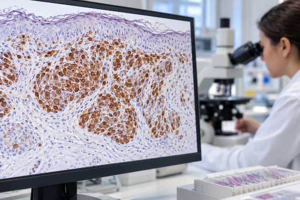
HMB-45 immunohistochemistry (IHC) is a tissue stain used to identify melanocytic differentiation in tumors and skin lesions. The antibody recognizes a melanosome-associated protein related to PMEL, a protein involved in pigment-organelle formation. HMB-45 can support melanoma or another melanocytic lesion when staining appears in the correct cells and fits the microscopic findings. It is not a stand-alone melanoma test. Some melanomas are HMB-45-negative, especially certain desmoplastic or poorly differentiated tumors, while benign nevi and several non-melanoma tumors can be positive. Pathologists therefore interpret HMB-45 with morphology and other melanocytic markers, commonly S100, SOX10, and Melan-A. In difficult lesions, additional stains such as PRAME or molecular tests may be used. The most useful question is not simply whether HMB-45 is positive, but how its distribution, intensity, and relationship to the lesion’s architecture fit the overall diagnosis.
- HMB-45 positivity supports melanocytic differentiation, but positive staining alone does not prove melanoma.
- True HMB-45 staining is usually cytoplasmic, often with a granular pattern in melanocytic cells.
- Many conventional melanomas are positive, but sensitivity is incomplete, so a negative stain cannot exclude melanoma.
- Benign nevi may also express HMB-45, often with a maturation-related pattern that differs from many melanomas.
- HMB-45 is best read as part of a panel, especially with S100, SOX10, Melan-A, morphology, and clinical context.
Table of Contents
- What HMB-45 IHC Measures
- When HMB-45 Is Used
- How Positive HMB-45 Staining Is Interpreted
- HMB-45 With Other Melanocytic Markers
- HMB-45 in Nevi and Melanoma
- Non-Melanoma Positivity and Pitfalls
- Questions After an HMB-45 Result
What HMB-45 IHC Measures
HMB-45 is an antibody used on fixed tissue sections. It recognizes an epitope associated with PMEL, historically called gp100, in immature melanosomes. Melanosomes are specialized structures involved in melanin production. Because melanocytes and many melanocytic tumors contain this machinery, HMB-45 can highlight cells showing melanocytic differentiation.
The test is performed in a pathology laboratory on tissue from a shave, punch, excisional, core, or surgical biopsy. It does not measure a substance in the blood, so there is no normal HMB-45 serum level and no fasting requirement. A patient generally does not need to prepare for the IHC stain itself; the clinically important step is obtaining an adequate tissue sample.
Under the microscope, true HMB-45 positivity is generally cytoplasmic and may look granular. Pathologists assess whether staining is present in the lesional cells, how many cells are involved, whether the pattern is superficial or deep, and how it relates to the architecture of the lesion. There is no single universal percentage cutoff that separates benign from malignant melanocytic lesions.
HMB-45 is sometimes described as a melanoma marker, but “melanocytic marker” is more accurate. Benign melanocytes and nevi can stain, and not every melanoma does. The marker is therefore used to answer a lineage and pattern question: do these cells show evidence of melanocytic differentiation, and is the pattern expected for the lesion being considered?
That distinction matters because melanoma can mimic carcinoma, sarcoma, lymphoma, or nerve-sheath tumors, especially in metastatic or poorly differentiated samples. HMB-45 can contribute to a tumor IHC panel, but the final diagnosis requires the entire histologic and immunophenotypic picture.
When HMB-45 Is Used
HMB-45 is commonly ordered when a pathologist is evaluating a pigmented or nonpigmented lesion that may be melanocytic. It is also useful in metastatic tumors when melanoma is part of the differential diagnosis. Melanoma does not always contain visible melanin, so an amelanotic tumor may need IHC to establish melanocytic lineage.
Typical uses include:
- distinguishing a melanocytic proliferation from a non-melanocytic mimic;
- supporting a diagnosis of melanoma in a primary skin lesion or metastasis;
- comparing the staining pattern of a nevus with a lesion suspicious for melanoma;
- assessing an unusual spindle-cell, epithelioid, or clear-cell tumor;
- evaluating tumors in which perivascular epithelioid cell differentiation is considered.
In a metastatic lesion, HMB-45 is rarely ordered alone. S100 and SOX10 are sensitive markers for many melanomas, while Melan-A and HMB-45 provide complementary melanocytic evidence. The exact panel depends on morphology and competing diagnoses.
One reason multiple markers are needed is that sensitivity and specificity differ. S100 is very sensitive for conventional melanoma but also stains many non-melanocytic tumors and normal cell types. SOX10 is a nuclear marker expressed in melanocytic and Schwannian lineages. HMB-45 is generally more restricted but is less sensitive in some melanoma subtypes. Melan-A is useful but can also stain certain steroid-producing and other tumors.
The lesion’s site also guides interpretation. A skin lesion with a junctional melanocytic component raises different questions from an undifferentiated mass in a lymph node, lung, liver, or soft tissue. Prior history of melanoma can increase suspicion for metastasis, but pathology still needs to establish that the current tumor fits that diagnosis rather than assuming recurrence from history alone.
HMB-45 may also be used in lesions that are not melanoma, particularly tumors in the PEComa family. This is an important reason a positive stain must be interpreted rather than translated directly into “melanoma positive.”
How Positive HMB-45 Staining Is Interpreted
A positive HMB-45 result means the antibody has bound to antigen in the cells being evaluated. The diagnostic meaning depends on which cells stain and how they stain. Pathologists first confirm that the staining is in the lesional cells rather than normal melanocytes, macrophages containing pigment, or another nearby cell population.
There is no universal scoring system such as 0, 1+, 2+, and 3+ for melanoma diagnosis. Reports may instead describe HMB-45 as positive or negative and may add qualifiers such as diffuse, patchy, focal, strong, or weak.
| Pattern | What it may suggest | Why context matters |
|---|---|---|
| Diffuse cytoplasmic positivity | Strong support for melanocytic differentiation in a compatible lesion | PEComas and some other tumors can also be positive |
| Patchy or focal positivity | May still support a melanocytic lesion | Limited expression reduces confidence if other markers disagree |
| Superficial positivity with loss in deeper nevus cells | Can accompany maturation in a benign nevus | Pattern must be assessed with architecture and cytology |
| Negative staining | No detectable HMB-45 in sampled tumor cells | Does not exclude melanoma, especially desmoplastic melanoma |
A positive stain also has to make biologic sense with the morphology. For example, HMB-45 in an epithelioid tumor that is also SOX10- and S100-positive may strongly support melanoma. The same HMB-45 result in a tumor with smooth-muscle marker expression and a different clinical setting may point toward a PEComa instead.
HMB-45 intensity alone does not establish malignancy. Benign nevi can show strong staining, particularly in superficial or activated melanocytes. Likewise, weak staining does not establish benign behavior. The pathologist relies on lesion symmetry, maturation, pagetoid spread, mitotic activity, cytologic atypia, ulceration, growth pattern, and other features appropriate to the specimen.
Because small biopsies may not show the full architecture, the significance of an HMB-45 pattern can be limited in a partial sample. An excision may be recommended when the distinction between nevus and melanoma depends on features that cannot be assessed reliably in the original biopsy.
HMB-45 With Other Melanocytic Markers
Melanocytic IHC works best as a panel because no single marker has perfect sensitivity and specificity. HMB-45, S100, SOX10, and Melan-A answer overlapping but not identical questions. Their combined pattern helps establish lineage and can reveal when an apparent result does not fit.
S100 IHC is highly sensitive for many melanomas. Its weakness is broad expression in other tissues, including Schwannian, myoepithelial, dendritic, and some mesenchymal lesions. SOX10 is nuclear and is also highly useful for melanocytic differentiation, but it shares expression with nerve-sheath and some other tumors.
HMB-45 and Melan-A are often viewed as differentiation markers. HMB-45 may be lost in more mature nevus cells and in some melanoma variants. Melan-A can highlight melanocytes well, which is useful for assessing distribution, but it also has recognized non-melanocytic expression. Using both lineage-sensitive and differentiation-associated markers can therefore be more informative than relying on either group alone.
PRAME has become an important adjunct in selected melanocytic lesions. Diffuse nuclear PRAME expression can support melanoma in the right setting, while many benign nevi are negative or show only limited staining. PRAME is not perfectly sensitive or specific and does not replace morphology. Its main value is as another piece of evidence when routine histology and established melanocytic markers leave a difficult borderline question.
A pathologist may also use MITF IHC in selected cases. Additional markers are chosen to exclude the most realistic mimic, such as cytokeratins for carcinoma or hematolymphoid markers for lymphoma. The goal is a coherent immunophenotype: the diagnosis should explain both the positive and negative stains.
In a clearly melanocytic lesion, a marker panel can also help characterize where abnormal cells are located. However, IHC should not be used to manufacture diagnostic certainty when the architecture remains ambiguous. Some challenging melanocytic tumors still require expert dermatopathology review, genomic testing, or clinicopathologic correlation.
HMB-45 in Nevi and Melanoma
The difference between a benign nevus and melanoma cannot be reduced to HMB-45 positive versus negative. Both may stain. What can be useful is the distribution of staining through the lesion, particularly when it is interpreted alongside morphologic maturation.
In many conventional acquired nevi, superficial melanocytes may express HMB-45 more strongly, while deeper dermal melanocytes become smaller and lose HMB-45 as they mature. This decreasing pattern can support benign maturation. Melanomas may retain HMB-45 expression deeper in the lesion or show a more irregular distribution. These are tendencies, not absolute rules.
Several factors limit the pattern. Some benign nevi do not show textbook maturation, and some melanomas lose HMB-45. Spitz-family lesions, blue nevi, recurrent nevi, congenital nevi, and other special settings can produce patterns that require specialized interpretation. A pathologist should never call melanoma only because HMB-45 extends to the base of a lesion.
Desmoplastic melanoma is a particularly important exception. It often shows strong S100 and SOX10 staining but may be negative or only focally positive for HMB-45 and Melan-A. If HMB-45 negativity were treated as an exclusion test, this melanoma subtype could be missed. Morphology and the more sensitive lineage markers are essential.
For established melanoma, HMB-45 is primarily diagnostic rather than a staging marker. It does not determine Breslow thickness, ulceration, mitotic features, margin status, lymph-node stage, or distant metastatic stage. Those variables come from histologic measurements, surgery, imaging, and clinical evaluation.
HMB-45 expression also is not the same as a treatment-predictive mutation. When systemic therapy is being considered, molecular testing for actionable melanoma alterations may be far more relevant. The pathology report may therefore contain both IHC findings that establish melanoma lineage and molecular findings that influence treatment. They answer different questions.
Non-Melanoma Positivity and Pitfalls
HMB-45 positivity outside melanoma is a central interpretive pitfall. Perivascular epithelioid cell tumors, or PEComas, can express HMB-45 and Melan-A while also showing smooth-muscle differentiation. The PEComa family includes lesions arising in several organs and includes angiomyolipoma. In the appropriate morphologic and clinical setting, this combined melanocytic and myogenic phenotype points away from melanoma.
Other uncommon tumors may also express melanocytic markers. Clear cell sarcoma of soft tissue can show melanocytic differentiation and may be HMB-45-positive, but it has distinct molecular biology and clinical behavior. Some meningeal melanocytic tumors and other rare lesions also require site-specific differential diagnosis.
Technical and interpretive problems include:
- Melanin versus chromogen. Brown pigment in a melanocytic lesion can make it harder to distinguish true stain from endogenous melanin without careful microscopic assessment.
- Normal melanocytes. Epidermal or follicular melanocytes can stain and should not be mistaken for invasive tumor cells.
- Macrophages. Pigment-laden macrophages can sit close to melanoma cells and complicate visual interpretation.
- Tumor heterogeneity. One area may stain while another does not, particularly in metastases or treated tumors.
- Small or damaged samples. Crush artifact, necrosis, cautery, or scant tissue can reduce confidence.
- Overreliance on a single marker. HMB-45 positivity without a compatible panel can misclassify non-melanoma tumors.
A negative stain presents the opposite danger. Poorly differentiated melanoma can lose conventional differentiation markers. When melanoma remains plausible, sensitive markers such as SOX10 and S100, clinical history, morphology, and sometimes molecular tests may keep the diagnosis in consideration despite HMB-45 negativity.
The safest approach is pattern-based interpretation. A pathologist asks whether the full set of findings behaves like the suspected entity. Unexpected positivity or negativity should trigger review of controls, tumor-cell identity, and alternate diagnoses rather than being forced into a binary rule.
Questions After an HMB-45 Result
For most patients, the final diagnosis is more important than the individual HMB-45 result. A pathology report may list multiple stains because each helped narrow the differential diagnosis. Useful questions include:
- Was HMB-45 positive in the tumor cells, and was the staining diffuse, patchy, or focal?
- Were S100, SOX10, Melan-A, or other melanocytic markers also tested?
- Does the morphology support melanoma, a benign nevus, a PEComa, or another diagnosis?
- If melanoma is suspected, could the subtype have reduced HMB-45 expression?
- Was PRAME or another adjunct marker used in a difficult melanocytic lesion?
- Is the sample large enough to assess lesion architecture and maturation reliably?
- Does the diagnosis require molecular testing or specialist dermatopathology review?
If the final diagnosis is melanoma, ask about the features that guide staging and management rather than focusing only on HMB-45. For a primary cutaneous melanoma, these can include Breslow thickness, ulceration, margins, and other report elements. For metastatic melanoma, the site of disease and treatment-relevant molecular findings may be more important.
If the report says HMB-45 is negative but still diagnoses melanoma, that is not automatically contradictory. Melanoma is a diverse disease, and some variants lack this marker. Conversely, a positive HMB-45 result does not convert a benign lesion into melanoma. The stain must be interpreted in the larger pattern.
When the diagnosis remains uncertain, the most productive next step may be review of the original slides by a dermatopathologist or soft-tissue pathologist, depending on the lesion. Additional tissue, a targeted IHC panel, or molecular testing can resolve cases in which HMB-45 provides useful but incomplete evidence.
HMB-45 can also help show how cells are distributed within a lesion because the stain makes positive melanocytes easier to see against surrounding tissue. In selected cases, this can draw attention to an abnormal deep population or an uneven pattern that deserves closer morphologic review. The stain should not be used as a substitute for measuring Breslow thickness or defining the invasive front, however. Those tasks require careful examination of routine sections and, when needed, correlation with other melanocytic markers.
Prior treatment can further complicate interpretation. Immunotherapy, targeted therapy, regression, or scarring may leave sparse viable melanoma cells mixed with inflammation and fibrosis. A panel of sensitive melanocytic markers can help identify residual cells, while HMB-45 may be absent in a subset. Pathologists weigh the treated morphology and marker pattern together rather than expecting the original tumor’s immunophenotype to remain unchanged.
References
- Immunohistochemistry for Skin Cancers: New Insights into Diagnosis and Treatment of Melanoma. 2025 (Review)
- The Markers Auxiliary in Distinguishing Between Nodal Nevi and Melanoma Metastases. 2025 (Review)
- Diagnostic utility of combining PRAME and HMB-45 stains in primary melanocytic tumors 2023
- Immunohistochemistry for PRAME in Dermatopathology 2023 (Review)
- The use of immunohistochemical staining in Mohs micrographic surgery for melanoma: a systematic review 2023 (Systematic Review)
- Immunohistochemistry in melanocytic lesions: Updates with a practical review for pathologists. 2022 (Review)
Disclaimer
HMB-45 IHC is a pathology tool and cannot diagnose or exclude melanoma by itself. Results must be interpreted with the lesion’s microscopic features, other stains, clinical history, and any needed molecular testing. Discuss the final pathology diagnosis and next steps with your treating clinician or pathologist.





