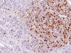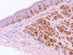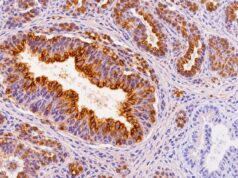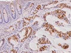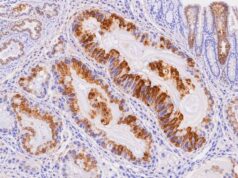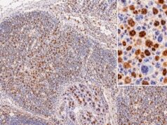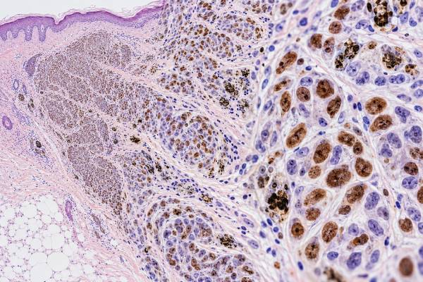
MITF immunohistochemistry (IHC) is a tissue stain used to identify microphthalmia-associated transcription factor, a nuclear protein that helps control melanocyte development and pigment-related genes. In pathology, nuclear MITF staining can support melanocytic differentiation in a suspected melanoma or other melanocytic tumor. Its nuclear pattern is often useful because it outlines the actual cells being assessed rather than filling the cytoplasm. Still, a positive MITF result is not equivalent to a melanoma diagnosis: benign nevi and normal melanocytes can also express MITF, and some non-melanocytic cells or tumors may stain depending on the antibody and setting. In addition, melanoma can lose MITF expression, especially in desmoplastic, spindle-cell, dedifferentiated, or phenotypically altered tumors. For those reasons, pathologists rarely rely on MITF alone. They interpret it with the tumor’s morphology and a panel that may include SOX10, S100, Melan-A/MART-1, HMB-45, PRAME, cytokeratins, and other markers tailored to the differential diagnosis.
- MITF IHC is a nuclear melanocytic marker: convincing nuclear staining supports melanocytic differentiation in the right morphologic setting.
- Positive MITF does not prove melanoma: benign melanocytes and nevi commonly express the protein as well.
- A negative result does not rule out melanoma: desmoplastic, spindle-cell, metastatic, or dedifferentiated melanomas may show reduced or absent MITF.
- MITF is best interpreted as part of a panel: SOX10, S100, Melan-A, HMB-45, PRAME, and lineage-exclusion stains often provide essential context.
- No special preparation is needed: the test is performed on biopsy, excision, cytology cell-block, or other tumor tissue submitted to pathology.
Table of Contents
- What MITF IHC Measures
- What Positive and Negative Staining Mean
- MITF in Melanoma Diagnosis
- MITF in Benign and Other Melanocytic Tumors
- How MITF Compares With Other Melanoma Markers
- Limitations and Diagnostic Pitfalls
- How to Read an MITF Pathology Result
What MITF IHC Measures
MITF IHC detects a transcription factor inside cell nuclei and is used mainly to show that cells are following a melanocytic differentiation program. MITF stands for microphthalmia-associated transcription factor. It belongs to the MiT family of transcription factors and has a central role in melanocyte survival, differentiation, and regulation of genes involved in melanin production.
In routine pathology, an antibody binds MITF in formalin-fixed tissue. A positive reaction appears primarily in the nucleus. That nuclear localization is diagnostically useful because the pathologist can count or map individual positive cells without the broad cytoplasmic staining produced by markers such as Melan-A or HMB-45.
MITF IHC may be requested when a tumor is difficult to classify on routine sections. Common reasons include:
- an amelanotic tumor that lacks visible pigment;
- a metastatic malignancy with an uncertain primary site;
- a small focus of possible melanoma in a lymph node or other organ;
- a melanocytic lesion in which the distribution of lesional cells needs clarification;
- an unusual ocular, central nervous system, or soft-tissue melanocytic neoplasm;
- a tumor with a partial or conflicting immunophenotype.
The result is usually reported qualitatively—positive, negative, focal, diffuse, weak, or strong—rather than as a standardized percentage with a universal cutoff. Unlike hormone-receptor tests or some predictive biomarkers, there is no clinically accepted MITF “positive threshold” that applies to every melanoma.
MITF is also different from a mutation test. It does not determine whether a melanoma carries a BRAF, NRAS, KIT, or other genomic alteration. It shows protein expression and differentiation state in the cells present on the stained slide.
What Positive and Negative Staining Mean
A positive MITF stain means that the nuclei of the cells being evaluated contain detectable MITF protein. In a tumor with melanoma-like morphology, that finding can strengthen evidence for melanocytic differentiation. The interpretation is strongest when the staining occurs in the atypical tumor cells and agrees with other melanocytic markers.
A report may describe MITF as diffusely positive when many tumor nuclei stain, focally positive when only a subset stains, or negative when no convincing nuclear reaction is present. These descriptions are more useful than a simple plus sign because melanomas can be heterogeneous.
Positive MITF does not answer whether a melanocytic lesion is benign or malignant. Ordinary nevi generally contain MITF-positive melanocytes, so a benign nevus can be just as convincingly positive as a melanoma. Morphologic features—such as asymmetry, poor maturation, pagetoid spread, cytologic atypia, mitotic activity, and destructive growth—remain central to distinguishing melanoma from nevus.
A negative stain also has limits. Melanoma cells can shift their differentiation state and reduce MITF expression. This is particularly important in spindle-cell and desmoplastic melanomas, in which classic melanocytic differentiation markers may be weak or absent. A pathologist facing suspicious morphology should not exclude melanoma solely because MITF is negative.
The practical interpretation is therefore asymmetric: positive staining can be supportive, while negative staining must be weighed against the known sensitivity of the marker in that tumor subtype.
MITF in Melanoma Diagnosis
MITF is most useful when the pathologist needs another line of evidence that a tumor is melanocytic. It can be especially helpful in conventional epithelioid melanoma because many such tumors retain nuclear expression. The stain may also aid the detection of small melanocytic deposits because individual positive nuclei can stand out against surrounding tissue.
In a metastatic tumor, the pathologist usually starts with morphology and clinical information, then selects a broad panel. If a malignant tumor is MITF positive and also expresses SOX10 or another melanocytic marker while epithelial and hematolymphoid markers are negative, metastatic melanoma becomes more likely. If the tumor is MITF positive but the remaining panel points elsewhere, the pathologist must consider a non-melanoma explanation.
Primary cutaneous melanoma
In a primary skin lesion, MITF can highlight the extent and distribution of melanocytes. This may help in an amelanotic lesion or in a proliferation obscured by inflammation. However, because normal epidermal melanocytes and benign nevi also stain, MITF cannot independently define the edge of melanoma in situ or prove that a junctional proliferation is malignant.
The interpretation becomes more difficult in chronically sun-damaged skin. Such skin can show increased numbers of background melanocytes. A nuclear melanocytic stain may make these cells easier to see, but visibility is not the same as malignancy. The pathologist must distinguish the architecture of the lesion from the normal or reactive melanocyte population.
Metastatic and dedifferentiated melanoma
Metastatic melanoma can show striking variation from a patient’s original tumor. Some metastases become amelanotic or lose conventional differentiation antigens. MITF may remain positive in a subset of cells, but complete loss is possible. When the phenotype is unusual, comparison with the prior melanoma and a broader IHC or molecular workup may be necessary.
Modern research also recognizes that MITF expression is biologically dynamic. Melanoma cells can occupy higher-MITF differentiated states or lower-MITF invasive and therapy-adapted states. That biology helps explain why a marker that is fundamental to melanocytes is not uniformly present in every malignant melanoma cell.
MITF in Benign and Other Melanocytic Tumors
MITF is a marker of melanocytic differentiation, not a marker exclusive to malignant melanoma. Benign melanocytic nevi typically express MITF because the cells retain the same core lineage program as normal melanocytes. This is why the stain must not be interpreted as a “cancer-positive” test.
In challenging melanocytic tumors, MITF can contribute to lineage confirmation while other stains address malignancy or molecular subtype. A pathologist may pair it with PRAME, p16, Ki-67, HMB-45, or molecular tests depending on the question. None of those tests substitutes for the histologic pattern.
MITF expression can also occur in extracutaneous melanocytic neoplasms. Uveal melanoma is an important example. Studies of uveal tumors show that MITF expression varies with differentiation and molecular features, so the stain can have biologic meaning without serving as a stand-alone prognostic test.
Some tumors outside conventional melanoma can show melanocytic differentiation or MiT-family biology. Clear cell sarcoma, historically called “malignant melanoma of soft parts,” may express MITF along with S100 and HMB-45. PEComa-family tumors can express melanocytic markers, although their exact profiles vary. These possibilities become relevant when the tumor arises in deep soft tissue or an internal organ rather than in a typical cutaneous setting.
The anatomic site therefore changes the differential diagnosis. Nuclear MITF positivity in a pigmented epidermal lesion, a deep tendon-associated soft-tissue mass, and a visceral neoplasm cannot be interpreted identically even if the stain looks similar.
How MITF Compares With Other Melanoma Markers
MITF adds value because it is a nuclear melanocytic marker, but SOX10 is often favored when maximum sensitivity is needed, especially for desmoplastic melanoma. Different markers answer overlapping but not identical questions.
| Marker | Staining location | Main advantage | Important limitation |
|---|---|---|---|
| MITF | Nuclear | Clearly outlines melanocytic nuclei in many conventional lesions | May be lost in spindle/desmoplastic melanoma; some non-tumor cells can stain |
| SOX10 | Nuclear | High sensitivity for melanocytic lineage and many desmoplastic melanomas | Also expressed in Schwannian and selected other tumors |
| S100 | Nuclear and cytoplasmic | Very sensitive in melanoma | Low specificity; many other cell types and tumors are positive |
| Melan-A/MART-1 | Cytoplasmic | Strong, easy-to-see staining in many melanocytic lesions | Benign nevi stain and desmoplastic melanoma may not |
| HMB-45 | Cytoplasmic | Useful melanocytic differentiation marker with characteristic maturation patterns in some nevi | Less sensitive in several melanoma subtypes |
| PRAME | Nuclear | Diffuse expression can support melanoma over nevus | Not all melanomas are positive and occasional benign lesions express it |
Panels work because the markers fail in different ways. A desmoplastic melanoma may be SOX10 positive but MITF, Melan-A, and HMB-45 negative. A conventional nevus may be MITF and Melan-A positive but lack diffuse PRAME. A metastatic carcinoma usually expresses epithelial markers that a melanoma lacks, even if one stain is unexpectedly reactive.
For that reason, the question “Which melanoma marker is best?” has no universal answer. The best stain is the one that addresses the diagnostic problem while the rest of the panel covers the most plausible mimics.
Limitations and Diagnostic Pitfalls
The largest MITF pitfall is treating nuclear positivity as melanoma-specific. The stain must be localized to the correct cells and interpreted with known cross-reactivity and subtype-related loss of expression.
Reactive histiocytes, multinucleated giant cells, and other background cells have been reported to show nuclear staining with some MITF antibodies. In an inflamed or scarred specimen, these cells can mimic scattered tumor cells if the slide is interpreted without careful morphologic correlation. This is one reason SOX10 may be preferred for evaluating suspected desmoplastic melanoma in scar tissue.
Another pitfall is the desmoplastic melanoma exception. Conventional melanoma markers associated with melanocytic differentiation can be reduced in this subtype. A negative MITF result therefore has a different meaning in a fibrosing spindle-cell lesion than in a typical epithelioid melanoma.
Technical factors can also affect results. Poor fixation, prolonged decalcification, scant tissue, crush artifact, and weak internal controls can make a negative stain less trustworthy. Conversely, nonspecific background or unexpected cell staining can make a positive reaction less specific.
The antibody clone and laboratory protocol matter as well. Published sensitivity and specificity figures from one reagent or setting should not automatically be applied to every laboratory. The pathologist interprets the actual validated assay used by that laboratory.
Finally, MITF expression itself can change with tumor biology. A metastatic lesion may have a lower expression level than the primary tumor, and treatment can select for phenotypically different tumor cells. Discordant IHC does not necessarily mean the original diagnosis was wrong; it can reflect tumor evolution, sampling, or technical differences.
How to Read an MITF Pathology Result
Read MITF as one piece of the immunophenotype, not as the final diagnosis. The most informative pathology reports connect the stain to a specific interpretation, such as “the tumor is positive for SOX10 and MITF, supporting melanocytic differentiation.”
When reviewing a report, focus on five questions:
- Is staining nuclear? True MITF interpretation is based mainly on nuclear labeling.
- Are the actual tumor cells positive? Background melanocytes or histiocytes should not be mistaken for the lesional population.
- Is staining diffuse or focal? Limited positivity may still be useful, but it carries different weight from widespread concordant staining.
- What do the companion stains show? A panel is more reliable than any single antibody.
- Does the result fit the morphology and specimen site? Unexpected staining should trigger reconciliation, not automatic reclassification.
If MITF is positive in a lesion already diagnosed as melanoma, the stain generally confirms differentiation rather than changing stage or treatment. If MITF is negative, the next step depends on the diagnostic suspicion. A pathologist may add SOX10 and S100, compare previous material, repeat the stain with controls, or use molecular methods if lineage remains uncertain.
Patients should also know what MITF cannot tell them. The stain does not provide Breslow thickness, ulceration status, lymph-node stage, or a genomic target for therapy. Those elements come from the rest of the pathology and staging workup. MITF IHC is primarily a diagnostic lineage tool.
The most useful summary is straightforward: nuclear MITF positivity can support a melanocytic tumor, but it does not distinguish a benign nevus from melanoma by itself and it is not present in every melanoma. Its strength comes from combining a recognizable nuclear pattern with morphology and complementary stains chosen for the exact diagnostic setting.
When a pathology result remains indeterminate after IHC, that uncertainty can be appropriate rather than a failure of testing. Some melanocytic tumors fall into biologically gray categories, and no stain can force a definitive benign-versus-malignant answer. In those cases, expert dermatopathology review, correlation with the lesion’s clinical appearance and prior biopsies, deeper sections, or targeted molecular testing may provide more useful information than repeating the same marker. The decision to add testing should be driven by whether the result could change diagnosis, margin assessment, staging, or management.
A few practical details can make an MITF result easier to interpret. Normal melanocytes in skin may provide an internal positive control because their nuclei can label even when the lesion itself is weak or negative. That is useful technically, but it also creates a trap: staining of scattered background melanocytes should not be counted as staining of the tumor. The pathologist compares the immunostain directly with the routine hematoxylin-and-eosin slide to make sure the labeled nuclei belong to the lesional cells.
Heterogeneous expression is another reason to avoid absolute conclusions from a small sample. A melanoma can contain strongly positive areas next to areas with little or no MITF, and a core biopsy may sample only one component. Spindle-cell, desmoplastic, or dedifferentiated areas are especially likely to lose conventional melanocytic markers. In a difficult case, preservation of SOX10 or S100 with loss of MITF and Melan-A may still fit melanoma, while broad loss of melanocytic markers may require comparison with an earlier specimen or molecular evidence of a shared tumor lineage.
The laboratory method also matters. Antibody clone, antigen retrieval, fixation, and the threshold used for calling a stain positive can affect apparent intensity. For that reason, a percentage from one laboratory should not be treated as a universal cutoff. The clinically useful question is whether the staining pattern, controls, morphology, and companion markers form a coherent diagnosis. That integrated approach is more reliable than ranking MITF as simply “positive” or “negative.”
References
- MITF Is an Essential and Functionally Multifaceted Transcription Factor in Cutaneous Melanoma 2026 (Review)
- Immunohistochemistry for Skin Cancers: New Insights into Diagnosis and Treatment of Melanoma 2025 (Systematic Review)
- Immunohistochemistry in melanocytic lesions: Updates with a practical review for pathologists 2022 (Review)
- Microphthalmia-Associated Transcription Factor: A Differentiation Marker in Uveal Melanoma 2023
- Immunohistochemistry for PRAME in Dermatopathology 2023 (Review)
Disclaimer
MITF IHC must be interpreted by a qualified pathologist in the context of morphology, specimen site, technical controls, and other immunostains. A positive or negative MITF result alone cannot confirm or exclude melanoma, determine cancer stage, or select treatment. This article provides general educational information and is not a substitute for individualized pathology or medical advice.