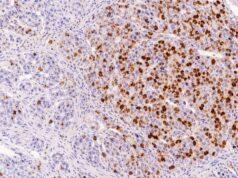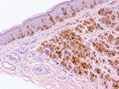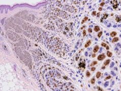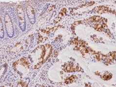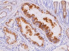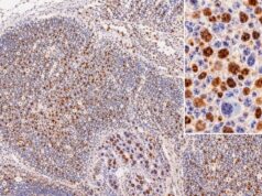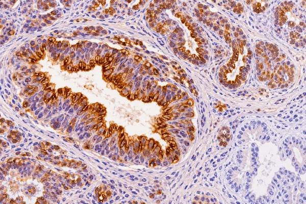
MUC1 immunohistochemistry (IHC) detects mucin 1, a large transmembrane glycoprotein normally found on the apical surface of many glandular and ductal epithelial cells. In cancer, MUC1 is often overexpressed, abnormally glycosylated, and redistributed from a neat apical border to broader membranous or cytoplasmic staining. These changes make MUC1 useful as a supportive epithelial and carcinoma-associated marker, but not as a stand-alone test for cancer or tumor origin. Many adenocarcinomas express MUC1, including tumors of the breast, pancreas, lung, ovary, endometrium, kidney, and other organs. Normal tissues and some non-carcinoma tumors can express it as well. The exact result also depends on which MUC1 antibody or epitope the laboratory tests, because different antibodies may recognize the protein core or tumor-associated glycoforms. Pathologists therefore interpret the location, intensity, and distribution of MUC1 staining with morphology and a broader immunohistochemical panel rather than treating “MUC1 positive” as a diagnosis by itself.
- MUC1 IHC detects a cell-surface mucin commonly expressed by epithelial tumors, especially adenocarcinomas.
- Normal epithelium often shows polarized apical staining, while carcinoma may show diffuse membranous and/or cytoplasmic staining with loss of normal polarity.
- MUC1 positivity is not specific for one organ or for malignancy: many normal epithelia and several tumor types express the protein.
- The antibody clone matters: different MUC1 assays can recognize different epitopes or glycosylation states and may produce different staining patterns.
- No special patient preparation is required: MUC1 IHC is performed on biopsy or surgical tissue already collected for pathology.
Table of Contents
- What MUC1 IHC Detects
- What Positive MUC1 Staining Means
- MUC1 Staining Patterns
- Which Tumors Express MUC1
- How MUC1 Is Used in Diagnostic Panels
- Limitations and Pitfalls
- How to Read a MUC1 Pathology Result
What MUC1 IHC Detects
MUC1 IHC shows where mucin 1 protein is present in a tissue section. MUC1 is a transmembrane mucin: one part projects outside the cell and carries dense carbohydrate chains, while another part spans the cell membrane and connects to a cytoplasmic tail involved in signaling. In healthy polarized epithelium, MUC1 is concentrated mainly on the apical or luminal surface, where it contributes to a protective barrier.
Cancer changes that organization. Tumor cells often lose normal polarity, increase MUC1 production, and alter the carbohydrates attached to the protein. As a result, MUC1 may become visible across the entire membrane or within the cytoplasm rather than staying confined to the luminal edge. Cancer-associated glycosylation can also expose protein or carbohydrate epitopes that are less accessible in normal tissue.
This biology explains why the term “MUC1 stain” can be broader than it sounds. Different antibodies target different portions or glycoforms of the molecule. Two laboratories can both test MUC1 yet use reagents with somewhat different sensitivity and tissue distribution. A pathology report should therefore be interpreted according to the validated assay used locally.
MUC1 is also related to familiar serum tumor markers. CA 15-3 and CA 27.29 assays detect circulating epitopes associated with MUC1, mainly in the context of breast cancer monitoring. Tissue MUC1 IHC is a different test: it examines protein expression in cells on a slide rather than measuring a concentration in blood.
What Positive MUC1 Staining Means
A positive MUC1 stain means that the cells being examined contain detectable MUC1 antigen, but the result does not prove carcinoma or identify a single primary site. The pathologist first determines whether the staining is truly within the tumor cells and then evaluates its pattern.
MUC1 positivity can support epithelial differentiation when it agrees with morphology and other markers. For example, an adenocarcinoma with cytokeratin expression and strong MUC1 may fit a gland-forming epithelial tumor. However, because MUC1 is widely distributed, the stain is usually less useful for pinpointing whether the tumor came from breast, pancreas, lung, or another organ.
The clinical meaning also depends on why the stain was ordered. In one case, MUC1 may help characterize a poorly differentiated carcinoma. In another, it may be part of research or a therapeutic-biomarker assay designed to detect a particular tumor-associated MUC1 epitope. A generic positive result from a diagnostic antibody should not automatically be translated into eligibility for a MUC1-targeted drug or clinical trial.
A negative result has similar limitations. Some carcinomas have little or no detectable MUC1, and expression can be heterogeneous. A small biopsy may sample a negative area even when another part of the tumor is positive. Technical factors and the selected antibody can also influence sensitivity.
MUC1 Staining Patterns
The distribution of MUC1 staining can be more informative than positivity alone. Pathologists may describe the result as apical, luminal, membranous, circumferential, cytoplasmic, diffuse, focal, or heterogeneous.
| Pattern | What it looks like | Typical interpretation |
|---|---|---|
| Apical/luminal | Staining is concentrated at the surface facing a gland or lumen | Often reflects preserved epithelial polarity and can be seen in normal or well-differentiated epithelium |
| Membranous | Cell borders stain beyond the luminal surface | May reflect loss of polarity in neoplastic epithelium |
| Cytoplasmic | Stain fills part of the cell body | Common in many carcinomas and may accompany altered processing or trafficking of MUC1 |
| Diffuse | Most tumor cells stain | Provides stronger evidence that MUC1 is a consistent tumor phenotype |
| Focal/heterogeneous | Only subsets or regions stain | May limit the marker’s diagnostic or therapeutic usefulness |
In normal glandular tissue, an orderly apical pattern makes physiologic sense because MUC1 faces the lumen. In carcinoma, loss of polarity can expose MUC1 around the cell and in the cytoplasm. That change is common enough to be biologically important, but it is not a universal malignancy rule. Some cancers retain polarized staining, and inflamed or regenerating epithelium can also alter its expression.
Intensity can be described as weak, moderate, or strong, but there is no universal MUC1 scoring system for routine diagnosis. Research studies may use percentage cutoffs or composite scores, while clinical assays developed for a specific drug may use their own validated algorithm. Results from one assay should not be substituted for another without evidence that the methods are equivalent.
Which Tumors Express MUC1
MUC1 is broadly expressed across epithelial malignancies, which makes it a common tumor-associated antigen but a relatively nonspecific site-of-origin marker. Expression is particularly frequent in adenocarcinomas.
Breast carcinomas commonly express MUC1, and the molecule is the source of epitopes measured by serum CA 15-3 and CA 27.29 assays. Pancreatic ductal adenocarcinoma also often shows aberrant MUC1, in contrast with some mucin-producing pancreatic precursor lesions that may favor other mucin profiles. Lung adenocarcinomas, ovarian and endometrial carcinomas, gastric cancers, renal cell carcinomas, and a range of other epithelial tumors can express MUC1 to varying degrees.
MUC1 expression may become broader or more cytoplasmic as epithelial cells lose polarity and acquire invasive behavior. Many studies have linked high or aberrant MUC1 expression with aggressive features in particular cancer types. Those associations, however, are not interchangeable across organs. A prognostic relationship found in one cancer cannot be applied automatically to another, and routine pathology reports do not usually use MUC1 alone to calculate prognosis.
Why tumor-associated MUC1 is different
Normal MUC1 is heavily glycosylated, meaning that carbohydrate chains cover much of its extracellular region. Cancer cells frequently make shorter or abnormal glycans. This can expose new epitopes and create forms often described as tumor-associated MUC1. These altered forms are important because they can change cell adhesion and signaling and can be recognized by therapeutic antibodies or immune approaches.
A tumor-associated MUC1 assay may therefore target a more cancer-restricted epitope than a conventional antibody that detects total MUC1. That distinction is crucial when reading research papers or clinical-trial eligibility criteria. “MUC1 positive” is not one standardized result across all antibody clones.
MUC1 expression is not the same as tumor grade
MUC1 biology is connected with invasion, signaling, immune interactions, and treatment resistance in many experimental systems, but routine IHC does not convert those complex mechanisms into a universal grade. A low-grade tumor can be strongly MUC1 positive, and a high-grade tumor can show only patchy staining. Histologic grade is assigned using the rules established for the specific cancer, such as gland formation, nuclear features, mitotic activity, or other disease-specific criteria.
The same caution applies to prognosis. Researchers often divide tumors into “high” and “low” MUC1 groups using study-specific scores. One study may count the percentage of positive cells, another may combine percentage and intensity, and a third may focus on a tumor-associated glycoform. Those categories cannot be assumed to mean the same thing. If a pathology report mentions that MUC1 expression is prognostically unfavorable, the clinically useful question is whether that association has been validated for the patient’s exact tumor type and assay.
MUC1 also illustrates why tumor markers can change during cancer progression. Loss of epithelial polarity, clonal selection, metastatic spread, and therapy can alter both the amount and cellular location of the protein. A metastasis can therefore stain differently from the primary tumor. When tissue from two sites is being compared, differences in fixation and antibody method should be considered alongside true biologic evolution.
How MUC1 Is Used in Diagnostic Panels
In routine surgical pathology, MUC1 is usually a supporting marker rather than the deciding stain. Pathologists combine it with markers that provide stronger lineage or site specificity.
For a poorly differentiated tumor, broad-spectrum cytokeratins may first establish epithelial differentiation. Organ-associated markers can then narrow the origin: examples include TTF-1 and Napsin A for lung adenocarcinoma, GATA3 and mammaglobin-related markers for breast or urothelial differentiation, PAX8 for selected renal, thyroid, and Müllerian tumors, and CDX2/SATB2 for intestinal differentiation. MUC1 can complement this pattern but rarely overrules it.
Mucin panels can be particularly useful in gastrointestinal and pancreatobiliary pathology. MUC1 is a membrane-associated mucin, while MUC2 and MUC5AC are secreted gel-forming mucins with different normal tissue distributions. Their combined profiles can help describe intestinal, gastric, pancreatic, or other differentiation patterns. Even then, morphology and disease-specific criteria remain more important than a single stain.
MUC1 can also enter differential diagnosis when distinguishing certain epithelial tumors from mimics, but its broad expression means a positive result usually needs a second, more specific marker. The diagnostic value comes from patterns across a panel, not from collecting as many positive stains as possible.
Limitations and Pitfalls
MUC1’s greatest limitation is its lack of specificity: normal tissue and many unrelated tumors can be positive. A strong stain can look convincing while still providing little information about the exact diagnosis.
Several pitfalls deserve attention:
- Antibody-dependent results: antibodies to different MUC1 epitopes or glycoforms may not stain the same tissues in the same way.
- Normal epithelial staining: background ducts and glands can be strongly positive and should not be mistaken for tumor.
- Heterogeneity: a biopsy may not represent the full range of MUC1 expression in a larger tumor.
- No universal cutoff: routine diagnostic MUC1 lacks one percentage or intensity threshold that defines a clinically meaningful positive result across cancers.
- Therapy mismatch: positivity with a standard diagnostic antibody may not satisfy the companion or trial assay required for a MUC1-directed treatment.
- Prognostic overreach: studies linking MUC1 with worse outcomes are often tumor-specific and assay-specific; they do not turn a routine positive stain into an individual prognosis.
Preanalytic tissue handling can also influence staining. Fixation quality, decalcification, tissue age, and processing conditions affect antigen preservation. Good internal controls help the pathologist decide whether an unexpectedly negative result is believable.
Interpretation should also account for mucin-rich material outside cells. Secreted mucin, luminal debris, or necrotic material can show background reaction with some assays. The diagnostically relevant question is whether viable tumor cells display the expected membrane or cytoplasmic pattern. Careful localization is particularly important in tiny biopsies, where only a few intact glands may be present.
The specimen itself matters too. A tiny metastatic focus, necrotic sample, or post-treatment biopsy may show a different phenotype from the untreated primary tumor. If MUC1 expression would affect a major diagnostic or therapeutic decision, additional tissue or a validated repeat assay may be appropriate.
How to Read a MUC1 Pathology Result
Start by asking what diagnostic question MUC1 was meant to answer. A line that says “MUC1: positive” has limited meaning without the final diagnosis, staining pattern, and companion markers.
A useful reading sequence is:
- Identify the tumor and specimen site. The same stain has different significance in breast, pancreas, lung, kidney, ovary, or soft tissue.
- Look for the staining pattern. Apical, membranous, cytoplasmic, diffuse, and focal patterns convey different biologic information.
- Check the other IHC markers. Site-specific and lineage-specific stains usually carry more diagnostic weight.
- Determine whether the assay measures total or tumor-associated MUC1. This is especially important in research and therapeutic settings.
- Ask whether the result changes management. Many routine MUC1 stains are diagnostic support only and do not independently determine treatment.
For example, a pancreatic mass that is MUC1 positive may fit pancreatic ductal adenocarcinoma, but MUC1 cannot establish pancreatic origin on its own because adenocarcinomas from several organs share that phenotype. Conversely, a negative MUC1 result does not exclude carcinoma if morphology and other epithelial markers are convincing.
If a report mentions MUC1 overexpression or a percentage score, it is reasonable to ask which antibody and scoring system were used and whether that system has a validated clinical cutoff. This prevents a research-style number from being interpreted as though it were a universally standardized companion diagnostic.
The central takeaway is that MUC1 is a widely expressed epithelial mucin whose abnormal distribution is common in carcinoma. Positive staining can support an epithelial tumor and provide useful information about differentiation, but it is rarely specific enough to establish tumor origin or malignancy by itself. The most reliable interpretation combines staining pattern, morphology, anatomic site, and a focused panel of other markers.
MUC1 results can also vary because different antibodies recognize different parts or glycosylation states of the molecule. Some assays highlight broadly expressed epithelial MUC1, while others are designed to recognize tumor-associated forms or altered carbohydrate structures. This distinction matters when comparing a routine pathology stain with a research study, clinical trial, or experimental targeted therapy. Two tests labeled “MUC1” may therefore answer different questions and should not be assumed to be interchangeable.
The location of staining often carries as much information as its intensity. In non-neoplastic glandular epithelium, MUC1 commonly has an orderly apical or luminal distribution. Carcinomas may show stronger, more diffuse membranous and cytoplasmic staining as cell polarity is lost. That abnormal distribution can support malignant epithelial differentiation, but it still does not prove cancer because reactive or metaplastic epithelium can express MUC1 as well. Conversely, some carcinomas are weak or negative.
It is also important not to confuse tissue MUC1 IHC with serum tumor-marker tests. Blood assays such as CA 15-3 or CA 27.29 detect circulating antigens related to MUC1 biology, but they are different tests with different indications, cutoffs, and limitations. A tissue MUC1 stain should not be used to predict what a serum marker will show, and a serum value cannot substitute for microscopic localization in a biopsy. In routine diagnosis, MUC1 is most informative when its pattern is interpreted beside morphology and more site-specific markers.
References
- Expression of MUC1 in different tumours and its clinical significance (Review) 2022 (Review)
- Mucin-1: a promising pan-cancer therapeutic target 2025 (Review)
- Tumor-associated carbohydrate antigens of MUC1 – Implication in cancer development 2024 (Review)
- The Oncoprotein Mucin 1 in Pancreatic Cancer Onset and Progression: Potential Clinical Implications 2025 (Review)
- Expression of the cell surface mucin gene family in adenocarcinomas 2004
Disclaimer
MUC1 IHC must be interpreted by a qualified pathologist together with tissue morphology, specimen site, antibody method, and other immunostains. A positive MUC1 result alone cannot diagnose carcinoma, establish the primary organ, predict an individual outcome, or determine eligibility for a MUC1-targeted therapy. This article is for general education and does not replace individualized medical advice.