
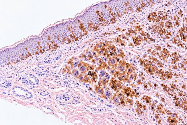
Melan-A immunohistochemistry (IHC), also called MART-1 IHC, is a tissue stain that helps pathologists identify melanocytic differentiation. A positive result means the tested cells contain Melan-A protein, which is commonly expressed by normal melanocytes, benign melanocytic nevi, and many melanomas. That makes Melan-A useful for confirming that a tumor may be melanocytic, especially when the cells are poorly differentiated or a metastasis has an uncertain origin. However, Melan-A positivity does not by itself diagnose melanoma because benign nevi also stain and several non-melanocytic tumors can express the marker. Likewise, some melanomas—particularly desmoplastic and spindle-cell variants—may show weak, focal, or absent Melan-A staining. Pathologists therefore interpret Melan-A with the tumor’s microscopic appearance and a panel that may include SOX10, S100, HMB-45, MITF, PRAME, and other markers chosen for the differential diagnosis. The staining pattern, location of positive cells, and clinical setting are as important as the word “positive.”
- Melan-A/MART-1 is mainly a melanocytic differentiation marker: positive cytoplasmic staining supports melanocytic lineage but is not specific for melanoma.
- Most conventional melanomas are Melan-A positive, but desmoplastic and spindle-cell melanomas can be negative or only focally positive.
- Benign nevi usually stain with Melan-A too: the test cannot reliably separate a nevus from melanoma by positivity alone.
- A positive stain outside the skin needs context: adrenal cortical, steroid-producing, and some PEComa-family tumors can also express Melan-A.
- No special patient preparation is needed: Melan-A IHC is performed on biopsy or surgical tissue already submitted to pathology.
Table of Contents
- What Melan-A IHC Detects
- What Positive Melan-A Staining Means
- How Melan-A Is Used in Melanoma Diagnosis
- Melan-A in Benign and Atypical Melanocytic Lesions
- Non-Melanoma Tumors That Can Stain
- Limitations and Common Pitfalls
- How to Read a Melan-A Pathology Result
What Melan-A IHC Detects
Melan-A IHC detects a melanocyte-associated protein in tissue sections and is used primarily to show melanocytic differentiation. Melan-A and MART-1 refer to the same antigen in routine diagnostic pathology. The commonly used A103 antibody produces predominantly cytoplasmic staining in cells that express the protein.
Melanocytes are pigment-producing cells normally present in the epidermis, hair follicles, mucosal sites, the eye, and other locations. Because Melan-A is part of the melanocytic differentiation program, it is often retained when melanocytes form benign nevi or malignant melanoma. The stain can make small or inconspicuous melanocytic cells easier to see than they are on routine hematoxylin-and-eosin sections.
This makes Melan-A useful in several situations. A pathologist may use it to:
- confirm that an atypical tumor has melanocytic differentiation;
- highlight the distribution of melanocytes in a difficult skin lesion;
- assess melanoma at a surgical margin in selected settings;
- help evaluate a suspected melanoma metastasis;
- distinguish melanocytic cells from many epithelial, hematolymphoid, and mesenchymal tumors as part of an antibody panel.
Melan-A is not a blood test and does not produce a serum level or reference range. The report usually describes staining as positive or negative and may add terms such as diffuse, patchy, focal, strong, or weak. Those descriptors explain how many cells stain and how convincing the pattern is.
The key limitation is that lineage and malignancy are separate questions. Melan-A can answer “Do these cells show melanocytic-type differentiation?” more readily than “Is this lesion malignant?” A benign nevus and a melanoma may both be strongly positive. To decide whether a lesion is melanoma, the pathologist relies first on architecture and cytology, then uses immunostains to solve specific diagnostic problems.
What Positive Melan-A Staining Means
Positive Melan-A staining means that tumor cells contain detectable Melan-A protein; it supports, but does not prove, a melanocytic diagnosis. In a typical melanoma, positive cells show granular or diffuse cytoplasmic staining. The pathologist confirms that staining is located in the cells of interest rather than in nearby normal melanocytes.
The meaning changes with the specimen. In a skin biopsy containing an obvious melanocytic proliferation, Melan-A may simply help outline the lesion. In a lymph node containing an undifferentiated malignant tumor, strong Melan-A positivity can shift the differential toward metastatic melanoma—but the same result must still be checked against morphology and other markers.
A positive result does not establish any of the following on its own:
- that a lesion is malignant;
- that a melanoma is invasive rather than in situ;
- that a positive lymph-node focus is definitely metastatic melanoma rather than a benign nodal nevus;
- the Breslow thickness, stage, or recurrence risk;
- a specific melanoma mutation such as BRAF V600;
- eligibility for a particular systemic therapy.
Melan-A is also not a quantitative proliferation marker. Strong staining does not mean the tumor is growing rapidly, and weak staining does not necessarily indicate less aggressive behavior. Proliferative activity is assessed by morphology, mitotic activity, and sometimes markers such as Ki-67, while prognosis in cutaneous melanoma depends heavily on established factors such as Breslow thickness, ulceration, nodal status, and distant spread.
Diffuse, focal, and negative results
A diffuse positive result means a large proportion of the target cells stain. This is common in many conventional melanomas and nevi. Focal or patchy positivity means only part of the lesion stains; this may occur because the tumor is heterogeneous or has partially lost melanocytic differentiation. A negative result means no convincing staining is identified under the laboratory’s conditions, but it does not exclude melanoma when the morphology remains suspicious.
That final point is especially important in desmoplastic melanoma. These tumors often have spindle-shaped cells in a fibrotic background and may lose Melan-A and HMB-45 expression. SOX10 and S100 are generally more useful melanocytic markers in that setting.
How Melan-A Is Used in Melanoma Diagnosis
Melan-A works best as one member of an IHC panel selected to answer a specific differential diagnosis. No single stain captures every melanoma while excluding every mimic. Pathologists combine markers because each has a different balance of sensitivity, specificity, staining pattern, and known pitfalls.
| Marker | Typical staining | Practical strength | Important limitation |
|---|---|---|---|
| Melan-A/MART-1 | Cytoplasmic | Highlights many melanocytic lesions clearly | Benign nevi are positive; desmoplastic melanoma may be negative |
| SOX10 | Nuclear | Highly sensitive for melanocytic lineage, including many spindle/desmoplastic melanomas | Also marks Schwannian and some other tumors |
| S100 | Nuclear and cytoplasmic | Very sensitive for melanoma | Low specificity because many non-melanocytic cells and tumors stain |
| HMB-45 | Cytoplasmic | Supports melanocytic differentiation and may show maturation patterns | Less sensitive in some melanomas |
| MITF | Nuclear | Can identify melanocytic nuclei when cytoplasmic stains are difficult to interpret | Not entirely specific and may be lost in some melanomas |
| PRAME | Nuclear | Can support melanoma over nevus when expression is appropriately diffuse | Not all melanomas are positive and occasional benign lesions stain |
In a metastatic tumor of unknown origin, the panel may be broader. Cytokeratins can evaluate carcinoma, lymphoid markers can evaluate lymphoma, and lineage-specific markers can assess sarcoma or other tumors. Melan-A becomes persuasive when its staining fits the morphology and agrees with another melanocytic marker such as SOX10.
Melan-A in margin assessment
Melan-A is also used in some forms of margin-controlled surgery for melanoma, particularly MART-1 immunostaining on frozen sections. The stain can reveal melanocytes that are difficult to recognize on routine frozen sections. This can help surgeons and pathologists assess peripheral margins while preserving tissue in anatomically sensitive areas.
The tradeoff is that Melan-A highlights both normal and abnormal melanocytes. Sun-damaged skin may contain increased or irregular melanocytes, and inflammatory or technical changes can complicate interpretation. Margin assessment therefore requires experience with normal melanocyte density, the original tumor pattern, and the limitations of frozen-section morphology.
Melan-A in Benign and Atypical Melanocytic Lesions
Melan-A commonly stains benign nevi, so the presence of staining should never be equated with melanoma. This is one of the most important points for patients reading a pathology report. The marker tells the pathologist what type of differentiation the cells show; it does not independently decide whether they are benign or malignant.
In ordinary nevi, Melan-A can sharply outline nests and single melanocytes. That may help the pathologist evaluate symmetry, lateral spread, depth, and maturation. In some lesions it can also make the melanocyte population appear more crowded than it looks on routine sections. This is useful when interpreted correctly but can be misleading if stain density is mistaken for biologic aggression.
For difficult lesions, the pathologist may combine Melan-A with stains that address different questions. PRAME can be helpful because diffuse nuclear PRAME expression occurs in many melanomas while most conventional nevi are negative or only focally positive. Ki-67 may help show an unexpectedly proliferative dermal component. HMB-45 can contribute information about maturation. p16 expression patterns can add context in selected cases.
None of these markers is perfect. A benign lesion can show an unusual immunophenotype, and melanoma can lack an expected marker. Spitzoid tumors, blue-nevus-family tumors, heavily pigmented lesions, recurrent nevi, and lesions on chronically sun-damaged skin can be particularly challenging. Molecular testing may be added when morphology and IHC remain inconclusive.
Nodal nevi versus melanoma metastasis
Benign melanocytic cells can occasionally be found in lymph nodes as nodal nevi. Because both nodal nevi and melanoma metastases may express Melan-A, a positive Melan-A stain in a lymph node is not automatically evidence of metastatic disease.
Pathologists evaluate where the cells are located in the node, their cytologic features, relationship to the capsule, growth pattern, and staining with additional markers. PRAME, HMB-45, Ki-67, and other studies can help in selected cases, but the final diagnosis depends on the complete pattern rather than one positive stain.
Non-Melanoma Tumors That Can Stain
Melan-A is not restricted to melanomas, and positive staining in an unknown tumor has several important non-melanocytic alternatives. The best-known examples involve steroid-producing tissues and tumors.
Adrenal cortical cells can express Melan-A, and the marker is commonly used in panels for adrenal cortical neoplasms. A metastatic adrenal cortical carcinoma may therefore be Melan-A positive even though it has no melanocytic origin. The distinction usually becomes clear when the stain is interpreted with markers such as inhibin, calretinin, SF-1, cytokeratins, and the clinical imaging pattern.
Melan-A can also be expressed in tumors from the perivascular epithelioid cell tumor (PEComa) family. These tumors often coexpress melanocytic markers and smooth-muscle markers. A Melan-A-positive soft-tissue or visceral tumor should therefore not be called melanoma without considering PEComa and other site-specific possibilities.
Some sex-cord/stromal or steroidogenic tumors may also express Melan-A. The practical lesson is that the marker is most specific when the differential diagnosis has already been narrowed by morphology and anatomic site.
For example, a Melan-A-positive liver mass in a patient with an adrenal primary has a different interpretation from a Melan-A-positive dermal tumor beneath a suspicious pigmented lesion. The antibody result is identical, but the diagnostic probability is not.
Limitations and Common Pitfalls
The main pitfalls are assuming that positive means malignant, assuming that negative excludes melanoma, and ignoring staining in non-tumor cells. Avoiding those errors requires attention to both the slide and the clinical context.
First, Melan-A can highlight background melanocytes and benign nevi. In sun-damaged skin, an increased number of junctional melanocytes can be conspicuous on MART-1 staining and may complicate assessment of melanoma in situ at a margin.
Second, melanoma can lose melanocytic antigens as it becomes dedifferentiated. Metastatic or treatment-altered tumors may show an incomplete marker profile. If morphology strongly suggests melanoma but Melan-A is negative, the pathologist may add SOX10, S100, MITF, PRAME, or molecular studies rather than dismiss the diagnosis.
Third, melanin pigment itself can be brown and may visually resemble some chromogenic IHC reactions. Laboratories and pathologists use appropriate controls and microscopic features to distinguish endogenous pigment from true antibody staining.
Fourth, tissue quality matters. Poor fixation, crush artifact, decalcification, tiny biopsies, and scant tumor can reduce confidence. A negative stain in badly preserved tissue is less informative than a negative stain with strong internal controls and well-preserved tumor.
Finally, sensitivity and specificity depend on the tumor subtype. Conventional epithelioid melanoma is often strongly Melan-A positive, while desmoplastic melanoma is a classic exception. A test that performs well in one morphologic subtype should not be assumed to perform equally well in another.
How to Read a Melan-A Pathology Result
Start with the final diagnosis, then use the Melan-A line to understand how the stain supported that diagnosis. A pathology report may list several antibodies in a compact format such as “SOX10 positive, Melan-A positive, pancytokeratin negative.” The meaning comes from the pattern across the panel.
When reviewing the report, look for these details:
- Which cells are positive? The report should refer to tumor cells, lesional melanocytes, or another defined population.
- How extensive is staining? Diffuse, patchy, or focal staining can affect how strongly the result supports a diagnosis.
- Which other markers agree or disagree? Concordant SOX10 and Melan-A staining in the right morphology is more useful than either marker alone.
- What is the specimen site? Skin, lymph node, adrenal gland, soft tissue, and visceral organs have different differential diagnoses.
- Is the question lineage, malignancy, or margin status? Melan-A performs different jobs in each setting.
If the result is surprising—such as a suspected melanoma that is Melan-A negative—the next step is not to reinterpret the stain in isolation. The pathologist can review the routine sections, examine controls, add complementary antibodies, compare with a previous tumor, or request molecular testing when appropriate.
For patients, a phrase such as “Melan-A positive” should be treated as one line in a larger diagnostic argument. It does not tell you the melanoma stage, whether lymph nodes are involved, or whether immunotherapy or targeted therapy is needed. Those decisions require the final histologic diagnosis, tumor thickness or extent, staging information, and molecular or predictive biomarkers when indicated.
The practical takeaway is that Melan-A is a strong marker of melanocytic differentiation but an imperfect marker of melanoma itself. Its value comes from showing where melanocytic-type cells are and how their pattern fits with morphology and other stains. Positive staining can strongly support melanoma in the right context, yet the same antibody can stain a benign nevus or a non-melanocytic tumor. The panel and the tissue context determine the diagnosis.
There are also practical slide-level pitfalls. Normal epidermal melanocytes and cells in a benign nevus commonly express Melan-A, so positive cells at the edge of a lesion can be an internal control without proving that an atypical population is melanoma. In heavily pigmented tumors, brown melanin can also make a brown chromogen difficult to judge. Pathologists can use the routine slide, a different chromogen, bleaching methods in selected laboratories, or complementary nuclear markers to clarify which signal represents immunostaining.
Sensitivity depends on melanoma subtype. Conventional epithelioid melanoma is frequently Melan-A positive, whereas desmoplastic and markedly dedifferentiated melanomas may lose Melan-A even when the clinical and morphologic evidence favors melanoma. A negative stain therefore has less exclusionary power in those settings. Conversely, positive staining outside melanoma is a recognized diagnostic pitfall, including in adrenal cortical tumors, PEComa-family tumors, and occasional non-melanocytic carcinomas.
For that reason, unexpected Melan-A positivity should lead to a panel rather than a shortcut. SOX10 and S100 can assess melanocytic or neural-crest differentiation, broad-spectrum cytokeratins can support carcinoma, and organ-specific markers may be added according to the anatomic site. The goal is not to collect the largest possible number of stains, but to test competing diagnoses efficiently while preserving tissue for molecular studies when needed.
References
- Immunohistochemistry in melanocytic lesions: Updates with a practical review for pathologists 2022 (Review)
- Immunohistochemistry for Skin Cancers: New Insights into Diagnosis and Treatment of Melanoma 2025 (Systematic Review)
- Melan-A expression in non-melanocytic carcinoma: A potential diagnostic pitfall 2024
- Immunohistochemistry for PRAME in Dermatopathology 2023 (Review)
- Cutaneous Melanoma: A Review of Multifactorial Pathogenesis, Immunohistochemistry, and Emerging Biomarkers for Early Detection and Management 2023 (Review)
Disclaimer
Melan-A IHC must be interpreted by a qualified pathologist together with the tissue morphology, specimen site, and other immunostains. A positive or negative Melan-A result alone cannot confirm or exclude melanoma, determine stage, or select treatment. This information is for general education and does not replace review of an individual pathology report by the treating medical team.





