Arginase-1 immunohistochemistry (IHC) is a tissue stain used mainly to show hepatocellular differentiation—evidence that tumor cells resemble liver hepatocytes. It is one of the most useful markers for supporting hepatocellular carcinoma (HCC), especially when a liver mass must be distinguished from metastatic cancer or intrahepatic cholangiocarcinoma. Normal hepatocytes also express arginase-1 strongly, so a positive stain does not by itself mean cancer.
Pathologists interpret arginase-1 by asking whether the abnormal tumor cells, rather than only the surrounding liver, show the expected cytoplasmic and sometimes nuclear staining. The result is usually combined with morphology and other markers such as HepPar-1, glypican-3, canalicular CD10 or polyclonal CEA, and sometimes albumin RNA in situ hybridization. Arginase-1 is highly specific for hepatocellular differentiation, but no single marker is perfect. Poorly differentiated HCC can lose expression, while rare non-HCC tumors—including hepatoid carcinomas—can be positive.
- Arginase-1 IHC detects a liver-associated enzyme in tissue and is used to identify hepatocellular differentiation.
- Positive staining in tumor cells strongly supports HCC in the right setting, but normal hepatocytes are also naturally positive.
- Arginase-1 is often more sensitive than HepPar-1 in poorly differentiated HCC, although either marker can be negative in individual tumors.
- Rare non-liver tumors can express arginase-1, so the stain should be interpreted as part of a panel rather than as proof of liver origin.
- There is no blood reference range or numeric “positive level” for this IHC test; the pattern, tumor-cell distribution, and morphology determine its meaning.
Table of Contents
- What Arginase-1 IHC Shows
- How Arginase-1 Staining Looks
- Arginase-1 in Hepatocellular Carcinoma
- Distinguishing HCC From Other Tumors
- What Positive and Negative Results Mean
- Limitations and Common Pitfalls
- What Happens After Arginase-1 Testing
What Arginase-1 IHC Shows
Arginase-1 is an enzyme in the urea cycle, the liver pathway that helps convert nitrogen waste into urea. In normal tissue, hepatocytes are the main cells with strong arginase-1 expression. That restricted pattern makes the protein a useful marker when a pathologist needs to decide whether a tumor is showing hepatocellular differentiation.
IHC does not measure the amount of arginase-1 in blood. A laboratory applies an antibody to a thin section of preserved biopsy tissue. If the protein is present, a colored reaction appears in the cells. The pathologist then compares staining in the tumor with normal liver and with the rest of the marker panel.
The most common clinical questions are:
- Is a liver mass hepatocellular carcinoma or a metastasis from another organ?
- Is a poorly differentiated carcinoma in the liver showing hepatocellular differentiation?
- Does a tumor have a phenotype that fits HCC rather than intrahepatic cholangiocarcinoma?
- Does a metastasis at another site look like it could have originated from an HCC?
Arginase-1 is a lineage marker more than a malignancy marker. Benign hepatocytes are strongly positive, and some benign hepatocellular lesions can also retain staining. Therefore, it helps identify what type of cell a tumor resembles; it does not independently distinguish benign from malignant hepatocytes.
For that reason, the stain is usually ordered as part of a broader tumor immunohistochemistry panel. The pathologist integrates tissue architecture, cell shape, vascular pattern, background liver disease, imaging findings, and several complementary markers before naming the tumor.
How Arginase-1 Staining Looks
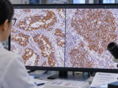
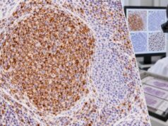
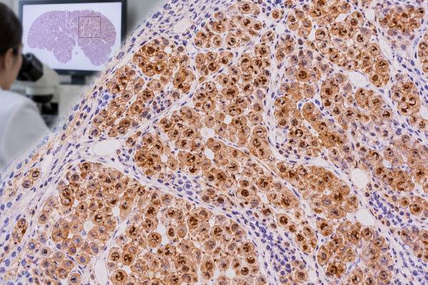
Arginase-1 usually produces strong cytoplasmic staining in hepatocytes and often also shows nuclear staining. The combination can make hepatocellular tissue conspicuous on a biopsy slide. Bile duct epithelium, endothelial cells, and many other nonhepatocellular tissues generally lack the same strong diffuse pattern.
In HCC, staining may be diffuse across most tumor cells or patchy. Well and moderately differentiated tumors commonly retain expression. Poorly differentiated HCC can still be positive, which is one reason arginase-1 became valuable in surgical pathology, but loss of expression becomes more likely as tumors dedifferentiate.
The pathologist pays close attention to which cells are staining. This is especially important in a liver biopsy because normal hepatocytes around a metastatic tumor can be intensely positive. A casual glance could create the false impression that the tumor is positive when the stain is actually highlighting only entrapped or adjacent liver cells.
Reports may use language such as:
- “Arginase-1: positive in tumor cells.”
- “Strong diffuse cytoplasmic and nuclear staining.”
- “Focal positive staining.”
- “Negative in tumor; internal control hepatocytes are positive.”
That last type of statement is useful because the surrounding normal liver serves as an internal control. If hepatocytes stain normally but the tumor does not, a true negative result is more credible. If both tumor and normal hepatocytes fail to stain, the pathologist may suspect a technical problem and repeat the assay.
No universal percentage cutoff is required to interpret arginase-1 across all situations. Distribution, intensity, cell identity, and the competing diagnoses matter more than a single number.
Arginase-1 in Hepatocellular Carcinoma
Arginase-1 is one of the strongest routine markers of hepatocellular differentiation. Classic studies found high sensitivity for HCC, including better retention than HepPar-1 in many poorly differentiated tumors. More recent reviews continue to place arginase-1 among the core stains used when morphology alone cannot confidently establish hepatocellular origin.
HCC can range from well differentiated tumors that closely resemble normal liver plates to high-grade carcinomas with little obvious hepatocellular morphology. In a well-differentiated lesion, the challenge may be separating HCC from hepatocellular adenoma or dysplastic nodules. In a poorly differentiated lesion, the challenge is often determining whether the carcinoma is hepatic at all. Arginase-1 helps most directly with the second problem: lineage.
A common companion is HepPar-1 IHC. HepPar-1 shows granular cytoplasmic staining related to mitochondrial antigen expression. It is highly useful but can lose sensitivity in poorly differentiated HCC and can stain some nonhepatic tumors. Arginase-1 often performs better in that difficult high-grade setting.
Another companion is glypican-3 IHC. Glypican-3 differs from arginase-1 because it is more closely associated with malignant hepatocellular phenotype rather than simply normal hepatocellular lineage. A tumor that is arginase-1 positive and glypican-3 positive can therefore provide two complementary clues: liver-type differentiation and a marker commonly expressed in HCC.
Canalicular markers add another layer. CD10 can show a canalicular staining pattern that supports hepatocellular differentiation. Polyclonal CEA can show a similar canalicular pattern. These patterns are specific when well developed but may be difficult to appreciate in poorly differentiated tumors.
Serum markers are a separate issue. The AFP blood test may be elevated in HCC, but AFP can be normal even when tissue confirms HCC. Tissue arginase-1 and serum AFP answer different questions and should not be expected to match one another.
Distinguishing HCC From Other Tumors
One of the most important uses of arginase-1 is distinguishing primary HCC from a carcinoma that has metastasized to the liver. The liver is a common site of metastasis from colon, pancreas, stomach, lung, breast, kidney, and other organs. A high-grade metastasis can sometimes mimic HCC on a small core biopsy.
A positive arginase-1 stain in the actual tumor cells strongly favors hepatocellular differentiation, especially when it is diffuse and accompanied by other supportive markers. A negative stain makes HCC less likely but does not eliminate it, particularly in a poorly differentiated tumor.
The pathologist often combines positive liver-lineage markers with markers directed at alternative primaries. For example, CK7, CK20, CDX2, PAX8, TTF-1, GATA3, or other stains may be selected based on morphology and imaging. The goal is not to run every possible stain but to test the most plausible competing origins efficiently.
HCC versus intrahepatic cholangiocarcinoma
Intrahepatic cholangiocarcinoma arises from biliary epithelium and typically lacks diffuse hepatocellular markers. Arginase-1 is usually negative, while biliary-type keratins such as CK7 and CK19 are more often expressed. However, rare cholangiocarcinomas can show focal arginase-1 staining, so a small focus of positivity should not override the rest of the phenotype.
Combined hepatocellular-cholangiocarcinoma can contain both hepatocellular and biliary components. In such tumors, different areas may show different marker profiles. Sampling becomes especially important because a small core may capture only one component.
HCC versus clear cell tumors
Clear cell change occurs in HCC but also in renal cell carcinoma, adrenal cortical tumors, and other neoplasms. Recent work has shown that arginase-1 can be particularly helpful in HCC with cytoplasmic clearing, a setting where HepPar-1 may be less reliable. PAX8, carbonic anhydrase IX, inhibin, Melan-A, and other markers may be added depending on the differential.
Hepatoid adenocarcinoma
A key exception is hepatoid adenocarcinoma. These extrahepatic carcinomas develop liver-like morphology and can express hepatocellular markers, including arginase-1 and AFP. They most often arise in the gastrointestinal tract but can occur elsewhere. Clinical imaging and organ-specific markers become crucial because a positive arginase-1 stain cannot distinguish true HCC from every hepatoid carcinoma.
Small biopsies deserve special caution because hepatocellular tumors can be heterogeneous. A core may sample a well-differentiated area that retains liver markers or, by chance, a high-grade focus that has lost them. If imaging and morphology strongly support HCC but arginase-1 is negative, testing another block or adding complementary markers can be more informative than treating the first stain as decisive. Conversely, a tiny focus of positivity in an otherwise nonhepatic phenotype should be interpreted conservatively.
The background liver also provides clinically useful context. Cirrhosis, steatohepatitis, chronic viral hepatitis, or other liver disease can make benign hepatocytes architecturally abnormal, yet those cells normally remain arginase-1 positive. IHC cannot substitute for the morphologic criteria used to distinguish dysplastic nodules, hepatocellular adenoma, and well-differentiated HCC. Reticulin pattern, stromal invasion, cytologic atypia, vascular changes, and lesion-specific markers may be needed in that difficult low-grade differential.
What Positive and Negative Results Mean
A positive arginase-1 result means the stained cells contain detectable arginase-1 protein. In a tumor, that usually supports hepatocellular differentiation. It does not automatically mean the tumor is malignant, and it does not prove that a cancer originated in the liver without considering morphology and clinical location.
| Result pattern | Typical meaning | Important caution |
|---|---|---|
| Strong diffuse tumor-cell positivity | Strongly supports hepatocellular differentiation | Hepatoid and rare non-HCC tumors can occasionally stain |
| Focal tumor-cell positivity | May support HCC but is less definitive | Use a broader panel and verify that tumor cells truly stain |
| Tumor negative, normal hepatocytes positive | Argues against hepatocellular differentiation | Poorly differentiated HCC can lose arginase-1 |
| Only background liver is positive | Does not support HCC in the unstained tumor | Entrapped hepatocytes can mimic tumor positivity |
| No staining in tumor or normal liver | Result may be technically unreliable | Controls and assay performance should be reviewed |
A negative result therefore lowers the probability of HCC but cannot be used as an absolute exclusion test. The most aggressive HCCs may lose recognizable liver markers as they dedifferentiate. In that setting, glypican-3, albumin in situ hybridization, canalicular stains, and the clinical-radiologic picture can become more important.
A mixed marker result is not necessarily contradictory. HCC can be arginase-1 positive but glypican-3 negative, or vice versa, because the proteins reflect different aspects of tumor biology. HepPar-1 may be strong in one area and lost in another. Pathologists therefore look for convergence across several markers instead of requiring every hepatocellular stain to be positive.
This panel approach is particularly useful in metastatic disease. A known HCC that spreads to bone, lung, lymph node, or adrenal gland can become less differentiated and may not reproduce every stain from the liver primary. Retained arginase-1 can be very helpful, but comparison with the original pathology and current imaging often provides the decisive context.
Limitations and Common Pitfalls
Arginase-1 is highly useful precisely because its expression is restricted, but the restriction is not absolute. Large tissue studies have documented occasional staining in non-HCC tumors, usually focal rather than the strong diffuse pattern common in hepatocellular lesions. Strong staining should therefore be persuasive, not magical.
Several practical pitfalls recur:
- Confusing normal liver with tumor: benign hepatocytes can surround or become trapped within a metastasis and stain intensely.
- Assuming positivity equals HCC: benign hepatocellular lesions and some hepatoid carcinomas can also be positive.
- Assuming negativity excludes HCC: poorly differentiated HCC may lose arginase-1.
- Ignoring the stain pattern: diffuse convincing tumor-cell staining carries more weight than rare isolated cells.
- Using one marker in isolation: difficult liver tumors often require a panel that tests hepatocellular, biliary, and metastatic possibilities.
- Overlooking sample limitations: necrosis, crush artifact, small cores, or poor fixation can reduce the amount of interpretable tumor.
The best diagnostic use of arginase-1 is comparative. A pathologist asks whether the stain fits the leading diagnosis better than the alternatives and whether other markers tell a coherent story.
Another practical limitation is that “liver origin” and “hepatocellular differentiation” are related but not identical concepts. A metastatic HCC outside the liver may remain strongly arginase-1 positive, while an extrahepatic hepatoid carcinoma can acquire a liver-like phenotype without arising from hepatocytes. The anatomic distribution on imaging and the rest of the immunophenotype therefore remain essential even when the stain is impressive.
Arginase-1 also does not replace molecular or clinical classification of primary liver cancer. Once HCC is established, decisions about resection, transplantation, ablation, embolization, radiation, systemic therapy, or surveillance depend on stage and liver reserve rather than on the intensity of the stain. In that sense, arginase-1 is most powerful at the diagnostic fork in the road—deciding what the tumor is—rather than at later treatment-selection steps.
When a liver lesion is well differentiated, arginase-1 often confirms hepatocyte identity but may not settle whether the lesion is benign or malignant. In that setting, the pathologist shifts attention to architecture, reticulin framework, stromal invasion, cytologic atypia, and lesion-specific molecular or immunohistochemical findings. The question changes from “Is this hepatocellular?” to “Which hepatocellular lesion is it?”
What Happens After Arginase-1 Testing
If arginase-1 supports HCC, the pathologist usually correlates it with the microscopic pattern and additional stains before finalizing the diagnosis. The clinical team then uses imaging, liver function, tumor size and number, vascular involvement, extrahepatic disease, performance status, and underlying liver disease to stage the cancer and select treatment.
If the stain is negative or equivocal, the next step depends on the differential. The pathologist may add HepPar-1, glypican-3, albumin RNA in situ hybridization, CK7, CK19, organ-specific markers, or other studies. A second tissue block may be tested if the first contains little viable tumor.
When the biopsy comes from outside the liver and arginase-1 is positive, the result can suggest metastatic HCC, but imaging is needed to identify a liver primary and to exclude a hepatoid carcinoma elsewhere. Prior pathology is also valuable because comparing the new lesion with a known tumor may establish whether they share the same morphology and phenotype.
For patients, the most useful interpretation is the final integrated diagnosis rather than the stain in isolation. Ask whether the tumor cells themselves were arginase-1 positive, which companion markers agreed with the result, and whether the pathology favors HCC, another primary liver cancer, or a metastasis. Those answers explain what the stain actually contributed.
References
- An Update of Immunohistochemistry in Hepatocellular Carcinoma 2025 (Review)
- Arginase-1 is More Specific Than Hepatocyte Paraffin 1 for Differentiating Hepatocellular Carcinomas With Cytoplasmic Clearing from Nonhepatocellular Clear Cell Tumors in Liver Biopsies 2024
- Advances in Histological and Molecular Classification of Hepatocellular Carcinoma 2023 (Review)
- Diagnostic value of glypican-3, arginase-1 and hepatocyte paraffin antigen -1 in differentiating hepatocellular carcinoma from intrahepatic cholangiocarcinoma 2022
- Arginase-1: a new immunohistochemical marker of hepatocytes and hepatocellular neoplasms 2010
Disclaimer
Arginase-1 IHC is a tissue-based pathology marker and cannot diagnose liver cancer by itself. Results must be interpreted with tumor morphology, other stains, imaging, and clinical information, especially when the tumor is poorly differentiated or could represent a metastasis. Discuss the complete pathology report with the treating clinician or pathologist.