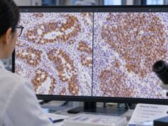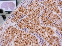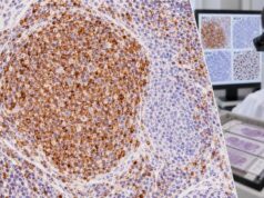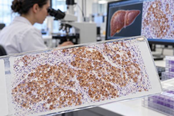
HepPar-1 immunohistochemistry is a tissue stain used to show hepatocellular differentiation and support the diagnosis of hepatocellular carcinoma (HCC). The antibody recognizes a mitochondrial enzyme, carbamoyl phosphate synthetase 1, and typically produces a granular cytoplasmic staining pattern in normal hepatocytes and many HCCs. A positive HepPar-1 result can be very helpful when a liver tumor may represent HCC, but it is not completely specific for liver origin. Some gastric, intestinal, pulmonary, and other carcinomas—especially tumors with hepatoid differentiation—can also express HepPar-1. In addition, poorly differentiated HCC may lose the marker. Pathologists therefore interpret HepPar-1 with tumor morphology and companion stains such as arginase-1, Glypican-3, canalicular markers, cytokeratins, and organ-specific markers. The most useful question is not simply whether HepPar-1 is positive, but whether the complete immunophenotype supports hepatocellular differentiation strongly enough to distinguish HCC from cholangiocarcinoma, metastatic adenocarcinoma, or another mimic.
- HepPar-1 positivity usually supports hepatocellular differentiation, especially when the tumor has compatible liver morphology.
- The expected staining pattern is granular cytoplasmic, reflecting the mitochondrial protein detected by the antibody.
- A negative HepPar-1 stain does not exclude HCC, particularly in poorly differentiated tumors.
- HepPar-1 is not fully liver-specific; some non-hepatic carcinomas and hepatoid tumors can stain positive.
- The stain is best interpreted with arginase-1, Glypican-3, and morphology, not as a stand-alone diagnosis.
Table of Contents
- What HepPar-1 IHC Measures
- How HepPar-1 Supports HCC Diagnosis
- How Positive Staining Is Interpreted
- HepPar-1 With Other Liver Markers
- HepPar-1 in Metastatic Tumor Workups
- Limitations and Pitfalls
- What to Ask After the Result
What HepPar-1 IHC Measures
HepPar-1 stands for hepatocyte paraffin 1. The antibody was developed to recognize an antigen strongly expressed in hepatocytes, and its target was later identified as carbamoyl phosphate synthetase 1, a mitochondrial enzyme involved in the urea cycle. Because hepatocytes contain abundant mitochondria, staining usually appears as coarse or granular cytoplasmic reactivity.
The test is performed on formalin-fixed, paraffin-embedded tissue from a liver biopsy, surgical resection, cell block, or metastatic-site specimen. A thin section is exposed to the HepPar-1 antibody, followed by a detection system that creates visible color where the antibody binds.
HepPar-1 is not a blood test. It has no serum concentration, reference interval, or patient preparation requirement. Pathologists describe whether tumor cells are positive or negative and may add the extent and intensity of staining. There is no universal percentage threshold that independently makes a tumor HCC.
Normal liver is strongly HepPar-1-positive, which can be both helpful and challenging. Native hepatocytes provide an internal positive control, but entrapped normal hepatocytes in a tumor can also be mistaken for positive malignant cells if the slide is not carefully correlated with the routine hematoxylin-and-eosin section.
The marker identifies hepatocellular differentiation, not malignancy itself. Benign hepatocytes, hepatocellular adenomas, and focal nodular hyperplasia can also be positive. Therefore, HepPar-1 is not designed to distinguish every benign hepatocellular lesion from HCC. The diagnostic question determines which additional stains and morphologic features matter most.
How HepPar-1 Supports HCC Diagnosis
HCC often resembles hepatocytes and may form trabeculae, pseudoglands, solid nests, or compact sheets. In a typical case, eosinophilic granular cytoplasm, bile production, endothelial wrapping, and characteristic architecture can strongly suggest HCC. HepPar-1 then serves as supportive evidence of hepatocellular lineage.
The stain is especially useful in moderate or well-differentiated tumors. These cancers often retain the metabolic machinery of hepatocytes and therefore preserve HepPar-1 expression. Sensitivity can decline as HCC becomes poorly differentiated, so a negative result in a high-grade tumor should not end the workup.
In a poorly differentiated liver mass, the main alternatives may include intrahepatic cholangiocarcinoma, metastatic adenocarcinoma, neuroendocrine carcinoma, renal cell carcinoma, adrenocortical carcinoma, melanoma, or another epithelioid neoplasm. HepPar-1 can narrow that differential when it is convincingly positive, but companion markers are essential because several extrahepatic tumors can express the antigen.
A useful principle is to separate two questions: Is the tumor hepatocellular? and Is the tumor malignant? HepPar-1 mainly helps with the first. The second may require morphology, stromal invasion, reticulin framework, vascular invasion, molecular findings, and other IHC markers.
For example, well-differentiated hepatocellular lesions in noncirrhotic liver can include focal nodular hyperplasia, hepatocellular adenoma, and HCC. HepPar-1 may be positive in all three. A different panel involving glutamine synthetase, liver fatty acid-binding protein, serum amyloid A, C-reactive protein, beta-catenin pathway markers, or Glypican-3 may be more informative depending on the lesion.
How Positive Staining Is Interpreted
A positive HepPar-1 result means the pathologist sees specific granular cytoplasmic staining in tumor cells. The diagnostic weight depends on how much of the tumor stains, how strong the reaction is, and whether the morphology fits hepatocellular differentiation.
| Pattern | General meaning | Important caution |
|---|---|---|
| Strong, diffuse granular cytoplasmic staining | Strong support for hepatocellular differentiation | Some non-hepatic tumors can show similar staining |
| Focal positive staining | Supportive but less decisive | Could reflect heterogeneity or a non-hepatic mimic |
| Negative staining | HepPar-1 expression is not detected | Poorly differentiated HCC can be negative |
| Scattered positive cells within an otherwise negative tumor | May represent entrapped hepatocytes | Must correlate with tumor-cell morphology |
Strong diffuse staining in a liver tumor with classic HCC morphology is highly supportive. By contrast, focal staining in a gland-forming adenocarcinoma should be interpreted cautiously because some gastric and intestinal tumors can express HepPar-1.
The absence of a numeric cutoff is important. HepPar-1 is not scored like HER2 IHC or breast ER. Its value is diagnostic pattern recognition rather than treatment eligibility based on a specific percentage.
A positive HepPar-1 stain also does not indicate stage or aggressiveness. A small localized HCC and a metastatic HCC can both be strongly positive. Tumor stage depends on size, number of lesions, vascular invasion, nodal or distant spread, and the staging system used.
HepPar-1 With Other Liver Markers
Modern HCC diagnosis rarely relies on HepPar-1 alone. Pathologists combine markers with complementary strengths.
Arginase-1 is one of the most useful hepatocellular markers and often has higher sensitivity than HepPar-1 in poorly differentiated HCC. It commonly shows cytoplasmic staining with or without nuclear reactivity. The arginase-1 IHC test is therefore a frequent partner when hepatocellular differentiation is being assessed.
Glypican-3 is an oncofetal marker expressed in many HCCs and usually absent from normal adult hepatocytes. Unlike HepPar-1, it can help support malignancy in some hepatocellular settings. The Glypican-3 IHC test is particularly useful when a malignant hepatocellular lesion is suspected.
Polyclonal CEA and CD10 can show a canalicular staining pattern in HCC. This thin branching pattern outlines bile canaliculi between tumor cells and is more informative than simple diffuse positivity.
CK7 and CK19 support biliary differentiation when cholangiocarcinoma is considered. However, some HCCs can express CK19 or CK7, especially progenitor-like or aggressive subtypes, so these keratins are not absolute.
Albumin RNA in situ hybridization may support primary hepatic origin and can be useful in difficult HCC-versus-cholangiocarcinoma or metastasis cases. It detects albumin messenger RNA rather than protein.
The best panel depends on tumor appearance. A classic trabecular HCC may need only a small confirmatory panel. A poorly differentiated tumor with no clear lineage may require epithelial, melanocytic, neuroendocrine, renal, adrenal, and hepatocellular markers before a final diagnosis is secure.
HepPar-1 in Metastatic Tumor Workups
HepPar-1 is often ordered when a metastatic tumor could represent HCC. For example, a lung, bone, adrenal, or lymph-node mass in a patient with a liver lesion may be sampled. Strong HepPar-1 and arginase-1 positivity can support metastatic HCC if the morphology is compatible.
The reverse situation also occurs: an adenocarcinoma metastatic to the liver may show unexpected HepPar-1 staining. Gastric adenocarcinoma is a well-known example, especially tumors with hepatoid morphology. Some esophageal, intestinal, pancreatic, pulmonary, and gynecologic carcinomas may also show focal or even diffuse positivity.
This is why organ-specific markers matter. A liver mass positive for HepPar-1 but also strongly CDX2- and SATB2-positive may raise colorectal or intestinal differentiation. A tumor positive for PAX8 may suggest renal, thyroid, or Müllerian origin depending on morphology. A CDX2 IHC test can therefore help resolve a gastrointestinal mimic.
Hepatoid adenocarcinoma deserves special mention because it can look and stain like HCC. These tumors arise outside the liver, commonly in the stomach but also in other organs, and may express HepPar-1, Glypican-3, and AFP. Clinical imaging and organ-specific morphology become decisive.
When the differential remains unresolved, molecular profiling, radiologic correlation, and review of prior specimens can be more informative than adding many nonspecific stains. The pathologist’s final wording may appropriately state that the immunophenotype “supports” rather than “proves” a particular origin.
Limitations and Pitfalls
The main limitation of HepPar-1 is that neither sensitivity nor specificity is perfect. A meaningful subset of poorly differentiated HCCs is negative, while several non-hepatic carcinomas can be positive.
Common pitfalls include:
- mistaking entrapped normal hepatocytes for positive tumor cells;
- interpreting focal granular staining as proof of HCC without other hepatocellular markers;
- ruling out HCC solely because HepPar-1 is negative;
- assuming any HepPar-1-positive metastasis came from the liver;
- ignoring tumor morphology and clinical imaging;
- using necrotic or poorly fixed tissue that may have reduced antigen preservation.
Another limitation is that HepPar-1 does not reliably separate benign from malignant hepatocellular lesions. Benign hepatocytes and hepatocellular adenomas can stain strongly. In a well-differentiated nodule, pathologists rely on architecture, reticulin, stromal invasion, and lesion-specific panels rather than on HepPar-1 alone.
Small biopsies introduce sampling error. HCC can be heterogeneous, and a core may capture only a negative portion. Conversely, a metastatic carcinoma may contain only a focal HepPar-1-positive area that is overrepresented in a small sample.
Technical quality also matters. Fixation time, antibody clone, detection platform, and laboratory validation can change staining strength. A pathologist evaluates controls and the expected pattern before calling a result truly positive or negative.
What to Ask After the Result
If your pathology report mentions HepPar-1, focus on the integrated diagnosis. Useful questions include:
- Is the tumor strongly and diffusely HepPar-1-positive, or only focally positive?
- Are arginase-1 and Glypican-3 also positive?
- Does the microscopic appearance support hepatocellular carcinoma?
- Were cholangiocarcinoma and metastatic adenocarcinoma considered?
- Could the positive cells be entrapped normal hepatocytes?
- Does imaging identify a likely primary liver tumor or another primary site?
- Is additional molecular testing or expert liver-pathology review needed?
If HepPar-1 is negative, ask what other evidence supports HCC. A negative result is not a definitive exclusion. A tumor may still be HCC if arginase-1, Glypican-3, canalicular staining, morphology, and clinical features align.
It is also useful to separate diagnosis from treatment. HepPar-1 is not a marker that directly selects immunotherapy, targeted therapy, transplant, or surgery. HCC treatment depends on stage, liver function, performance status, vascular involvement, prior therapy, and multidisciplinary assessment. In at-risk patients, current HCC guidelines also emphasize imaging-based diagnostic criteria, with biopsy used when imaging is indeterminate or when tissue is clinically needed.
The most important information in a pathology report is the final diagnosis and the reasoning that supports it. HepPar-1 is one piece of that reasoning, often a very useful one, but it works best as part of a carefully chosen panel.
HepPar-1 can be especially helpful when a liver mass is poorly characterized clinically and the main question is HCC versus metastatic carcinoma. Metastases to the liver are common, so location inside the liver is not proof of hepatic origin. Strong granular HepPar-1 staining in tumor cells, together with arginase-1 and compatible morphology, can move the diagnosis toward hepatocellular differentiation. Conversely, an adenocarcinoma pattern with organ-specific markers may support a metastasis even if a small subset of cells shows HepPar-1 reactivity.
The distinction between hepatocellular differentiation and malignancy is also important. Normal hepatocytes are strongly HepPar-1-positive, and many benign hepatocellular lesions retain staining. HepPar-1 therefore does not function as a simple “cancer on/cancer off” marker. In a well-differentiated nodule, the pathologist may need reticulin, Glypican-3, heat-shock protein 70, glutamine synthetase, beta-catenin-related patterns, or other stains selected for the differential diagnosis. Architectural evidence and clinical imaging may be more decisive than HepPar-1 itself.
Poorly differentiated HCC creates the opposite problem. As tumor cells lose mature hepatocyte features, HepPar-1 sensitivity can fall. Arginase-1 may retain better sensitivity in some settings, while Glypican-3 can add evidence of HCC even though it is not a pure lineage marker. A negative HepPar-1 result should therefore lead to a broader panel rather than an automatic diagnosis of metastasis. This is especially important in needle biopsies, where only a small and potentially unrepresentative tumor area is available.
HepPar-1 also has recognized non-hepatic expression. Some gastrointestinal adenocarcinomas, especially tumors with hepatoid differentiation, can show positivity. Other carcinomas may stain less commonly. When a tumor outside the liver is HepPar-1-positive, the differential includes metastatic HCC but also a non-hepatic tumor that has acquired hepatocellular-like features. Arginase-1, Glypican-3, site-associated markers, serum studies, and imaging can help resolve that distinction.
Preanalytic factors matter in difficult cases. Heavily necrotic tissue may lose granular staining, while entrapped normal hepatocytes can create a false impression of tumor positivity at the edge of an infiltrating lesion. The pathologist needs to map the stain back to the cells seen on routine hematoxylin-and-eosin sections. When there is uncertainty, testing another block or obtaining additional tissue can be more reliable than forcing a result from a compromised sample.
The marker’s target helps explain its characteristic appearance. HepPar-1 recognizes carbamoyl phosphate synthetase 1, a mitochondrial enzyme involved in the urea cycle. Because hepatocytes contain abundant mitochondria, staining often appears as coarse or finely granular cytoplasmic reactivity. This pattern is different from a nuclear marker and helps pathologists judge whether the observed signal is biologically plausible.
Hepatocellular tumors can also contain clear, fatty, steatohepatitic, fibrous, or other morphologic patterns that make lineage less obvious. In those variants, a coordinated panel can prevent one unusual feature from dominating the diagnosis. Arginase-1 and HepPar-1 provide evidence of hepatocellular differentiation, while Glypican-3 can support HCC in the right setting. Canalicular staining with polyclonal CEA or CD10 may provide additional evidence, although interpretation can be technically demanding.
Cholangiocarcinoma is another major primary liver cancer in the differential. It usually follows a biliary/adenocarcinoma immunophenotype rather than a hepatocellular one, but mixed or combined hepatocellular-cholangiocarcinoma can contain areas with different differentiation. Sampling only one component may produce an incomplete picture. When imaging or morphology suggests a mixed tumor, additional blocks or a larger specimen can be important for accurate classification.
For patients, a HepPar-1-positive report should therefore be read together with the final diagnostic wording. “Positive for HepPar-1” means the sampled cells express a marker associated with hepatocellular differentiation; “hepatocellular carcinoma” is a diagnosis reached after that result is integrated with morphology and other evidence. This distinction becomes especially important when the tumor is outside the liver or when the patient has a history of another cancer.
This integrated approach is particularly useful in small biopsies, where preserving tissue for additional IHC or molecular studies may be as important as repeating a single marker.
References
- An Update of Immunohistochemistry in Hepatocellular Carcinoma 2025 (Review)
- Pathologic assessment of hepatocellular carcinoma in the era of immunotherapy: a narrative review 2024 (Review)
- Advances in Histological and Molecular Classification of Hepatocellular Carcinoma 2023 (Review)
- The Role of Immunohistochemistry in the Differential Diagnosis between Intrahepatic Cholangiocarcinoma, Hepatocellular Carcinoma and Liver Metastasis, as Well as Its Prognostic Value 2023 (Review)
- Evaluating Liver Biopsies with Well-Differentiated Hepatocellular Lesions 2023 (Review)
- Diagnostic utility of the HepPar1 antibody to differentiate hepatocellular carcinoma from metastatic carcinoma in fine-needle aspiration samples 2002
Disclaimer
HepPar-1 IHC is an ancillary tissue test and cannot diagnose hepatocellular carcinoma or establish liver origin by itself. Results must be interpreted with morphology, other markers, imaging, and clinical information. Discuss the complete pathology report and its implications with your pathologist or treating clinician.