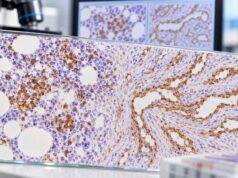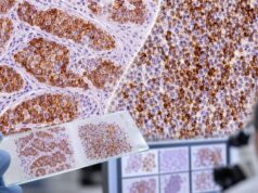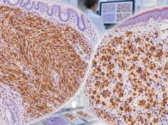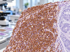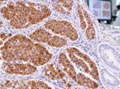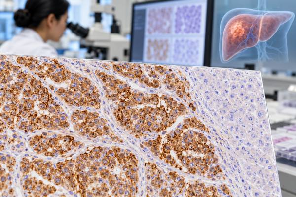
Glypican-3 (GPC3) immunohistochemistry is a tissue stain used mainly to support the diagnosis of hepatocellular carcinoma (HCC), particularly when a liver tumor must be distinguished from benign hepatocellular nodules or from other malignant tumors. GPC3 is an oncofetal cell-surface proteoglycan that is highly expressed in many HCCs but is usually absent from normal adult hepatocytes and most benign liver tissue. A positive result can therefore strengthen an HCC diagnosis, especially when it is interpreted with tumor morphology and other hepatocellular markers such as arginase-1 and HepPar-1. The stain is not perfectly sensitive or specific: some HCCs are GPC3-negative, and several non-HCC tumors—including certain germ cell tumors and other malignancies—can express GPC3. For that reason, pathologists use GPC3 as part of a panel rather than as a stand-alone cancer test. The pattern, extent, and intensity of staining all matter, but there is no single universal percentage cutoff that independently establishes HCC.
- Glypican-3 positivity supports hepatocellular carcinoma when the tumor’s morphology and other liver markers fit.
- A negative GPC3 stain does not rule out HCC, especially in well-differentiated or biologically heterogeneous tumors.
- GPC3 is usually absent from normal adult hepatocytes, which helps distinguish malignant hepatocellular lesions from many benign liver nodules.
- GPC3 is not specific only to liver cancer; some germ cell tumors and other cancers can also stain positive.
- GPC3 IHC is a tissue test, not a blood level, so there is no normal serum range or special patient preparation.
Table of Contents
- What Glypican-3 IHC Measures
- Why GPC3 Is Used in HCC Diagnosis
- How Positive Staining Is Interpreted
- GPC3 With Other Liver Markers
- Differential Diagnosis and Pitfalls
- What GPC3 Does Not Tell You
- Questions After a GPC3 Result
What Glypican-3 IHC Measures
Glypican-3 is a heparan sulfate proteoglycan attached to the cell surface. It has an important role during embryonic development and is strongly expressed in fetal tissues, including fetal liver. In most normal adult tissues, GPC3 expression is low or absent. Many hepatocellular carcinomas re-express the protein, which is why it is described as an oncofetal marker.
GPC3 IHC is performed on a thin section of formalin-fixed, paraffin-embedded tissue from a liver biopsy, surgical resection, or metastatic tumor. An antibody binds the GPC3 protein, and a detection system produces visible color under the microscope. Pathologists evaluate staining in the malignant cells rather than simply reading an automated numeric value.
The staining pattern can be membranous, cytoplasmic, or both. Some HCCs show a strong coarse or granular cytoplasmic pattern, and membrane accentuation may be visible. The percentage of positive tumor cells can vary widely. A report may therefore describe the stain as focal or diffuse and weak, moderate, or strong.
There is no universal “positive if greater than X%” rule that applies to every laboratory and diagnostic setting. Unlike a treatment-linked biomarker such as ER in breast cancer, GPC3 is mainly an ancillary diagnostic marker. Its value comes from how the pattern fits the morphology and the rest of the panel.
GPC3 IHC should also not be confused with experimental or research measurements of soluble GPC3 in blood. The routine pathology test discussed here evaluates tissue expression. For circulating liver-cancer markers, clinicians may use tests such as alpha-fetoprotein (AFP) or des-gamma-carboxy prothrombin in selected settings, but those blood tests answer different questions.
Why GPC3 Is Used in HCC Diagnosis
Hepatocellular carcinoma can often be diagnosed from its architecture and cytology, especially in a patient with cirrhosis and a liver mass showing characteristic imaging. However, tissue diagnosis becomes challenging when the lesion is small, well differentiated, poorly differentiated, or found in a patient without classic risk factors.
GPC3 is helpful because benign adult hepatocytes generally do not express it. A malignant hepatocellular lesion with convincing GPC3 positivity is therefore more concerning for HCC than a regenerative nodule with no GPC3 staining.
In small or well-differentiated lesions, the main differential may include high-grade dysplastic nodule, hepatocellular adenoma, focal nodular hyperplasia, or well-differentiated HCC. No single IHC marker solves every case. Pathologists integrate reticulin architecture, stromal invasion, cytologic atypia, vascular changes, and panels that can include GPC3, heat-shock protein 70, glutamine synthetase, and other markers depending on the specific problem.
In poorly differentiated cancer, the question can be different: is this an HCC that has lost some typical morphology, an intrahepatic cholangiocarcinoma, or a metastasis from another organ? GPC3 can support HCC, but a broader lineage panel becomes essential.
The arginase-1 IHC test is highly useful for hepatocellular differentiation and is often paired with GPC3. A HepPar-1 IHC test provides another hepatocellular marker with a different sensitivity and specificity profile.
The combination matters because marker expression changes with tumor differentiation. A poorly differentiated HCC may lose HepPar-1 or arginase-1 expression yet retain GPC3, while another tumor may show the opposite pattern. Using several complementary stains reduces the chance of over-relying on one biologically variable marker.
How Positive Staining Is Interpreted
A “GPC3-positive” report means the pathologist sees specific staining in tumor cells. The strength of that evidence depends on distribution, intensity, localization, and the tumor’s microscopic appearance.
| Pattern | General meaning | Important limitation |
|---|---|---|
| Strong, diffuse membranous/cytoplasmic staining in a liver tumor | Strong support for HCC when morphology is compatible | Non-HCC tumors can occasionally show similar staining |
| Focal positive staining | May support HCC but carries less weight | Sampling and tumor heterogeneity can affect the result |
| Negative staining | GPC3 expression is not detected | HCC is not excluded |
| Positive staining in an extrahepatic tumor | GPC3-expressing tumor is possible | Do not assume metastatic HCC without other hepatocellular evidence |
A strong positive stain is usually more persuasive than a few weak cells, but there is no standard equation that converts intensity into a probability of HCC. Pathologists also check for internal controls and for nonspecific background staining.
Tumor differentiation affects sensitivity. GPC3 is commonly expressed in moderately and poorly differentiated HCC, while a subset of well-differentiated HCCs may be negative. This is one reason a negative result should not be used as a veto when morphology and other markers support hepatocellular carcinoma.
Positive GPC3 also does not mean “advanced HCC.” The stain can be positive in a small localized tumor as well as metastatic disease. Stage is determined by tumor burden, vascular invasion, lymph nodes, metastases, liver function, and the staging system used—not by IHC intensity.
GPC3 With Other Liver Markers
The most reliable HCC workups combine markers that answer different questions. GPC3 is useful because it often marks malignant hepatocellular differentiation, while arginase-1 and HepPar-1 are more directly associated with hepatocellular lineage.
Arginase-1 is a cytoplasmic and sometimes nuclear marker of hepatocytes. It is often more sensitive than HepPar-1 in poorly differentiated HCC and has high specificity for hepatocellular differentiation in the right panel.
HepPar-1 recognizes carbamoyl phosphate synthetase 1 and produces a granular cytoplasmic staining pattern. It is highly useful but can be positive in some non-hepatic adenocarcinomas, especially hepatoid tumors.
Canalicular markers such as polyclonal CEA or CD10 can show a characteristic canalicular pattern in HCC. The pattern, rather than simple positivity, is what supports hepatocellular differentiation.
CK7 and CK19 may be used when cholangiocarcinoma is considered. Many cholangiocarcinomas express these keratins, whereas conventional HCC is often negative or only focally positive. CK19-positive HCCs do occur, so the distinction cannot be made from one keratin stain.
Albumin RNA in situ hybridization may support primary hepatic origin in difficult cases and can be useful for both HCC and intrahepatic cholangiocarcinoma, depending on the assay and context. Molecular testing may also help if a tumor remains unclassified.
The panel should match the clinical question. A well-differentiated nodule in cirrhosis needs a different set of stains from a poorly differentiated metastasis in the liver. GPC3 is valuable in both settings, but its partners and interpretive weight change.
Differential Diagnosis and Pitfalls
The most important pitfall is assuming GPC3 is liver-specific. It is not. GPC3 can be strongly expressed in yolk sac tumor and may appear in other germ cell tumors, some squamous carcinomas, a subset of lung cancers, ovarian tumors, and other malignancies. Tumors with hepatoid morphology outside the liver can also express GPC3.
This creates a particular challenge when a GPC3-positive tumor is found in the liver of a younger patient. A metastatic germ cell tumor must be considered when the morphology and clinical context fit. Markers such as SALL4, AFP, OCT4, and other germ-cell markers may be added. The SALL4 IHC test can be especially helpful because diffuse nuclear SALL4 supports germ-cell differentiation in the correct setting.
Hepatoid adenocarcinoma is another mimic. These extrahepatic carcinomas can look like HCC and can express AFP, GPC3, and even HepPar-1. Clinical imaging and organ-specific markers are essential before calling a GPC3-positive tumor metastatic HCC.
Technical issues can also mislead. Poor fixation, necrosis, small biopsy size, treatment effect, and antibody/platform differences can change staining. Because HCC can be heterogeneous, a tiny biopsy may under-sample a positive component.
Background staining should be distinguished from true tumor-cell membrane or cytoplasmic labeling. Pathologists compare the IHC slide with the matching hematoxylin-and-eosin section to ensure the stained cells are actually malignant.
A second expert opinion can be useful when a liver tumor remains ambiguous after imaging and IHC. Misclassifying HCC as cholangiocarcinoma or metastasis can substantially change systemic treatment, transplant considerations, and surgical planning.
What GPC3 Does Not Tell You
GPC3 IHC does not measure liver function, determine HCC stage, or establish whether a patient can undergo surgery, ablation, embolization, transplant, or systemic therapy. Those decisions depend on tumor distribution, vascular invasion, extrahepatic disease, liver reserve, performance status, and multidisciplinary assessment.
The stain is also not a validated stand-alone prognostic score. Higher GPC3 expression has been associated with aggressive features in some studies, but routine patient-specific risk prediction does not rely on staining intensity alone.
GPC3 is an active therapeutic research target. Antibodies, bispecific agents, vaccines, CAR-T cells, radiopharmaceuticals, and other GPC3-directed strategies are under investigation. However, diagnostic GPC3 positivity on a pathology slide does not automatically make a patient eligible for a GPC3-targeted therapy. Eligibility depends on the exact clinical trial or approved treatment criteria.
For routine HCC management, current practice relies heavily on imaging criteria in at-risk patients, pathology when needed, and clinical staging. AASLD guidance emphasizes that biomarkers alone should not establish HCC diagnosis when imaging or histology is required.
This distinction is important for patients who also have an elevated blood AFP. A high AFP and positive tissue GPC3 can point in the same direction, but they are still different tests with different false-positive and false-negative patterns. Neither should be interpreted without the whole clinical picture.
Questions After a GPC3 Result
If a pathology report says Glypican-3 is positive, useful questions include:
- Does the final diagnosis favor hepatocellular carcinoma?
- How strong and widespread is GPC3 staining?
- Were arginase-1, HepPar-1, or canalicular markers also positive?
- Were cholangiocarcinoma, metastatic carcinoma, or germ cell tumor considered?
- Does the tumor’s morphology look hepatocellular?
- Is additional molecular testing or imaging needed to establish the primary site?
- Is GPC3 being used only for diagnosis, or is a clinical trial using GPC3 expression as an eligibility criterion?
If GPC3 is negative, ask what other evidence supports or argues against HCC. A negative stain is not equivalent to a negative cancer diagnosis. In difficult tumors, the pattern across several markers is often more informative than any single result.
For example, an arginase-1-positive, HepPar-1-positive, GPC3-negative tumor with classic trabecular architecture may still be HCC. Conversely, a GPC3-positive tumor that is SALL4-positive and lacks convincing hepatocellular markers may lead the pathologist toward a germ-cell or hepatoid differential. The integrated diagnosis is the clinically meaningful endpoint.
The diagnostic challenge is often greatest in a small biopsy of a well-differentiated hepatocellular lesion. Benign hepatocytes and low-grade HCC can look similar, and no single stain perfectly separates them. GPC3 can add evidence of malignancy, particularly when it is combined with other markers used in hepatocellular-lesion panels and with architectural findings such as thickened trabeculae, loss of the normal reticulin framework, stromal invasion, or increased unpaired arteries. A negative GPC3 stain in this setting leaves the morphologic question open rather than settling it as benign.
GPC3 is also different from markers whose main job is to establish liver lineage. Arginase-1 and HepPar-1 more directly support hepatocellular differentiation, whereas GPC3 is often used because expression is increased in many HCCs and is uncommon in normal adult hepatocytes. That means a useful panel may contain both lineage and malignancy-associated markers. A metastatic carcinoma can be excluded more confidently when the entire pattern, not just one stain, fits HCC.
The patient’s underlying liver disease provides another layer of context. HCC commonly arises in cirrhosis or chronic liver injury, but it can also occur without cirrhosis. Imaging patterns, serum alpha-fetoprotein, liver function, viral-hepatitis history, and the presence of other lesions may all shape the pretest probability. Tissue IHC is especially valuable when imaging is atypical, the patient has another known cancer, or the sample comes from an extrahepatic metastasis.
GPC3 is being investigated as a therapeutic target because it is expressed on the surface of many HCC cells and has limited expression in most normal adult tissues. Trials have explored antibody-based strategies, cellular therapies, and other GPC3-directed approaches. This research role should not be confused with routine diagnostic use: a positive pathology stain does not by itself mean an approved GPC3-targeted treatment is indicated. Trial eligibility can require a specific assay, threshold, disease stage, and prior-treatment history.
A GPC3 result can be especially helpful when a metastatic lesion is sampled outside the liver. If a bone, lung, adrenal, or lymph-node metastasis has hepatocellular morphology and expresses GPC3 together with hepatocellular lineage markers, metastatic HCC becomes a strong possibility. The same stain is less persuasive when the tumor has gland-forming morphology or a marker profile that clearly supports another organ. In other words, the anatomic site of the biopsy changes the question but not the need for a panel.
Germ cell tumors are an important non-hepatic source of GPC3 positivity. Yolk sac tumor is a classic example and can arise in gonadal or extragonadal locations. Patient age, serum markers, SALL4, AFP-related staining, and other germ-cell markers help separate this diagnosis from HCC when morphology overlaps. Recognizing this differential is particularly important in a young patient or a midline mass, where assuming that GPC3 automatically means liver cancer could be misleading.
Pathology reports may describe GPC3 as membranous, cytoplasmic, or both. Unlike HER2, there is no broadly used 0-to-3+ clinical scoring system that determines routine HCC treatment. Extent and intensity can support interpretation, but the final diagnosis depends on the whole pattern. A small focus of positivity deserves less weight than diffuse convincing expression when other findings are equivocal.
When results remain borderline, comparing another tumor block can help show whether the apparent GPC3 pattern is truly representative of the lesion rather than a small heterogeneous focus.
References
- An Update of Immunohistochemistry in Hepatocellular Carcinoma 2025 (Review)
- The role of glypican-3 in hepatocellular carcinoma: Insights into diagnosis and therapeutic potential 2024 (Review)
- Exploring Glypican-3 as a Molecular Target in Hepatocellular Carcinoma: Perspectives on Diagnosis and Precision Immunotherapy Strategies 2024 (Review)
- AASLD Practice Guidance on prevention, diagnosis, and treatment of hepatocellular carcinoma 2023 (Guideline)
- Progress and challenges in glypican-3 targeting for hepatocellular carcinoma therapy 2024 (Review)
- Glypican-3: a novel serum and histochemical marker for hepatocellular carcinoma 2003
Disclaimer
Glypican-3 IHC is an ancillary pathology test and cannot diagnose hepatocellular carcinoma by itself. Positive and negative results must be interpreted with tumor morphology, other liver and non-liver markers, imaging, and the clinical setting. Discuss your complete pathology and liver-cancer evaluation with your treating team.