
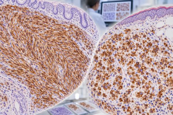
CD56 immunohistochemistry (IHC) detects neural cell adhesion molecule (NCAM) in tissue. Pathologists most often use CD56 as part of a panel for neuroendocrine tumors and for selected natural killer (NK)-cell and T-cell neoplasms, but the marker is not specific to either group. Many small cell neuroendocrine carcinomas are strongly positive, yet other cancers and normal tissues can also express CD56. Likewise, CD56 supports an NK-cell phenotype in the right hematolymphoid setting but does not by itself establish an NK-cell lymphoma. The result must be interpreted with the tumor’s microscopic appearance, other lineage markers, and—depending on the suspected disease—tests such as synaptophysin, chromogranin, INSM1, cytokeratins, CD45, cytotoxic markers, or Epstein-Barr virus testing. A positive stain therefore answers “is CD56 present?” rather than “what cancer is this?”
- CD56-positive staining can support neuroendocrine or NK-cell differentiation, but it is not specific enough to diagnose either one alone.
- High-grade neuroendocrine carcinomas often express CD56 even when chromogranin is weak, which is one reason panels are used.
- NK/T-cell lymphomas require hematolymphoid markers and usually EBV evaluation; CD56 positivity alone is insufficient.
- CD56 staining is generally membranous, and reports may describe its intensity and distribution rather than use a universal cutoff.
- Unexpected CD56 positivity occurs in several non-neuroendocrine tumors, so morphology and companion markers determine its meaning.
Table of Contents
- What CD56 IHC detects
- CD56 in neuroendocrine tumors
- CD56 in NK- and T-cell neoplasms
- What positive staining means
- Markers used with CD56
- Pitfalls and limitations
- How to read the report
- Practical CD56 interpretation scenarios
What CD56 IHC detects
CD56 is the common pathology name for NCAM1, a cell-surface adhesion molecule expressed in natural killer cells, subsets of T cells, neural and neuroendocrine tissues, skeletal muscle lineage, and several neoplasms. IHC visualizes the protein in a fixed tissue section, so the pathologist can see both the stained cells and their architecture.
The stain is usually membranous. In some specimens there may be cytoplasmic accentuation or technical background, but crisp membrane staining in the target cell population is the classic pattern. There is no single percentage threshold that defines a “CD56-positive cancer” across diagnoses. Some laboratories record diffuse versus focal staining, while others simply report positive or negative in the context of the panel.
CD56 is best viewed as a sensitive but relatively nonspecific differentiation marker. That makes it useful in cases where the pathologist already has a focused differential diagnosis. It is less useful as an isolated screen because many unrelated tumors can express it.
For a small blue-cell tumor, for example, CD56 positivity could fit a high-grade neuroendocrine carcinoma, an NK-cell neoplasm, certain acute leukemias, plasma-cell neoplasms, or other entities. The decisive information comes from morphology and markers that establish lineage. If the cells are keratin positive and express neuroendocrine markers, an epithelial neuroendocrine carcinoma is favored. If they are CD45 positive with cytoplasmic CD3 epsilon, cytotoxic molecules, and appropriate EBV findings, an NK/T-cell process becomes more plausible.
CD56 in neuroendocrine tumors
CD56 has long been used as a general neuroendocrine marker alongside synaptophysin and chromogranin A. It is particularly sensitive in many high-grade neuroendocrine carcinomas, including small cell carcinoma. The tradeoff is lower specificity: CD56 can be expressed by tumors without true neuroendocrine differentiation.
Modern neuroendocrine diagnosis therefore emphasizes morphology plus a panel. Synaptophysin IHC is highly sensitive across many neuroendocrine neoplasms. Chromogranin IHC is generally more specific for dense-core secretory granules but may be weak or focal in poorly differentiated neuroendocrine carcinoma. INSM1, a nuclear transcription-factor marker, has become a valuable additional test and can be especially helpful in high-grade disease.
A well-differentiated neuroendocrine tumor typically has organoid, trabecular, nested, or gland-like architecture and relatively uniform cells with finely stippled “salt-and-pepper” chromatin. In that setting, diffuse synaptophysin and chromogranin support the diagnosis. CD56 may be positive but is often not the key deciding stain.
Poorly differentiated neuroendocrine carcinoma has more marked atypia, necrosis, and high proliferative activity. Chromogranin can be reduced because the cells may contain fewer mature secretory granules, while CD56 and synaptophysin can remain strong. This explains why a panel prevents false negatives when only one traditional marker is used.
The 2022 WHO neuroendocrine framework also separates differentiation from grade. CD56 supports phenotype but does not grade the tumor. Mitotic activity, necrosis, morphology, and in many sites the Ki-67 proliferation index are more relevant to grading or classification.
CD56 in NK- and T-cell neoplasms
Normal NK cells commonly express CD56, so the marker is also important in hematopathology. However, the term “NK-cell marker” can be misleading if interpreted too literally. CD56 is not unique to NK cells, and some NK-cell neoplasms can have variable expression.
Extranodal NK/T-cell lymphoma, nasal type, is a classic example in which CD56 often contributes to the diagnostic phenotype. These tumors commonly express cytoplasmic CD3 epsilon and cytotoxic proteins such as TIA1, granzyme B, or perforin, while lacking surface CD3. Demonstration of EBV, typically by EBER in situ hybridization, is central to diagnosis in the appropriate clinicopathologic setting. CD56 alone cannot substitute for this combination.
Aggressive NK-cell leukemia and other NK-cell proliferations also require integrated diagnosis. Peripheral blood, bone marrow, organ involvement, flow cytometry, cytotoxic phenotype, EBV status, and molecular data may all matter. The 2022-era WHO and International Consensus classifications refine several T- and NK-cell categories, which is why a modern pathology report may use multiple ancillary studies rather than a small legacy panel.
CD56 can also be aberrantly present in some T-cell lymphomas, plasma-cell neoplasms, and acute myeloid leukemias. In myeloma, for example, abnormal plasma cells frequently express CD56, while normal plasma cells generally do not. That finding can be useful immunophenotypically, but the diagnostic context is entirely different from neuroendocrine carcinoma or NK-cell lymphoma.
What positive staining means
A positive CD56 stain means that the target cells express detectable NCAM protein. The significance depends on which cells are stained and what the specimen looks like. There is no universal interpretation such as “positive equals neuroendocrine cancer.”
| CD56 finding | Possible implication | Needed context |
|---|---|---|
| Diffuse membranous staining in a small cell carcinoma | Supports neuroendocrine differentiation | Synaptophysin, INSM1/chromogranin, keratins, morphology |
| CD56-positive atypical lymphoid cells | May support NK or aberrant T-cell phenotype | CD45, CD3 pattern, cytotoxic markers, EBER, flow cytometry |
| CD56-positive plasma cells | Can support an abnormal plasma-cell phenotype | CD138, light chains, marrow findings, flow cytometry |
| Focal CD56 in an otherwise non-neuroendocrine carcinoma | May be aberrant expression | Do not diagnose neuroendocrine differentiation without corroboration |
| CD56-negative suspected neuroendocrine tumor | Does not exclude neuroendocrine lineage | Synaptophysin, chromogranin, INSM1 and morphology |
Extent and intensity can help pathologists judge whether the result is convincing, but they are not universally tied to prognosis. A strongly positive stain does not automatically mean a more aggressive tumor. Clinical behavior is driven by the actual tumor type, differentiation, grade, stage, molecular profile, and treatment response.
Markers used with CD56
For neuroendocrine tumors, the most common companion markers are synaptophysin, chromogranin A, and INSM1. Broad-spectrum cytokeratins establish epithelial differentiation in carcinomas. Site-of-origin markers may then be used if the tumor is metastatic: TTF-1 can support pulmonary origin in some settings, CDX2 can support intestinal origin, and organ-specific transcription factors or hormones may be selected based on morphology and imaging.
The INSM1 IHC test is useful because its nuclear staining is easy to localize and large studies show high sensitivity for neuroendocrine neoplasms, particularly neuroendocrine carcinomas. Still, INSM1 can also be expressed in selected non-neuroendocrine tumors, so even this newer marker works best in context.
For suspected NK/T-cell neoplasia, pathologists may use CD45, CD2, CD3, CD5, CD7, CD4, CD8, CD30, TIA1, granzyme B, perforin, and EBER. Flow cytometry is valuable when fresh tissue is available, although some NK/T-cell lymphomas can be difficult to capture in suspension due to necrosis or limited viable tumor.
For a very poorly differentiated tumor, the first step may be broader. Keratin, CD45, SOX10/S100, and other lineage markers can establish the tumor family before CD56 is given much weight. This prevents a common error: calling any CD56-positive undifferentiated malignancy “neuroendocrine.”
Pitfalls and limitations
CD56’s major limitation is lack of specificity. Expression occurs in diverse tumors, including neuroendocrine neoplasms, NK/T-cell disorders, plasma-cell neoplasms, some myeloid leukemias, and selected carcinomas and sarcomas. An isolated positive result therefore has limited diagnostic power.
Another pitfall is overcalling focal or weak staining. Tiny positive areas may be real but biologically nonspecific. In a tumor without neuroendocrine morphology, focal CD56 does not establish neuroendocrine carcinoma. Current pathology practice increasingly relies on more specific markers and on the overall pattern rather than on CD56 alone.
Technical factors can also influence staining. Poor fixation, necrosis, crush artifact, and small biopsies can reduce interpretability. Small cell lung carcinoma biopsies, for instance, are often crushed, and the limited tissue must also be conserved for predictive molecular or immunotherapy testing. A focused panel is therefore preferable to ordering many redundant stains.
For NK/T-cell lesions, extensive necrosis and inflammation can make it difficult to identify which cells are truly positive. EBER in situ hybridization must be interpreted in the atypical cells, not just in scattered bystanders. The same principle applies to CD56: the pathologist must correlate the stain with the malignant population.
Finally, CD56 has no stand-alone treatment threshold. It is not analogous to HER2 or PD-L1 scoring. In most settings it is a diagnostic phenotype marker rather than a predictive biomarker for a specific therapy.
How to read the report
When CD56 appears in a pathology report, start with the final diagnosis rather than the isolated stain list. The report may say that tumor cells are positive for CD56 and synaptophysin, supporting neuroendocrine differentiation, or that CD56 is positive together with cytotoxic markers in an EBV-associated lymphoid lesion. Those are fundamentally different interpretations.
If the report describes “CD56 positive, chromogranin negative,” that is not necessarily contradictory. Poorly differentiated neuroendocrine carcinomas can lose or reduce chromogranin while retaining other markers. The pathologist may use synaptophysin and INSM1 to resolve the phenotype. Conversely, CD56 positivity without any other neuroendocrine evidence may be treated as nonspecific.
Patients can ask whether CD56 was used to establish lineage, to support a suspected subtype, or simply as part of a screening panel. If the diagnosis is a neuroendocrine neoplasm, it is also useful to ask about differentiation, grade, Ki-67, primary site, and whether additional molecular testing is relevant. If an NK/T-cell neoplasm is suspected, ask whether EBV studies, flow cytometry, and appropriate hematopathology review have been completed.
A repeat biopsy or expert review may be appropriate when the sample is tiny, necrotic, heavily crushed, or immunophenotypically discordant. The aim is a coherent diagnosis supported by morphology and multiple complementary data points—not the largest possible number of stains.
CD56 is most informative when it is treated as one clue among several. Its sensitivity makes it helpful; its broad expression makes disciplined interpretation essential.
Practical CD56 interpretation scenarios
A small lung biopsy is one of the most familiar situations in which CD56 enters the panel. A tumor composed of small cells with nuclear molding, high mitotic activity, necrosis, and crush artifact may be suspected to represent small cell carcinoma. Diffuse membranous CD56 can support that impression, but modern practice usually seeks corroboration with synaptophysin or INSM1 and confirms epithelial differentiation with keratin. TTF-1 may be positive in many pulmonary small cell carcinomas, but it is not completely specific for lung origin. The diagnosis therefore rests first on morphology and then on a focused set of supportive stains.
Now consider a destructive nasal lesion with angiocentric growth and necrosis. CD56-positive atypical cells may raise extranodal NK/T-cell lymphoma, but that diagnosis requires a very different evidentiary chain. The abnormal cells should show a compatible hematolymphoid and cytotoxic phenotype, and EBV must be demonstrated in the tumor cells using the appropriate method. If EBER is negative in a technically adequate specimen, the pathologist should strongly reconsider the diagnosis rather than allowing CD56 positivity to dominate.
A bone marrow specimen provides another example of CD56’s lack of specificity. Abnormal plasma cells in myeloma often express CD56, and myeloid blasts can sometimes express it aberrantly. In those settings, CD56 is useful because it contributes to an abnormal immunophenotypic pattern, not because it defines lineage. CD138 and light-chain restriction establish plasma-cell differentiation, while myeloid markers and flow cytometry establish an acute myeloid process.
CD56 negativity must also be interpreted with restraint. A well-differentiated neuroendocrine tumor that is strongly synaptophysin and chromogranin positive remains neuroendocrine even if CD56 is absent. Similarly, an NK-lineage neoplasm can have variable expression. The panel is designed so that no single marker carries the entire diagnosis.
Sampling and tumor preservation can change confidence. High-grade neuroendocrine carcinomas frequently arrive as tiny biopsies with substantial crush artifact. Excessive IHC can exhaust the block and leave too little tissue for predictive testing. A pathologist may therefore use only one or two high-yield neuroendocrine markers rather than the full historical panel. This is not an incomplete workup when the morphology is classic; it is tissue stewardship.
For patients, a report listing CD56 among many positive stains can be confusing because the same marker appears in very different diseases. The key is to identify the diagnostic sentence that interprets the panel. If the report says the profile supports neuroendocrine carcinoma, CD56 is being used in an epithelial context. If it says the profile supports an NK-cell neoplasm, hematolymphoid and EBV findings should also be present. If CD56 is merely described as aberrant expression, it may have little independent meaning.
CD56 is therefore most valuable when it agrees with a recognizable pattern. Strong staining in the wrong morphologic setting is a reason to investigate further, not a reason to force the tumor into a CD56-associated category.
Another useful distinction is between diagnostic and prognostic use. CD56 expression may be studied in research for associations with behavior, but routine pathology generally does not assign a universal risk category from CD56 intensity or percentage. A tumor that is CD56 diffuse is not automatically higher grade than one that is focal. Grade comes from the disease-specific system: mitoses, necrosis, Ki-67 where applicable, cytologic differentiation, genetics, and anatomic stage. Keeping those roles separate prevents a descriptive immunostain from being mistaken for a clinical risk score.
CD56 also has different diagnostic weight in different organs. In a lung biopsy with classic small-cell morphology, diffuse CD56 can support neuroendocrine differentiation, but synaptophysin, chromogranin, INSM1, keratin staining, and morphology provide the more complete classification. In a destructive extranodal lymphoid lesion, especially in the nasal region, CD56 becomes more meaningful when paired with cytoplasmic CD3 epsilon, cytotoxic markers, and Epstein-Barr virus testing. That context-dependent use prevents the same positive stain from being interpreted as the same disease in every specimen.
For follow-up decisions, CD56 is usually a classification marker rather than a stand-alone prognostic or treatment biomarker. A report that says “CD56 positive” should therefore be read together with the final diagnosis, tumor grade or proliferation rate, anatomic site, and the rest of the immunophenotype. Those features, not CD56 intensity alone, determine whether additional molecular tests, staging studies, or disease-specific therapy are appropriate.
The staining pattern can add a small amount of context as well. CD56 is a cell-surface adhesion molecule, so convincing tumor staining is typically membranous, although laboratories may describe combined membrane and cytoplasmic accentuation. The pathologist must separate tumor-cell labeling from CD56-positive normal NK cells or other background elements. In a mixed inflammatory specimen, scattered positive lymphocytes are not equivalent to diffuse staining of the atypical population. This localization step is simple but important because CD56 is used across both epithelial neuroendocrine tumors and hematolymphoid neoplasms.
References
- Overview of the 2022 WHO Classification of Neuroendocrine Neoplasms 2022 (Review)
- Comparison of INSM1 immunostaining with established neuroendocrine markers synaptophysin and chromogranin A in over 14,000 neuroendocrine and non-neuroendocrine tumors 2024
- INSM1, a Novel Biomarker for Detection of Neuroendocrine Neoplasms: Cytopathologists’ View 2022 (Review)
- Small Biopsy and Cytology of Pulmonary Neuroendocrine Neoplasms: Brief Overview of Classification, Immunohistochemistry, Molecular Profiles, and World Health Organization Updates 2022 (Review)
- Classification and diagnostic evaluation of nodal T- and NK-cell lymphomas 2023 (Review)
- Update from the 5th Edition of the World Health Organization Classification of Head and Neck Tumors: Overview of the 2022 WHO Classification of Head and Neck Neuroendocrine Neoplasms. 2022 (Review)
Disclaimer
CD56 IHC cannot diagnose a neuroendocrine tumor or NK-cell neoplasm by itself. Individual results require interpretation with morphology, companion immunostains, EBV studies, flow cytometry, molecular findings, and the clinical setting as appropriate.





