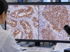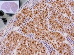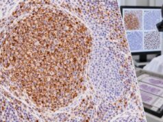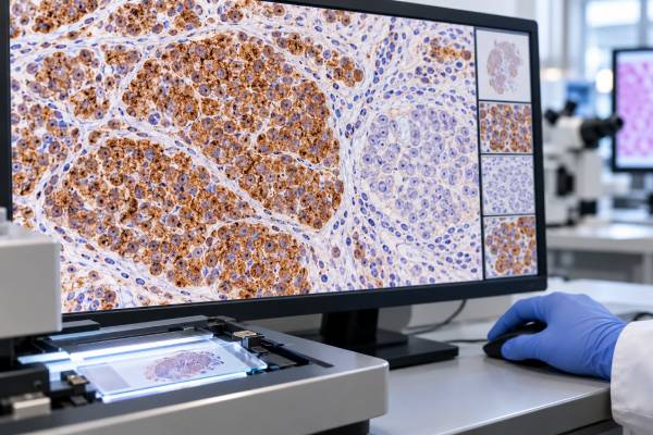
Synaptophysin immunohistochemistry (IHC) is one of the most widely used tissue stains for detecting neuroendocrine differentiation. Synaptophysin is a membrane protein found in small synaptic-like vesicles, so tumors with neuroendocrine features often show granular or diffuse cytoplasmic staining. The test is used across many organs, including the lung, pancreas, gastrointestinal tract, prostate, gynecologic tract, and skin. A positive result supports neuroendocrine differentiation, but it does not by itself prove that a tumor is a well-differentiated neuroendocrine tumor or identify where the cancer started. Some non-neuroendocrine tumors can show focal synaptophysin expression, while certain high-grade neuroendocrine carcinomas may have variable staining. Pathologists therefore combine synaptophysin with morphology and markers such as chromogranin A, INSM1, and Ki-67. Tumor site, differentiation, proliferative rate, and additional lineage markers are then used to reach the final classification.
- Synaptophysin-positive staining is usually cytoplasmic and often granular or diffuse in neuroendocrine cells.
- A positive stain supports neuroendocrine differentiation but does not identify a specific tumor type or primary site by itself.
- Synaptophysin is often more sensitive than chromogranin A, while newer nuclear marker INSM1 can add specificity in some settings.
- Ki-67 is used for grading many well-differentiated neuroendocrine tumors; synaptophysin is not a proliferation marker.
- There is no blood reference range or preparation for synaptophysin IHC; it is performed on tissue.
Table of Contents
- What the synaptophysin IHC test detects
- What positive synaptophysin staining means
- Neuroendocrine tumor versus neuroendocrine carcinoma
- Where synaptophysin-positive tumors occur
- False-positive, focal, and negative results
- How a neuroendocrine IHC panel is built
- What the result means clinically
What the synaptophysin IHC test detects
Synaptophysin is a vesicle-associated protein found in neurons and neuroendocrine cells. IHC detects the protein in fixed tissue, typically producing cytoplasmic staining in cells with neuroendocrine differentiation. The stain can be performed on biopsies, resections, and cytology cell blocks.
Pathologists commonly order synaptophysin when the microscopic appearance suggests a neuroendocrine neoplasm: cells may form nests, trabeculae, rosettes, or organoid patterns, or a high-grade tumor may show small-cell morphology. The stain may also be used in a broad workup of an undifferentiated carcinoma.
An IHC stain for chromogranin often accompanies synaptophysin because the two markers reflect related but distinct secretory machinery. Concordant positivity makes neuroendocrine differentiation more convincing.
What positive synaptophysin staining means
Positive staining means that the tumor cells contain detectable synaptophysin protein. Diffuse cytoplasmic staining in a morphologically typical neuroendocrine tumor is strong evidence of neuroendocrine differentiation. Focal staining can still be meaningful but should be interpreted more cautiously.
Synaptophysin is relatively sensitive, which is helpful in high-grade neuroendocrine carcinomas where chromogranin may be weak or patchy. The tradeoff is that synaptophysin can also be expressed focally in non-neuroendocrine neoplasms. The diagnosis therefore should not be changed to “neuroendocrine tumor” solely because a few tumor cells stain.
Pathologists increasingly use INSM1 IHC, a nuclear marker of neuroendocrine differentiation, as a companion or alternative. The relative sensitivity and specificity vary with tumor type and antibody clone.
Neuroendocrine tumor versus neuroendocrine carcinoma
A positive synaptophysin stain establishes phenotype, not grade. Current neuroendocrine classification separates well-differentiated neuroendocrine tumors (NETs) from poorly differentiated neuroendocrine carcinomas (NECs) because they differ in biology, morphology, molecular changes, and treatment.
Well-differentiated NETs often show classic organoid architecture and express synaptophysin and chromogranin. Their grade in many gastrointestinal and pancreatic sites uses mitotic activity and the Ki-67 proliferation index. A Ki-67 IHC test therefore answers a different question from synaptophysin.
Poorly differentiated NECs include small-cell and large-cell neuroendocrine carcinoma. They are high-grade by definition. They usually express one or more neuroendocrine markers, but morphology and additional markers such as p53 and RB can be important, especially in pancreatic and other organ-specific differentials.
Where synaptophysin-positive tumors occur
Synaptophysin-positive neuroendocrine neoplasms can arise throughout the body. Common examples include pulmonary carcinoid tumors and small-cell lung carcinoma; pancreatic and gastrointestinal NETs; medullary thyroid carcinoma; pheochromocytoma and paraganglioma; Merkel cell carcinoma; and neuroendocrine carcinomas of the prostate, bladder, cervix, endometrium, and other sites.
The same stain therefore cannot establish primary site. A liver metastasis that is strongly synaptophysin positive could represent a pancreatic NET, a small intestinal NET, a pulmonary neuroendocrine neoplasm, or another primary. Site-specific transcription factors, hormone stains, somatostatin receptor expression, imaging, and clinical history may help localize the source.
CD56 can support neuroendocrine differentiation but is less specific. An IHC test for CD56 must be interpreted carefully because NK-cell tumors and several non-neuroendocrine neoplasms can also express it.
False-positive, focal, and negative results
Focal synaptophysin positivity is a well-known pitfall. Prostate adenocarcinoma, adrenal cortical neoplasms, some sarcomas, and various carcinomas may contain scattered neuroendocrine-like cells or show partial neuroendocrine marker expression without meeting criteria for a neuroendocrine neoplasm.
Negative staining can also occur. Poor fixation, necrosis, scant tissue, treatment effect, or genuine biologic variability can reduce expression. If the morphology strongly suggests neuroendocrine differentiation but synaptophysin is negative, chromogranin and INSM1 may still be informative.
The pattern must match the cells of interest. Entrapped nerves and normal neuroendocrine cells can be synaptophysin positive and may serve as controls, but they should not be mistaken for tumor staining. Pathologists correlate the stain with the exact cells seen on the routine slide.
How a neuroendocrine IHC panel is built
A practical panel often starts with synaptophysin, chromogranin A, and/or INSM1 to establish neuroendocrine differentiation. Pancytokeratin helps show epithelial differentiation in many NETs and NECs. Ki-67 supports grading where applicable. Site-specific markers are then added based on the clinical question.
For example, TTF-1 may support pulmonary origin in some metastatic neuroendocrine carcinomas but is not perfectly specific. CDX2 can support intestinal origin in selected NETs. Islet-1 or other pancreatic markers may help in pancreatic primaries. Hormone stains can identify functional differentiation but are not always necessary.
The goal is a coherent profile. A broad tumor IHC panel is most effective when stains are added stepwise based on morphology rather than ordered indiscriminately.
What the result means clinically
Synaptophysin positivity often triggers more precise classification rather than ending the diagnostic workup. The pathology team determines whether the tumor is well differentiated or poorly differentiated, assesses grade when relevant, and investigates the likely primary site.
Those distinctions matter for treatment. A low-grade well-differentiated gastrointestinal NET is managed very differently from small-cell neuroendocrine carcinoma, even though both may stain strongly for synaptophysin. Somatostatin receptor imaging or therapy may be relevant to some well-differentiated NETs, while platinum-based systemic therapy is commonly considered for many NECs.
If a report says “synaptophysin positive,” look for the final diagnosis, Ki-67 or mitotic information, chromogranin/INSM1 results, and any site-of-origin comment. Those elements provide the clinically actionable interpretation.
References
- Comparison of INSM1 immunostaining with established neuroendocrine markers synaptophysin and chromogranin A in over 14,000 neuroendocrine and non-neuroendocrine tumors 2024 (Study)
- Synaptophysin and chromogranin A expression analysis in human tumors 2022 (Study)
- Overview of the 2022 WHO Classification of Neuroendocrine Neoplasms 2022 (Review)
- INSM1, a Novel Biomarker for Detection of Neuroendocrine Neoplasms: Cytopathologists’ View 2021 (Review)
- Second-Generation Neuroendocrine Immunohistochemical Markers: Reflections from Clinical Implementation. 2021 (Review)
Disclaimer
Synaptophysin IHC is an ancillary pathology test that shows neuroendocrine differentiation but does not determine tumor grade, primary site, or treatment by itself. Interpretation requires morphology, complementary IHC, proliferative assessment when relevant, and clinical findings. Discuss the integrated pathology diagnosis with the treating team.