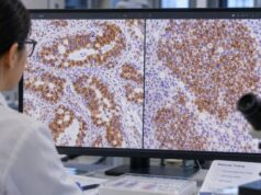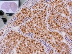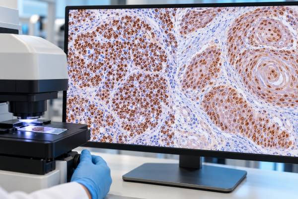
SOX2 immunohistochemistry (IHC) is a nuclear tissue stain used in several different diagnostic settings rather than as a marker for one specific cancer. In germ cell pathology, SOX2 is especially helpful for identifying embryonal carcinoma and separating it from seminoma, which typically follows a different transcription-factor pattern and is generally SOX2 negative. In epithelial tumors, SOX2 can be strongly expressed in squamous cell carcinomas, including many lung squamous cancers, but it is not sufficiently specific to replace established squamous markers such as p40. SOX2 is also expressed in some neuroendocrine and other neoplasms. Because the same positive stain can have very different meanings depending on the tissue, pathologists interpret SOX2 with morphology, tumor location, patient age, and a focused IHC panel. A report of “SOX2 positive” is therefore a phenotype clue—not a stand-alone diagnosis, cancer screen, or treatment marker.
- SOX2 IHC produces nuclear staining when tumor cells express the transcription factor.
- In testicular germ cell tumors, diffuse SOX2 supports embryonal carcinoma, while seminoma is usually SOX2 negative and SOX17 positive.
- Many squamous cell carcinomas can express SOX2, but p40 and other squamous markers are usually more specific for routine typing.
- SOX2 is not a blood test and has no normal serum range.
- The same SOX2-positive result can mean different things in different tumors, so panel interpretation is essential.
Table of Contents
- What the SOX2 IHC test detects
- SOX2 in germ cell tumors
- SOX2 in squamous cell carcinoma
- Other SOX2-positive tumors and normal tissues
- How positive and negative SOX2 results are read
- How SOX2 is combined with other IHC markers
- Clinical meaning and next steps after SOX2 IHC
What the SOX2 IHC test detects
SOX2 is a transcription factor involved in stem-cell biology and development. IHC detects the protein in fixed tissue, where meaningful staining is located in tumor-cell nuclei. The test may be performed on a biopsy, resection, or cytology cell block when the differential diagnosis makes SOX2 informative.
The marker is unusual because it crosses several diagnostic categories. It has a defined role in germ cell tumor subtyping, but it is also frequently expressed in squamous carcinomas and some pulmonary neuroendocrine carcinomas. For that reason, a positive result must be tied to the specific clinical problem.
In a young patient with a testicular, mediastinal, or retroperitoneal tumor, SOX2 may be part of a germ cell panel. In a lung biopsy, the pathologist may instead be trying to distinguish squamous carcinoma from adenocarcinoma. The stain’s meaning comes from that context.
SOX2 in germ cell tumors
In postpubertal-type testicular germ cell tumors, SOX2 is strongly associated with embryonal carcinoma. Embryonal carcinoma typically shows nuclear SOX2 and OCT4 and often expresses CD30. Seminoma, in contrast, typically expresses OCT4 and SOX17 but not SOX2. This reciprocal SOX2/SOX17 pattern can be useful in morphologically difficult areas of mixed germ cell tumors.
An OCT4 IHC stain alone cannot fully solve that distinction because both seminoma and embryonal carcinoma commonly express OCT4. Adding SOX2, CD30, CD117, and other markers makes the pattern much more informative.
SOX2 is not a universal germ cell marker. Yolk sac tumor is usually SOX2 negative, and teratomas can show expression in selected immature or epithelial components. A broad marker such as SALL4 can establish germ cell differentiation across a wider range of components, while SOX2 helps with specific subtyping.
SOX2 in squamous cell carcinoma
SOX2 expression is common in squamous cell carcinoma, particularly in the lung, where amplification and overexpression of the SOX2 locus have been linked to squamous lineage biology. Strong nuclear staining can therefore support a squamous phenotype when the morphology and other markers agree.
SOX2 is not the preferred stand-alone squamous marker because it is expressed in other tumor types. In lung pathology, p40 IHC is generally more specific for squamous differentiation, and CK5/6 or p63 may add support. TTF-1 and Napsin A are commonly used on the adenocarcinoma side of the differential.
This distinction matters on small biopsies because accurate non-small-cell lung cancer subtyping helps guide molecular testing and treatment. The pathologist aims to use the smallest effective panel so enough tissue remains for predictive biomarkers.
Other SOX2-positive tumors and normal tissues
SOX2 is not restricted to embryonal carcinoma or squamous cancer. It can be present in pulmonary neuroendocrine carcinomas and other tumors with stem-cell or developmental programs. Normal progenitor or basal-type epithelial compartments in some tissues can also express SOX2.
This broad distribution explains why the test is not used like a tumor-specific tag. A SOX2-positive metastatic carcinoma does not identify the primary site on its own. The pathologist must compare the result with lineage markers and the suspected organ of origin.
The same principle applies in germ cell pathology: a positive SOX2 stain is highly useful when the differential is seminoma versus embryonal carcinoma, but far less specific when the specimen is an undifferentiated tumor from an unrelated site.
How positive and negative SOX2 results are read
SOX2 positivity is judged by nuclear staining. Diffuse, strong staining in the appropriate tumor population carries more diagnostic weight than rare weak nuclei. Laboratories may report staining qualitatively or semiquantitatively, but there is no universal percentage cutoff across all diseases.
A negative result can be expected in some tumors, such as seminoma in the classic SOX2/SOX17 framework, but negative staining never works in isolation. Tissue quality, fixation, necrosis, treatment effect, and tumor heterogeneity can reduce expression.
Internal controls and the overall panel are therefore essential. If every expected control is negative, the stain may be technically uninterpretable. If controls are valid but the tumor is negative, the pathologist asks whether that result supports an alternative diagnosis or simply reflects a known limitation.
How SOX2 is combined with other IHC markers
For a suspected germ cell tumor, common companions include SALL4, OCT4, SOX17, CD30, PLAP, CD117, glypican-3, and AFP. The exact panel is tailored to the morphology. Embryonal carcinoma often produces the concordant pattern of OCT4/SOX2/CD30 positivity, while seminoma favors OCT4/SOX17 with CD117 or PLAP.
For a suspected lung squamous carcinoma, SOX2 has a supporting role behind p40 and other epithelial markers. A p63 IHC stain may also be positive, but p63 is broader than p40 and can label several non-squamous tumors.
When the tumor origin is unknown, SOX2 should be embedded in a broader, morphology-driven panel rather than ordered as an isolated tissue-of-origin marker.
Clinical meaning and next steps after SOX2 IHC
SOX2 IHC mainly improves diagnostic classification. In a germ cell tumor, correctly recognizing an embryonal carcinoma component can affect staging interpretation and treatment planning because mixed germ cell tumor composition matters clinically. In lung cancer, correct squamous versus adenocarcinoma classification helps determine which molecular and therapeutic pathways are relevant.
The stain itself is not a validated universal drug-selection test, and the intensity of SOX2 staining should not be translated directly into tumor aggressiveness for an individual patient. Prognosis depends on the final tumor diagnosis, stage, grade, molecular features, and response to therapy.
If the pathology report lists SOX2, ask why it was ordered, which tumor cells stained, which other markers were concordant, and whether the result changed the final diagnosis. That context is the clearest way to understand its significance.
References
- Pathogenesis and pathobiology of testicular germ cell tumours: a view from a developmental biological perspective with guidelines for pathological diagnostics. 2024 (Review)
- Testicular Germ Cell Tumors: Serological and Immunohistochemical Diagnosis 2021 (Review)
- Best practices recommendations in the application of immunohistochemistry in testicular tumors: report from the International Society of Urological Pathology consensus conference. 2014 (Consensus Statement)
- Differential expression of SOX2 and SOX17 in testicular germ cell tumors. 2009 (Study)
- Morphologic and clinicopathologic features of lung squamous cell carcinomas expressing Sox2 2012 (Study)
Disclaimer
SOX2 IHC is an ancillary pathology stain, not a stand-alone cancer diagnosis or treatment test. Its meaning differs substantially between germ cell tumors, squamous carcinomas, and other neoplasms. Interpretation requires the complete morphology, clinical setting, and complementary IHC results.