
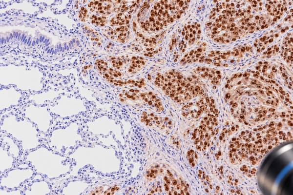
The p40 immunohistochemistry (IHC) test is a tissue stain that helps pathologists identify squamous differentiation, especially when classifying poorly differentiated lung cancers. p40 is an isoform of the TP63 protein family and produces a nuclear staining pattern. In lung pathology, strong tumor-cell nuclear p40 staining supports squamous cell carcinoma, while markers such as TTF-1 and Napsin A support adenocarcinoma. The test is useful because small biopsies may not show the classic architecture or keratinization needed for a confident diagnosis by microscope appearance alone. However, p40 is a lineage marker rather than an organ-specific marker: tumors from several sites can be p40 positive. The percentage, intensity, distribution, morphology, companion stains, and clinical location all matter. A p40 result therefore helps answer what type of cells a tumor resembles; it does not by itself determine where the cancer started, how advanced it is, or which treatment a person should receive.
- What p40 measures: p40 IHC detects a nuclear TP63 isoform associated with squamous and basal-cell differentiation.
- What positive staining usually means: convincing nuclear staining in tumor cells supports squamous differentiation, particularly in a compatible lung tumor.
- Why p40 is used in lung cancer: it is generally more specific for lung squamous cell carcinoma than broad p63 staining.
- What a negative result means: p40 negativity makes conventional squamous differentiation less likely but does not settle the diagnosis by itself.
- How results are interpreted: pathologists combine p40 with tumor shape and markers such as TTF-1, Napsin A, cytokeratins, or other site-specific stains.
Table of Contents
- What the p40 IHC Test Detects
- How p40 Helps Type Lung Cancer
- What Positive p40 Staining Means
- Negative, Focal, and Mixed p40 Results
- How p40 Fits Into IHC Marker Panels
- Limitations and Diagnostic Pitfalls
- What Happens After a p40 Result
What the p40 IHC Test Detects
p40 IHC detects ΔNp63, a major isoform produced from the TP63 gene. TP63 is related to TP53 but has a different role in tissue development and differentiation. p40 is normally expressed in nuclei of basal and squamous epithelial cells in several organs. Pathologists use that biology to recognize tumors that maintain a squamous or basal-type program.
The stain is performed on a thin section of formalin-fixed, paraffin-embedded tissue. The laboratory applies an antibody directed against p40, develops the reaction, and examines the slide under a microscope. A technically meaningful positive result appears in the nuclei of the cells being assessed. Cytoplasmic discoloration alone is not the expected diagnostic pattern.
p40 is sometimes confused with p63. The two tests are related, but they are not interchangeable. Traditional “pan-p63” antibodies detect more TP63 isoforms, while p40 antibodies target ΔNp63 more specifically. This narrower target is a major reason p40 usually has greater specificity for squamous cell carcinoma in the lung. The companion p63 IHC test remains useful in several settings, including urothelial and myoepithelial evaluation, but lung tumor classification often favors p40 when the question is squamous differentiation.
The test does not measure p40 in blood and does not produce a serum concentration. It also does not directly identify a TP63 gene mutation. The pathology report usually describes whether tumor cells stain, how much of the tumor is involved, and whether the pattern fits the morphologic impression.
A pathologist chooses p40 when the result can resolve a practical diagnostic question. Common examples include a non-small cell carcinoma in a small bronchoscopic biopsy, a metastatic tumor with squamoid features, or a poorly differentiated cancer in which the tissue lineage is uncertain. In each case, the goal is classification rather than screening.
How p40 Helps Type Lung Cancer
The most established use of p40 is distinguishing lung squamous cell carcinoma from lung adenocarcinoma, particularly in small biopsies and cytology specimens. This distinction matters because tumor classification guides downstream molecular testing and treatment planning.
When a lung tumor clearly forms glands or produces mucin, morphology may already support adenocarcinoma. Likewise, obvious keratin pearls and intercellular bridges can identify squamous cell carcinoma. The harder cases are poorly differentiated tumors that lack these classic features. In that setting, a limited immunostain panel can reveal the differentiation pattern while preserving tissue for biomarker testing.
p40 is a strong squamous marker. In a 2022 meta-analysis of lung squamous markers, pooled sensitivity for p40 was about 92% and specificity about 94%. p63 had similar sensitivity but lower specificity. Those numbers explain an important practical point: a strongly p40-positive lung non-small cell carcinoma with compatible morphology is highly supportive of squamous differentiation, but no stain is perfect.
Pathologists commonly compare p40 with TTF-1. TTF-1 supports pulmonary adenocarcinoma in the appropriate setting, while p40 supports squamous carcinoma. Napsin A can add evidence for adenocarcinoma, and keratin stains can confirm epithelial differentiation. A small panel often gives more reliable information than simply adding many stains.
The pancytokeratin IHC test answers a broader question—whether a poorly differentiated tumor shows epithelial differentiation—whereas p40 addresses a narrower subtype question. A tumor may be pancytokeratin positive yet p40 negative because many adenocarcinomas and other carcinomas do not have squamous differentiation.
p40 is not itself a drug-selection biomarker. A positive result does not mean a particular immunotherapy, chemotherapy, or targeted drug will work. For non-small cell lung cancer, treatment decisions can also depend on stage, PD-L1, genomic alterations, performance status, and other clinical factors. p40 helps establish the pathologic category that frames those later decisions.
What Positive p40 Staining Means
A positive p40 result means the antibody has labeled nuclei in the cells being examined. The clinical significance depends on which cells are positive, how extensive the staining is, and what the tumor looks like.
In a lung tumor, diffuse or convincing nuclear p40 staining favors squamous cell carcinoma when the morphology and remaining panel agree. Pathologists do not interpret any brown nuclear dot as equivalent evidence. Very focal staining, staining in non-neoplastic basal cells, or staining in an unexpected tumor requires more caution.
There is no universal “p40 score” used across all tumor types. Laboratories may describe staining as focal or diffuse, weak or strong, and may record an approximate percentage of positive tumor nuclei. The diagnostic threshold can depend on the specimen, antibody clone, laboratory validation, and clinical question. This differs from predictive assays that use a defined cutoff to determine treatment eligibility.
Positive p40 is also not specific to the lung. Squamous carcinomas arising in the head and neck, skin, cervix, anus, esophagus, and other organs may express p40. Urothelial carcinomas can express p40 because urothelium has basal/squamous-associated programs. Some salivary, thymic, and myoepithelial-type neoplasms may also show TP63-related staining. Therefore, p40 can support a type of differentiation without proving a primary site.
This distinction becomes crucial in metastatic disease. Suppose a p40-positive squamous carcinoma is found in a lymph node in a patient with both a lung mass and a head-and-neck lesion. p40 cannot by itself tell which lesion produced the metastasis. Imaging, clinical history, morphology, site-specific markers when available, viral studies in selected head-and-neck tumors, and comparison with prior specimens may be needed.
The word “positive” also should not be confused with “aggressive.” p40 expression is mainly diagnostic. Although research has explored TP63 biology and prognosis, routine p40 staining is not interpreted as a universal measure of tumor growth rate, stage, or survival.
Negative, Focal, and Mixed p40 Results
A negative p40 stain means the tumor cells do not show convincing nuclear labeling under the laboratory’s validated conditions. In an undifferentiated lung carcinoma, this lowers support for a conventional squamous phenotype, especially if TTF-1 or Napsin A is positive. It does not mean the tissue is normal, and it does not rule out cancer.
True squamous cell carcinomas are not invariably positive. Poor differentiation, limited viable tumor, treatment effect, tissue damage, or biologic variation can produce weak or absent staining. Technical factors such as delayed fixation, overfixation, decalcification, insufficient antigen retrieval, or a failed staining run can also reduce signal. Internal and external controls help distinguish a credible negative result from a technical failure.
Focal positivity deserves context. A few positive nuclei can occur in tumors that are not classic squamous carcinomas, and basal cells entrapped around glands can be mistaken for tumor if the architecture is difficult to read. Pathologists therefore correlate the stain with the exact cell population on the hematoxylin-and-eosin slide.
Mixed p40 and TTF-1 findings can be especially informative. In many routine tumors, a p40-dominant profile supports squamous carcinoma and a TTF-1-dominant profile supports adenocarcinoma. Rare tumors, however, can show genuine coexpression or different positive populations. Adenosquamous carcinoma, for example, contains morphologically distinct glandular and squamous components. A tiny biopsy may sample only one part, so the final resection can be classified differently.
Tumor heterogeneity also matters in metastases. A primary lesion may contain more than one phenotype, and a small metastatic biopsy may not reproduce every stain seen in the original specimen. This is one reason pathologists avoid turning p40 into a binary rule divorced from morphology.
When the result conflicts with the expected diagnosis, the appropriate response is not to force the tumor into a category. The pathologist may repeat the stain, review controls, use another squamous marker such as CK5/6, add adenocarcinoma markers, or broaden the panel to other lineages and organs.
How p40 Fits Into IHC Marker Panels
p40 is most useful when it answers one question within a carefully chosen panel. Excessive staining can exhaust a small biopsy, while an undersized panel can miss an important alternative diagnosis. The best panel is driven by morphology, tumor location, patient history, and the amount of tissue available.
For a poorly differentiated lung non-small cell carcinoma, p40 and TTF-1 are often high-value first-line markers. If one shows a convincing expected pattern and the other is negative, the pathologist may be able to subtype the tumor while conserving material. Napsin A, CK5/6, or mucin stains can be added when results are equivocal. Neuroendocrine markers are generally reserved for tumors with neuroendocrine morphology rather than used indiscriminately.
A practical interpretation looks like this:
| Typical pattern | What it may support | Why more context is needed |
|---|---|---|
| p40 strong/diffuse, TTF-1 negative | Squamous differentiation | p40 is not lung-specific |
| p40 negative, TTF-1 positive | Pulmonary adenocarcinoma phenotype | TTF-1 is not positive in every lung adenocarcinoma |
| p40 and TTF-1 both negative | Indeterminate by this pair | Other epithelial, neuroendocrine, metastatic, or non-epithelial possibilities may need testing |
| p40 and TTF-1 both convincingly positive | Unusual or mixed phenotype | Confirm staining in the same cells and consider uncommon entities or tumor heterogeneity |
When the origin is uncertain, broader testing may be needed. The tumor IHC panel approach can establish epithelial, hematolymphoid, melanocytic, or mesenchymal lineage before organ-specific markers are applied. In a suspected urothelial primary, GATA3 and urothelial markers can complement p40 or p63. In a head-and-neck squamous carcinoma, anatomic site and HPV- or EBV-related testing may be more informative than p40 for origin.
The panel should answer a diagnostic decision rather than merely accumulate positive markers. A stain that confirms what is already obvious may add little, while one that distinguishes two clinically meaningful possibilities can change the entire workup.
Limitations and Diagnostic Pitfalls
The biggest pitfall is treating p40 as an organ-specific marker. A p40-positive carcinoma may be squamous, urothelial, basal-like, or another tumor with TP63-lineage expression. If the clinical question is “Where did this metastasis start?” p40 usually needs companions rather than serving as the answer.
Another pitfall is equating p40 with p63. Because p40 recognizes ΔNp63, it tends to be more specific for squamous differentiation in lung cancer than pan-p63. A tumor that is p63 positive but p40 negative should not automatically be classified as squamous. The distinction is particularly important in poorly differentiated thoracic tumors, where p63 can be expressed in some nonsquamous neoplasms.
Technical quality can create false reassurance or false concern. Nuclear staining should be crisp enough to localize. Necrotic tissue, crushed cells, heavy cautery, scant tumor, decalcified bone specimens, and weak controls reduce confidence. A pathologist may request another block or specimen if the available tissue cannot support a reliable interpretation.
Interpretation can also be complicated by normal structures. Bronchial basal cells and squamous epithelium can be p40 positive. If a small biopsy contains only a few malignant cells mixed with benign epithelium, the observer must make sure the positive nuclei belong to tumor.
A rare but important problem is relying on p40 to exclude adenocarcinoma absolutely. Most conventional lung adenocarcinomas are p40 negative, but unusual tumors can have focal or aberrant expression. Conversely, some squamous carcinomas can be negative. The pathology diagnosis is a synthesis rather than a single-stain vote.
Finally, p40 does not substitute for molecular and predictive biomarker testing. Once a non-small cell lung cancer is classified, laboratories follow current clinical protocols for actionable genomic alterations and immune biomarkers when indicated. The diagnostic stain and the predictive tests answer different questions.
What Happens After a p40 Result
The next step depends on what the complete pathology report says. If p40 supports lung squamous cell carcinoma, the pathologist integrates that result with morphology and other stains, then issues a final tumor classification when the evidence is sufficient. If the result is equivocal or contradictory, additional stains, deeper tissue sections, repeat biopsy, or expert review may be appropriate.
For patients, the most useful part of the report is not the word “positive” by itself but the diagnostic conclusion and comment. A report may say that tumor cells show strong nuclear p40 expression and lack TTF-1, supporting squamous cell carcinoma. That statement carries more meaning than a standalone p40 result because it explains how the stain fits the whole case.
After diagnosis, staging and treatment evaluation proceed separately. Imaging and clinical findings determine disease extent. In lung cancer, molecular and immune biomarker testing may be performed according to tumor type, stage, specimen adequacy, and current treatment standards. p40 does not replace those tests.
If the suspected tumor could have come from more than one site, clinicians may compare the IHC profile with radiology, endoscopy, prior cancers, and other pathology specimens. A p40-positive metastasis in the lung is not automatically a primary lung cancer, especially in someone with a history of squamous cancer elsewhere.
Useful questions for the treating team include: Was the p40 staining nuclear and present in the malignant cells? Was it diffuse or only focal? Which markers were tested alongside it? Does the overall panel support a lung primary or only squamous differentiation? Is additional tissue needed for classification or molecular testing?
The main takeaway is simple: p40 is a highly useful squamous differentiation marker, particularly for lung tumor typing, but its strength comes from focused panel-based interpretation. It narrows the diagnosis while morphology, other stains, clinical information, and molecular studies complete the picture.
References
- Immunohistochemical markers to diagnose primary squamous cell carcinoma of the lung: a meta-analysis of diagnostic test accuracy 2022 (Systematic Review)
- Unravelling the diagnostic pathology and molecular biomarkers in lung cancer 2024 (Review)
- Rediscovering immunohistochemistry in lung cancer 2024 (Review)
- Current Biomarkers in Non-Small Cell Lung Cancer-The Molecular Pathologist’s Perspective 2025 (Review)
- Predictive immunohistochemistry in non-small cell lung cancer – Biomarkers, companion diagnostics, and molecular testing: A narrative review 2026 (Review)
Disclaimer
p40 IHC is a diagnostic pathology test that must be interpreted with tissue morphology, controls, other stains, and the clinical setting. A positive p40 result alone cannot prove the organ of origin, stage a cancer, or select treatment. Discuss the complete pathology report with the treating clinician or pathologist for case-specific meaning.





