
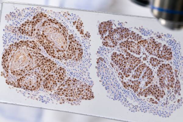
The p63 immunohistochemistry (IHC) test is a tissue stain used to identify a nuclear protein associated with basal cells, squamous differentiation, urothelial differentiation, and myoepithelial cells. Pathologists use p63 in several diagnostic settings, including poorly differentiated carcinomas, urothelial tumors, lung tumors, and breast or salivary lesions where the presence of a myoepithelial layer matters. A positive p63 result is not specific for one cancer or one organ. Instead, it shows that the stained cells express TP63-related protein, and the meaning comes from the tumor’s appearance, staining distribution, anatomic site, and companion markers. In lung cancer, p40 is often preferred when the narrow question is squamous differentiation because p40 is more specific than broad p63 antibodies. In urothelial and myoepithelial workups, p63 remains highly useful. The test does not determine stage, does not prove where a metastasis started by itself, and is not a treatment-response score.
- What p63 measures: p63 IHC detects TP63-family protein in cell nuclei and highlights several basal, squamous, urothelial, and myoepithelial cell populations.
- What positive staining can support: squamous cell carcinoma or urothelial differentiation when the morphology and marker panel agree.
- Why p63 is not organ-specific: many normal tissues and tumors from different sites can express p63.
- p63 versus p40: p40 targets a narrower TP63 isoform and is generally more specific for lung squamous cell carcinoma.
- How results are used: pathologists interpret the nuclear pattern with other stains rather than diagnosing a tumor from p63 alone.
Table of Contents
- What the p63 IHC Test Detects
- p63 in Squamous Cell Carcinoma
- p63 in Urothelial Cancer
- p63 in Breast and Myoepithelial Evaluation
- How p63 Fits Into Tumor-Origin Panels
- Limitations and Diagnostic Pitfalls
- What Happens After a p63 Result
What the p63 IHC Test Detects
p63 is a transcription factor encoded by the TP63 gene. The gene produces several protein isoforms that help regulate epithelial development and maintain basal or progenitor cell programs. In routine pathology, “p63 IHC” usually refers to a broad antibody that detects more than one TP63 isoform and produces nuclear staining.
Normal p63-positive cells are found in many sites. Examples include basal cells of stratified squamous epithelium, urothelial basal/intermediate cells, basal cells in the prostate, and myoepithelial cells surrounding many breast and salivary glands. This normal distribution explains both the usefulness and the limitations of the stain.
When a tumor retains a similar differentiation program, its nuclei may stain for p63. Squamous cell carcinomas are commonly positive. Many urothelial carcinomas express p63. Certain salivary, thymic, basal-like, and myoepithelial tumors can also be positive. Because this list crosses several organs, p63 should be viewed as a phenotype marker, not a geographic label.
The test is performed on a tissue section after biopsy or surgery. The laboratory uses a validated antibody and staining platform, and the pathologist compares the reaction with the tissue architecture. Nuclear localization is expected. The report may describe staining as diffuse or focal and strong or weak, sometimes with an estimated percentage of positive tumor cells.
p63 is related to p40 but is broader. The p40 IHC test targets ΔNp63 and generally provides greater specificity for lung squamous cell carcinoma. Broad p63 remains valuable when its wider sensitivity is useful, but the two names should not be treated as synonyms.
p63 in Squamous Cell Carcinoma
Squamous cell carcinomas arise in many organs and often show nuclear p63 expression. In a tumor with keratinization, intercellular bridges, or other squamous morphology, diffuse nuclear p63 can reinforce the diagnosis. It is especially useful when the tumor is poorly differentiated and classic features are limited.
In lung cancer, however, p63 has an important specificity limitation. Studies comparing markers have shown that p63 can stain some non-squamous lung cancers, including a subset of adenocarcinomas, more often than p40. A 2022 meta-analysis reported similar sensitivity for p40 and p63 in lung squamous carcinoma but higher specificity for p40. For this reason, many laboratories favor p40 as the primary squamous marker in small lung biopsies.
A p63-positive lung tumor still requires context. If TTF-1 and Napsin A support adenocarcinoma while p63 is only focal, the diagnosis should not be switched to squamous carcinoma simply because p63 is positive. Conversely, a strongly p63-positive and p40-positive tumor with compatible squamous morphology and absent adenocarcinoma markers gives much stronger evidence.
Outside the lung, p63 can support squamous differentiation in head-and-neck, cutaneous, cervical, esophageal, anal, and other squamous carcinomas. Yet p63 cannot tell these primary sites apart. A p63-positive metastatic squamous carcinoma in a lymph node therefore needs anatomic, clinical, radiologic, and sometimes viral or site-specific evidence to establish origin.
The stain also cannot distinguish benign squamous epithelium from cancer simply on positivity. Normal basal cells are p63 positive. The pathologist must identify the malignant population by morphology first and then determine whether those cells are labeled.
A positive p63 result is therefore best understood as supportive evidence for a squamous/basal phenotype rather than a definitive cancer label.
p63 in Urothelial Cancer
p63 is also an established marker in urothelial carcinoma, the most common cancer type arising from the urinary bladder lining. Normal urothelium expresses TP63-related proteins, and many conventional urothelial carcinomas retain nuclear p63 staining.
In a bladder tumor, p63 may support urothelial differentiation when interpreted with morphology and other markers such as GATA3, uroplakins, CK7, CK20, or high-molecular-weight keratins. The exact panel depends on whether the question is primary bladder tumor classification, distinction from a neighboring organ cancer, or identification of a metastasis.
The GATA3 IHC test is frequently useful in urothelial workups, but it also is not entirely organ-specific because breast and several other tumors can express GATA3. Combining markers with different strengths is more reliable than expecting one stain to identify the site.
Not every urothelial carcinoma is p63 positive. Expression can vary by tumor subtype and degree of differentiation, and some aggressive or variant morphologies may show reduced or lost staining. A negative result therefore does not automatically exclude urothelial origin.
Conversely, p63 positivity does not prove urothelial carcinoma. Squamous carcinomas from multiple organs, basal-type neoplasms, and several salivary or thymic tumors can be positive. In a pelvic mass involving bladder and adjacent organs, the pathologist may need PAX8, GATA3, p40, hormone receptors, prostate markers, or other stains depending on the differential diagnosis.
Urothelial pathology also uses morphology to distinguish in situ lesions, invasive carcinoma, and divergent differentiation. p63 can contribute to this assessment, but it is not a stage marker. Depth of invasion is determined from tissue anatomy, not staining intensity.
p63 in Breast and Myoepithelial Evaluation
A different but important use of p63 is highlighting myoepithelial cells. These contractile cells form a layer around many normal breast ducts and lobules and around certain salivary gland structures. Because their presence or absence can help distinguish in situ or benign processes from invasive carcinoma, a nuclear myoepithelial marker can be diagnostically valuable.
In breast pathology, p63 may outline myoepithelial nuclei around a duct involved by ductal carcinoma in situ. In a conventional invasive carcinoma, that continuous peripheral myoepithelial layer is lost. This makes p63 useful when the distinction between an in situ lesion and invasion is difficult on routine microscopy.
The interpretation is not simply “p63 present means benign” and “p63 absent means cancer.” Some benign glands may have patchy myoepithelial staining, tissue damage can reduce visibility, and certain tumors themselves can express p63. Myoepithelial-rich neoplasms and metaplastic or basal-like tumors may create particularly complex patterns.
Pathologists therefore often use more than one myoepithelial marker. p63 offers a relatively crisp nuclear signal, while markers such as smooth muscle myosin heavy chain, calponin, or smooth muscle actin highlight cytoplasmic features. Seeing concordant peripheral staining in the expected cells can strengthen interpretation.
In prostate pathology, related basal-cell applications also exist. Normal and benign prostatic glands contain basal cells that can be highlighted by p63, while typical acinar prostate adenocarcinoma lacks a basal layer. Laboratories often combine a basal marker with AMACR or use multiplex stains. Again, the architectural context matters more than a standalone positive or negative label.
This broad usefulness illustrates why p63 cannot be assigned one universal meaning. In one specimen, positive cells may support tumor differentiation; in another, they may be normal boundary cells that help determine whether a lesion is invasive.
How p63 Fits Into Tumor-Origin Panels
When the primary site of a poorly differentiated cancer is uncertain, p63 can narrow the possibilities but rarely identifies the origin by itself. The pathologist first asks what broad lineage the tumor belongs to, then applies markers that separate the most plausible organs or subtypes.
A common approach to an undifferentiated malignant tumor includes epithelial markers such as pancytokeratin, hematolymphoid markers such as CD45, and melanocytic/neural crest markers such as S100 or SOX10. Once carcinoma is established, p63 or p40 may indicate squamous/basal differentiation. The pancytokeratin IHC test is broader than p63 and is often used earlier in that decision tree.
Marker combinations can then address origin. A p63-positive, GATA3-positive carcinoma may fit urothelial carcinoma, but breast and other possibilities still require consideration. A p40/p63-positive thoracic tumor may be squamous carcinoma, yet imaging and other markers are needed to distinguish primary lung disease from metastasis. A p63-positive salivary tumor may represent myoepithelial or basal differentiation rather than conventional squamous carcinoma.
The distribution of staining is often as useful as the fact of staining. Diffuse tumor-cell nuclear positivity carries different weight from a few scattered positive nuclei. Peripheral p63-positive cells around glands may represent myoepithelium rather than the neoplastic epithelial cells themselves.
Prior pathology is highly valuable. If a patient has a known p63-positive urothelial carcinoma and later develops a morphologically matching p63/GATA3-positive lung nodule, comparison with the original tumor can support metastasis. But no marker profile should override strong contradictory clinical or morphologic evidence without review.
A good panel is focused. It tests genuine alternatives rather than every possible organ. This conserves tissue, reduces incidental positives, and makes the final interpretation easier to defend.
Limitations and Diagnostic Pitfalls
The principal limitation of p63 is lack of specificity. It is widely expressed across squamous, urothelial, basal, and myoepithelial lineages. A positive result can strongly support a phenotype while still leaving several possible primary sites.
Another pitfall is failing to distinguish p63 from p40. Pan-p63 detects more TP63 isoforms and can label nonsquamous tumors that p40 does not. In lung pathology, using p63 alone can therefore overcall squamous differentiation. If a report lists both stains, they should be interpreted as separate results.
Normal-cell staining can cause confusion. Basal or myoepithelial cells may be strongly positive next to tumor. In a small or crushed biopsy, the observer must confirm that the labeled nuclei actually belong to malignant cells. This is especially important in breast, prostate, salivary, bronchial, and cutaneous specimens.
Technical quality also matters. Poor fixation, decalcification, necrosis, scant viable tumor, or failed assay controls can reduce staining. A negative result is reliable only when the slide has adequate tissue and the laboratory controls behave as expected.
Tumor heterogeneity creates another limitation. One area can be p63 positive while another is negative, particularly in mixed or poorly differentiated cancers. A core biopsy may not represent the whole lesion. Recurrent or treated tumors can also change phenotype.
There is no single percentage of positive cells that carries the same meaning across every use of p63. Some diagnostic questions depend on a diffuse tumor-cell pattern, while others depend on identifying a thin layer of non-neoplastic myoepithelial cells. The pathologist must use criteria appropriate to the tissue and question.
Finally, p63 is not a direct molecular test for TP63 alterations and is not a general prognostic score. Expression of the protein does not automatically imply gene amplification, mutation, treatment sensitivity, or a particular survival outcome.
What Happens After a p63 Result
After p63 staining, the pathologist integrates the result into the differential diagnosis. If the tumor is clearly squamous and the p63 result is expected, no additional p63-related testing may be needed. In a lung biopsy, p40 and TTF-1 may provide more specific subtype information. In a suspected urothelial carcinoma, GATA3, uroplakins, cytokeratins, or other markers may be added.
If p63 is being used as a myoepithelial marker, the next question is architectural. Is there a continuous layer of positive cells around the suspicious structure, or are the positive cells part of the tumor? A second myoepithelial marker may help when the pattern is incomplete or difficult to interpret.
Discordant results often lead to targeted rather than indiscriminate follow-up. A p63-positive tumor that otherwise resembles adenocarcinoma may be tested with p40, TTF-1, Napsin A, or organ-specific markers. A p63-negative suspected urothelial carcinoma may still be diagnosed if morphology and other urothelial markers are convincing.
For patients reading a pathology report, useful questions include: Which cells were p63 positive? Was the staining diffuse or focal? Was p63 used to identify the tumor or to highlight a normal basal/myoepithelial layer? Which companion markers were tested? Does the result establish tumor type, or does it only support one possibility?
The final diagnosis is more important than any single marker line. p63 is a versatile stain because it can reveal several biologically related cell programs, but that versatility is also why it requires context. When used in a focused panel, it can meaningfully distinguish squamous, urothelial, basal, and myoepithelial patterns and guide the next diagnostic step.
Another useful application is the evaluation of lesions in which basal-cell preservation is itself diagnostic information. In prostate biopsies, for example, benign glands usually retain a basal layer that can be highlighted by p63, whereas typical acinar adenocarcinoma lacks that layer. Pathologists often combine p63 with another basal-cell marker and a positive carcinoma-associated marker in a cocktail. The pattern around individual glands matters more than the overall amount of staining. This use is conceptually different from calling a squamous carcinoma p63 positive because the cells being highlighted are non-neoplastic support cells rather than the malignant population.
Salivary gland tumors provide another example of why cell location matters. Myoepithelial and basal cells can be p63 positive in a range of benign and malignant salivary neoplasms, and some tumors have biphasic ductal and myoepithelial compartments. A pathologist may pair p63 with p40, S100, SOX10, smooth-muscle markers, or keratins to map those compartments. The same positive stain that supports squamous differentiation in one specimen can instead demonstrate myoepithelial differentiation in another.
These examples reinforce a practical rule: p63 should be interpreted as a map of a particular cell population, not as a generic “cancer stain.” The microscopic location of the positive nuclei, their relationship to glands or tumor nests, and the other markers in those same cells determine what the result means.
That cell-by-cell localization is often the difference between a useful p63 result and a misleading one.
References
- Tumour 63 protein (p63) in breast pathology: biology, immunohistochemistry, diagnostic applications, and pitfalls 2024 (Review)
- Scoring Systems for Immunohistochemistry in Urothelial Carcinoma 2023 (Review)
- Immunohistochemical markers to diagnose primary squamous cell carcinoma of the lung: a meta-analysis of diagnostic test accuracy 2022 (Systematic Review)
- Immunohistochemistry as a Cornerstone in Lung Cancer Diagnosis: Subtyping Based on the 2021 World Health Organization Classification and Its Practical Application 2026 (Review)
- Carcinoma of unknown primary (CUP): an update for histopathologists 2023 (Review)
Disclaimer
p63 IHC is a diagnostic tissue test whose meaning depends on the stained cell population, morphology, assay quality, and companion markers. A positive p63 result alone cannot prove a tumor’s organ of origin or determine stage or treatment. Discuss the complete pathology interpretation with the treating clinician or pathologist.





