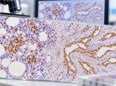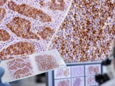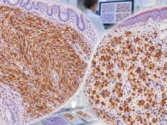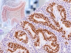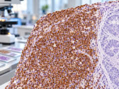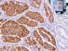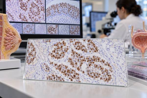
GATA3 immunohistochemistry is a tissue stain that helps pathologists identify breast and urothelial differentiation and narrow the likely origin of a carcinoma. GATA3 is a transcription factor, so true tumor staining is nuclear. Strong, diffuse nuclear positivity is common in many breast carcinomas and urothelial carcinomas, which makes the marker useful when a tumor is metastatic or its primary site is uncertain. The key limitation is that GATA3 is not exclusive to either organ. It can be expressed in salivary gland tumors, skin adnexal tumors, parathyroid lesions, some renal and gynecologic tumors, certain squamous carcinomas, and other neoplasms. A positive result therefore supports a lineage only when the tumor’s morphology, clinical setting, and companion stains point in the same direction. Pathologists often pair GATA3 with markers such as ER, PR, TRPS1, mammaglobin, p63, uroplakin, CK7, CK20, or PAX8 depending on the differential diagnosis.
- GATA3-positive nuclear staining commonly supports breast or urothelial differentiation, but it does not prove either origin by itself.
- Strong, diffuse staining is generally more supportive than weak or focal staining, especially in a metastatic carcinoma of uncertain origin.
- GATA3 can be positive in several non-breast and non-urothelial tumors, so a panel is more reliable than a single stain.
- A negative GATA3 result does not completely exclude breast or urothelial cancer, particularly in high-grade or poorly differentiated tumors.
- The test is performed on biopsy or surgical tissue, so there is no blood level, normal range, or patient preparation requirement.
Table of Contents
- What GATA3 IHC Measures
- GATA3 in Breast Cancer
- GATA3 in Urothelial Cancer
- Using GATA3 to Identify Tumor Origin
- How Positive Staining Is Interpreted
- Pitfalls and Limitations
- What Happens After the Result
What GATA3 IHC Measures
GATA3 IHC detects GATA-binding protein 3 in formalin-fixed tissue. GATA3 is a zinc-finger transcription factor involved in cell differentiation and gene regulation. Because the protein functions in the nucleus, the pathologist looks for nuclear staining in tumor cells. Cytoplasmic color without convincing nuclear labeling is not the classic specific pattern.
The stain can be performed on a core biopsy, surgical specimen, cell block from a fluid or fine-needle aspiration, or a metastatic-site sample. The laboratory applies an antibody to a thin tissue section and uses a chromogen to make antibody binding visible under the microscope.
GATA3 is not a quantitative laboratory test with a standard numeric range. Reports may describe the percentage of positive tumor cells and staining strength, but there is no universal cutoff comparable with the 1% estrogen-receptor threshold or the HER2 0-to-3+ system. The pathologist instead asks whether the pattern is sufficiently convincing and whether it fits the diagnostic question.
Normal tissues may also express GATA3. Breast luminal epithelial cells, urothelium, parathyroid tissue, subsets of lymphocytes, and other cell types can be positive. These normal patterns explain why GATA3 is best viewed as a lineage-associated marker rather than a cancer-specific marker.
When the primary site is uncertain, GATA3 is usually one part of a tumor immunohistochemistry panel. The rest of the panel is selected from the tumor’s microscopic appearance, location, and realistic clinical alternatives.
GATA3 in Breast Cancer
GATA3 is one of the most sensitive traditional immunohistochemical markers of breast differentiation, especially in estrogen receptor–positive and luminal-type breast carcinomas. Many invasive ductal carcinomas of no special type and invasive lobular carcinomas show strong nuclear labeling.
Its value becomes clear in metastatic disease. A patient may present with an adenocarcinoma in a lymph node, lung, liver, bone, pleura, or another organ years after treatment for breast cancer. If the metastatic tumor resembles the prior breast cancer and shows GATA3 positivity, the stain can support breast origin.
GATA3 is often more sensitive for breast carcinoma than older markers such as mammaglobin or GCDFP-15. However, it is less specific than a truly organ-exclusive marker would be. Modern panels may add TRPS1, which is highly sensitive for many breast cancers, particularly some triple-negative tumors. ER and PR also support breast differentiation in the appropriate setting, but many gynecologic tumors express hormone receptors, so they are not breast-specific.
The relationship between GATA3 and hormone receptors is clinically relevant. GATA3 tends to be retained in well-differentiated, hormone receptor–positive breast cancers and may be lost more often in high-grade or triple-negative disease. Therefore, GATA3 negativity does not rule out metastatic breast carcinoma, especially when the original tumor was triple-negative or poorly differentiated.
When the differential includes a Müllerian carcinoma such as ovarian or endometrial cancer, PAX8 and WT1 may be helpful alongside breast markers. A PAX8 IHC test that is strongly positive may support Müllerian, renal, or thyroid origin depending on morphology, while most conventional breast carcinomas are PAX8-negative.
GATA3 in Urothelial Cancer
GATA3 is also a major marker of urothelial differentiation. Many noninvasive and invasive urothelial carcinomas show nuclear positivity, which is useful in bladder tumors and in metastases where the urinary tract is a suspected primary site.
Expression is not identical across all urothelial carcinomas. Lower-grade and noninvasive tumors often retain strong GATA3 expression, while high-grade, muscle-invasive, poorly differentiated, or variant tumors may show weaker or absent staining. Published series report lower sensitivity in some aggressive subtypes than in conventional urothelial carcinoma.
Pathologists rarely use GATA3 alone to prove urothelial origin. Companion markers may include p63 or p40, uroplakin II or III, thrombomodulin, CK20, and CK7. The p63 IHC test can support urothelial or squamous differentiation, while uroplakin markers are more specific for urothelial lineage but can be less sensitive.
One common diagnostic problem is distinguishing metastatic urothelial carcinoma from squamous cell carcinoma. Both can express p63 and p40, and some squamous carcinomas can show GATA3 staining. Uroplakin expression, the overall staining pattern, tumor morphology, and clinical imaging become important in such cases.
Another problem is distinguishing urothelial carcinoma from metastatic breast carcinoma because both can be GATA3-positive. Sex, tumor location, history, ER/PR, mammaglobin or TRPS1, uroplakin, p63/p40, and the histologic pattern help separate them. The marker is therefore most useful when interpreted as part of a deliberately chosen differential-diagnosis panel.
Using GATA3 to Identify Tumor Origin
Tumor-origin workups are common when carcinoma is found in a metastatic site and the primary tumor is unknown or uncertain. GATA3 can quickly narrow the field, but its meaning depends on what other markers show.
Imagine a CK7-positive adenocarcinoma in a liver biopsy. Strong diffuse GATA3 staining might raise breast or urothelial origin. If the tumor is also ER-positive and TRPS1-positive and lacks uroplakin and p63, breast origin becomes more likely. If it is uroplakin-positive and p63-positive with appropriate morphology, urothelial origin becomes more likely.
Conversely, if the tumor is PAX8-positive, the pathologist may consider renal, thyroid, or Müllerian primary sites. If it is TTF-1 and Napsin A positive, lung adenocarcinoma becomes a stronger possibility. A TTF-1 IHC test therefore answers a different lineage question that can complement GATA3 in metastatic adenocarcinoma panels.
The best panels are not broad lists of every available stain. They are hypothesis-driven. Pathologists start from tumor architecture, cell shape, mucin, keratin pattern, clinical imaging, and history, then order markers that distinguish the most plausible alternatives. This approach preserves tissue for molecular testing and reduces confusing incidental positivity.
GATA3 is also used outside classic breast-versus-urothelial questions. It can appear in salivary duct carcinoma, secretory carcinoma, skin adnexal tumors, parathyroid lesions, mesothelioma subsets, trophoblastic lesions, and some soft-tissue neoplasms. These patterns are exactly why the marker should not be interpreted as an automatic organ label.
How Positive Staining Is Interpreted
A GATA3 report may state positive, negative, focal, diffuse, weak, moderate, or strong. The most diagnostically persuasive pattern is usually strong nuclear staining in a large proportion of viable tumor cells, but the meaning is always context-specific.
| Pattern | General interpretation | Next consideration |
|---|---|---|
| Strong, diffuse nuclear positivity | Strongly supports a GATA3-expressing lineage | Use companion markers to distinguish breast, urothelial, and other possibilities |
| Focal moderate positivity | Supportive but less specific | Weight morphology and the rest of the panel more heavily |
| Rare weak positive cells | May be nonspecific or biologically limited | Confirm that staining is truly in tumor nuclei |
| Negative | Does not support GATA3-associated differentiation | Breast or urothelial origin may still remain possible in selected high-grade tumors |
There is no universal percentage at which GATA3 becomes “positive” for every tumor type. Different studies and laboratories use different thresholds, and diagnostic practice relies on pattern recognition rather than a single treatment-linked cutoff.
The pathologist also checks staining localization. Because GATA3 is a nuclear transcription factor, crisp nuclear staining is expected. Diffuse cytoplasmic brown pigment or background staining should not be mistaken for true positivity.
A positive result does not measure how aggressive a tumor is. Although loss of GATA3 has been associated with higher grade or worse features in some breast and urothelial cancer studies, routine treatment and prognosis are not determined from GATA3 intensity alone.
Pitfalls and Limitations
The largest pitfall is over-specificity: assuming GATA3 equals breast or bladder cancer. Broad tumor studies have shown GATA3 expression across many neoplasms. The stain narrows the differential but does not replace a panel.
The second pitfall is over-reliance on a negative result. High-grade breast and urothelial carcinomas can lose lineage markers. Triple-negative breast cancer is particularly important because GATA3 sensitivity is lower than in ER-positive tumors. In that setting, TRPS1 or SOX10 may add useful information.
Technical factors can also produce misleading results. Poor fixation, decalcification, scant viable tumor, crush artifact, and treatment effect can reduce staining. Bone metastases require special caution because decalcification may damage antigens.
Some benign tissues stain strongly and can be mistaken for tumor in small samples. Lymphocytes, benign urothelium, and breast epithelium may provide background or internal-control staining. The pathologist must identify the actual malignant cell population on the hematoxylin-and-eosin slide and match it to the IHC section.
Finally, GATA3 is not a predictive treatment biomarker in the same way as ER or HER2. A breast cancer is not given endocrine or HER2-targeted therapy because it is GATA3-positive. Similarly, urothelial therapy is not selected solely from this stain. Its main role is diagnosis, classification, and support for tumor origin.
What Happens After the Result
What happens next depends on why the stain was ordered. If the diagnosis was already strongly suspected, GATA3 may simply confirm the expected lineage. If the primary site was unknown, the result can determine which additional stains, imaging studies, or molecular tests are most useful.
For a suspected breast primary, the pathology team may review ER, PR, HER2, TRPS1, mammaglobin, or SOX10. For suspected urothelial carcinoma, uroplakin, p63/p40, CK20, and morphology may be emphasized. The CK20 IHC pattern can help in selected urothelial and gastrointestinal differentials, but it is not specific by itself.
If the tumor is metastatic, the final report may use language such as “immunophenotype supports breast primary” rather than claiming certainty from one stain. That wording reflects good pathology practice: the result is integrated with clinical and radiologic evidence.
Useful questions to ask about a GATA3 result include:
- Is the staining strong and diffuse or only focal?
- Which tumor origins are being considered?
- What other markers support breast or urothelial differentiation?
- Could the known tumor type be GATA3-negative despite its origin?
- Does the pathology need comparison with a prior tumor specimen?
- Would additional molecular testing or expert pathology review change treatment?
The most important line in the report is the final diagnosis or interpretive comment, not the isolated “GATA3 positive” phrase. A well-constructed panel gives the marker its meaning.
GATA3 can be particularly useful when a metastasis has lost the classic appearance of its primary tumor. A breast carcinoma in a lymph node, bone, liver, or lung may be poorly differentiated and may not form obvious glands. Nuclear GATA3 in that setting can support breast origin, especially when it agrees with ER, mammaglobin, GCDFP-15, or newer breast-lineage markers. The same logic applies to urothelial carcinoma: GATA3 becomes more persuasive when it aligns with uroplakin, p63/p40, cytokeratin patterns, and a known urinary-tract lesion.
The degree of staining is not a validated stage or prognosis score. Strong diffuse GATA3 does not mean a breast or bladder cancer is more advanced, and weak staining does not automatically mean a tumor is more aggressive. Expression can vary by subtype and differentiation. Some triple-negative breast cancers and high-grade urothelial carcinomas have reduced or absent GATA3, so negative staining must be interpreted with markers that cover those less typical phenotypes.
Specimen quality can also change the result. Decalcification of bone metastases may reduce antigen preservation, and small biopsies may sample only a negative part of a heterogeneous tumor. Treatment can alter tumor morphology and marker expression. When the clinical stakes are high, a pathologist may repeat GATA3 on another block or add more lineage markers rather than treating one negative stain as definitive.
For a tumor of unknown primary, GATA3 is therefore best thought of as a probability-changing marker. It narrows the list of realistic origins but does not supply the answer in isolation. The pathologist combines the stain with sex, prior cancers, imaging, anatomic distribution, tumor morphology, and the rest of the immunophenotype. A diagnosis is strongest when several independent pieces of evidence point to the same site and plausible alternatives have been actively excluded.
In breast pathology, GATA3 is often most helpful as evidence of mammary differentiation when the usual hormone receptors are absent or the metastasis is morphologically unusual. It is not equivalent to estrogen receptor testing. A GATA3-positive breast cancer can be ER-negative, and a GATA3 result does not predict benefit from endocrine therapy. ER and progesterone receptor remain the predictive hormone-receptor assays used for that purpose.
In urothelial pathology, the same separation of roles applies. GATA3 helps support urothelial differentiation, but it does not replace assessment of tumor grade, depth of invasion, lymphovascular invasion, variant histology, or stage. Those features determine the clinical significance of a bladder or upper-tract urothelial carcinoma. A GATA3-negative high-grade carcinoma may still be urothelial if morphology and other markers support that origin.
Certain benign tissues also express GATA3, which is another reason the pathologist must identify the correct cells. Normal breast epithelium, urothelium, parathyroid tissue, and subsets of other cell types can show nuclear expression. In a biopsy containing both benign and malignant components, positive internal cells may be useful controls, but they must not be counted as evidence that the tumor itself is positive.
The practical takeaway is that GATA3 works best as a lineage clue, not a diagnosis label. Its value rises when the staining is convincingly nuclear, the tumor morphology fits, and companion markers tell the same story. When those pieces conflict, further testing is more reliable than choosing the diagnosis that matches GATA3 alone.
References
- Diagnostic and Prognostic Roles of GATA3 Immunohistochemistry in Urothelial Carcinoma 2023 (Systematic Review)
- Gene of the month: GATA3. 2023 (Review)
- GATA3 Expression in Human Tumors: A Tissue Microarray Study on 16,557 Tumors 2023
- Diagnostic utility and sensitivities of matrix Gla protein (MGP), TRPS1 and GATA3 in breast cancer: focusing on metastatic breast cancer, invasive breast carcinoma with special features, and salivary gland-type tumours 2024
- Immunohistochemical Markers for Distinguishing Metastatic Breast Carcinoma from Other Common Malignancies: Update and Revisit 2022 (Review)
- Immunohistochemical evaluation of GATA3 expression in tumors and normal tissues: a useful immunomarker for breast and urothelial carcinomas 2012
Disclaimer
GATA3 IHC is an ancillary pathology test and cannot identify a tumor’s primary site with certainty on its own. Its meaning depends on nuclear staining pattern, tumor morphology, clinical history, and companion markers. Discuss the complete pathology interpretation with your pathologist or treating clinician.