
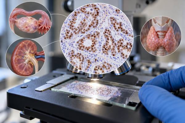
PAX8 immunohistochemistry (IHC) is a tissue stain that helps pathologists recognize tumors with Müllerian, renal, or thyroid follicular-cell differentiation. The PAX8 protein is a nuclear transcription factor involved in development of these organ systems, so convincing nuclear staining can provide a strong clue to tumor origin. PAX8 is commonly used when evaluating ovarian and other gynecologic carcinomas, renal cell carcinomas, thyroid carcinomas, and metastatic tumors whose primary site is uncertain. A positive result is not specific to one organ: ovarian, endometrial, kidney, and thyroid tumors can all be PAX8 positive, and staining varies by tumor subtype and antibody clone. A negative result also does not exclude every tumor from these sites. Pathologists therefore use PAX8 as part of a focused panel with morphology and other markers such as WT1, ER, thyroglobulin, TTF-1, CAIX, or GATA3. The test helps establish lineage and probable origin; it does not determine cancer stage or function as a treatment-response score.
- What PAX8 measures: PAX8 IHC detects nuclear expression of a transcription factor associated mainly with Müllerian, renal, and thyroid lineages.
- What positive staining can support: ovarian/gynecologic, kidney, or thyroid origin when the tumor morphology and companion markers agree.
- What positive PAX8 cannot prove: it cannot distinguish those organs by itself and does not establish stage or treatment sensitivity.
- Why antibody type matters: some polyclonal PAX8 antibodies can show cross-reactivity that is less common with well-validated monoclonal reagents.
- How results are interpreted: pathologists evaluate tumor-cell nuclear staining, its extent, specimen quality, and the complete IHC profile.
Table of Contents
- What the PAX8 IHC Test Detects
- PAX8 in Ovarian and Müllerian Tumors
- PAX8 in Kidney Cancer
- PAX8 in Thyroid Cancer
- How PAX8 Helps Determine Tumor Origin
- Limitations and Diagnostic Pitfalls
- What Happens After a PAX8 Result
What the PAX8 IHC Test Detects
PAX8 is a paired-box transcription factor with an important role in development and maintenance of the thyroid, kidneys, and Müllerian tract. In pathology, antibodies to PAX8 are used to detect the protein in cell nuclei. Because the staining is nuclear, convincing localization is usually easier to distinguish from nonspecific cytoplasmic background.
Normal tissues from these developmental lineages can express PAX8, and many tumors derived from them retain expression. This makes PAX8 a lineage-associated marker. It is particularly useful in metastatic cancer, where the microscope may show an adenocarcinoma but not reveal whether it began in the ovary, kidney, lung, breast, gastrointestinal tract, or another site.
Large tumor-series data illustrate both the strength and limits of the marker. A 2024 study of more than 17,000 tumors reported very high PAX8 expression in many thyroid, gynecologic, and renal cancers, while most breast, pulmonary, gastrointestinal, prostate, and hepatocellular carcinomas were negative. Those differences can be diagnostically powerful, but no percentage applies to every histologic subtype.
The stain is performed on formalin-fixed tissue from a biopsy or resection. The report may describe PAX8 as positive or negative and sometimes characterize the staining as focal or diffuse and weak, moderate, or strong. Laboratories must validate the antibody clone and platform because antibody choice can influence specificity.
PAX8 is not a blood tumor marker and does not measure tumor burden. It also is not a genetic test for PAX8 mutation or rearrangement. Its purpose is to show a protein-expression phenotype in the cells on the slide.
PAX8 in Ovarian and Müllerian Tumors
PAX8 is widely used to support Müllerian origin, which includes many tumors of the ovary, fallopian tube, endometrium, and related peritoneal sites. Serous, endometrioid, and clear cell carcinomas are commonly PAX8 positive, while expression is less consistent in mucinous ovarian tumors.
A 2023 meta-analysis of ovarian tumors found high PAX8 sensitivity in serous and transitional-type categories but much lower sensitivity in mucinous tumors. This is a useful reminder that a negative PAX8 stain does not have the same weight across all ovarian histologies.
In a metastatic adenocarcinoma, PAX8 positivity can help separate a Müllerian tumor from common mimics. For example, most breast and lung adenocarcinomas are PAX8 negative. However, PAX8 alone cannot distinguish ovarian from endometrial, tubal, renal, or thyroid origin. The panel must reflect the morphology and clinical distribution.
WT1 is often paired with PAX8 in suspected serous carcinoma. Diffuse WT1 expression strongly supports serous Müllerian differentiation in the right setting, whereas many endometrioid and clear cell carcinomas are WT1 negative. Estrogen receptor can add information, and p53 staining can help characterize high-grade serous carcinoma or molecular subgroups in endometrial cancer. The progesterone receptor IHC test may also contribute in hormone-receptor-expressing gynecologic tumors, although its interpretation differs from breast cancer scoring.
PAX8 can be especially valuable when a pelvic or peritoneal tumor is poorly differentiated. A positive result narrows the field toward Müllerian and other PAX8-positive lineages, allowing the pathologist to choose the next markers efficiently.
PAX8 in Kidney Cancer
Most major renal cell carcinoma subtypes express PAX8 because renal tubular epithelium belongs to a PAX8-associated developmental lineage. Clear cell, papillary, and chromophobe renal cell carcinomas commonly show nuclear positivity, although the frequency and intensity can vary.
This is particularly useful when renal cell carcinoma presents as a metastasis. Kidney cancers can spread to lung, bone, lymph nodes, adrenal gland, brain, skin, and other locations. A metastatic clear cell tumor can resemble adrenal cortical carcinoma, hepatocellular carcinoma, clear cell gynecologic carcinoma, or other clear-cell neoplasms. PAX8 provides an important clue when interpreted with the rest of the panel.
Renal origin is usually strengthened by additional markers chosen for the suspected subtype. CAIX is often strong in clear cell renal cell carcinoma. CD10 and other renal-associated markers can support the diagnosis, while morphology and imaging help locate a renal mass. PAX8 positivity alone is not enough because Müllerian and thyroid tumors can also stain.
A negative PAX8 result makes common renal cell carcinoma less likely but does not absolutely exclude renal origin. Dedifferentiated, sarcomatoid, treated, or poorly preserved tumors may lose lineage markers. Small metastases may also be heterogeneous.
The distinction between renal and urothelial origin can be clinically important in tumors involving the renal pelvis or urinary tract. Urothelial carcinomas are usually evaluated with p63/p40, GATA3, and uroplakin markers, while renal cell carcinomas more often express PAX8. The p63 IHC test can therefore provide complementary evidence in this differential.
PAX8 does not reveal the renal tumor stage or grade. Those are determined from size, anatomic extension, nodal or distant spread, morphology, and formal grading systems.
PAX8 in Thyroid Cancer
PAX8 is expressed in thyroid follicular cells and many tumors derived from them. Papillary thyroid carcinoma, follicular thyroid carcinoma, and several other follicular-cell-derived neoplasms can therefore show nuclear PAX8 staining.
In a metastatic tumor, PAX8 can support thyroid origin when combined with markers such as TTF-1 and thyroglobulin. Thyroglobulin is especially helpful because it is much more specific for differentiated thyroid follicular-cell lineage. TTF-1 is shared with lung adenocarcinoma, so a TTF-1-positive tumor cannot be assumed to be thyroid in origin. A PAX8-positive, thyroglobulin-positive, TTF-1-positive profile in the appropriate morphology strongly supports differentiated thyroid origin.
Poorly differentiated and anaplastic thyroid carcinomas can lose thyroglobulin and sometimes other differentiation markers. PAX8 may remain useful in a subset of such cases, depending on the antibody and tumor biology. In that setting, the pathologist may use a larger panel to separate thyroid carcinoma from metastatic lung carcinoma, squamous carcinoma, lymphoma, melanoma, and sarcoma.
Medullary thyroid carcinoma arises from parafollicular C cells rather than follicular cells and has a different marker profile, including calcitonin and neuroendocrine markers. PAX8 is therefore not used in the same way for every thyroid malignancy. Serum calcitonin testing addresses a different diagnostic and monitoring question from tissue PAX8 staining.
Antibody clone matters in thyroid pathology because some older polyclonal PAX8 reagents can cross-react with related PAX proteins. Modern monoclonal assays generally improve specificity, but every laboratory must validate its own method.
How PAX8 Helps Determine Tumor Origin
PAX8 is most powerful when it changes the probability of a proposed primary site. In an adenocarcinoma of unknown origin, convincing nuclear PAX8 positivity makes renal, Müllerian, and thyroid lineages substantially more plausible than common lung, breast, colorectal, pancreatic, prostate, or hepatocellular primaries.
That conclusion should be framed as probability, not proof. The next markers separate the PAX8-positive possibilities. A practical panel may look like this:
| Possible origin | Markers often considered with PAX8 | Interpretive idea |
|---|---|---|
| Müllerian/ovarian | WT1, ER, p53, Napsin A depending on morphology | PAX8 establishes lineage; companion markers refine subtype |
| Renal | CAIX, CD10, RCC-associated markers | Pattern and morphology help identify renal subtype |
| Thyroid follicular-cell | Thyroglobulin, TTF-1 | Thyroglobulin is highly supportive of differentiated thyroid origin |
| Urothelial or breast mimic | GATA3, p63/p40, uroplakins or breast markers | Most conventional examples are PAX8 negative |
| Lung adenocarcinoma mimic | TTF-1, Napsin A | PAX8 is usually negative in primary lung adenocarcinoma |
One common real-world problem is distinguishing a PAX8-positive ovarian or renal metastasis from a new primary lung tumor. A lung mass does not automatically mean lung primary. If the tumor is PAX8 positive and TTF-1/Napsin A negative, the clinical team should consider metastasis from a PAX8-lineage organ, especially when the patient has a relevant history.
The reverse is also true: a pelvic mass can represent metastasis from a non-Müllerian organ. A negative PAX8 stain may support that possibility, but the pathologist still needs appropriate positive markers for the alternative diagnosis.
A focused panel reduces false certainty. Rather than asking whether PAX8 “proves” an origin, pathologists ask whether it fits the proposed lineage and whether independent markers point in the same direction.
Limitations and Diagnostic Pitfalls
The largest limitation is that PAX8 is shared by several organ systems. A positive stain cannot by itself distinguish ovary from endometrium, kidney, or thyroid. Clinical location and companion markers are mandatory when more than one of these sites is plausible.
Antibody choice is another important issue. Some polyclonal PAX8 antibodies can cross-react with other PAX-family proteins and may label tumors or lymphoid cells that are not truly expressing PAX8. Well-validated monoclonal antibodies can reduce this problem, but assay performance still depends on clone, platform, retrieval, and interpretation.
Not every tumor from a PAX8-associated organ remains positive. Mucinous ovarian tumors are less consistently positive than serous tumors. Dedifferentiated or anaplastic cancers may lose lineage markers. Treatment can change antigen expression. A negative result should therefore be weighted according to the suspected histologic subtype rather than used as an absolute exclusion.
Normal tissues can also stain. Benign renal tubules, thyroid follicles, and Müllerian epithelium may be PAX8 positive. In a small biopsy, the pathologist must make sure the positive nuclei belong to the malignant cells rather than entrapped normal structures.
Weak or equivocal staining deserves caution. True diagnostic staining should have appropriate nuclear localization. Nonspecific background or scattered questionable cells should not be given the same weight as a convincing tumor-cell pattern.
Another pitfall is confusing PAX8 expression with a molecular alteration. IHC does not test for PAX8 gene mutation, amplification, or fusion. It simply detects protein expression.
Finally, PAX8 is not a prognostic or treatment-selection score in routine use. Its primary value is diagnostic lineage and origin. The treatment implications arise from the final tumor diagnosis and disease-specific biomarkers, not from the intensity of PAX8 staining itself.
What Happens After a PAX8 Result
After PAX8 staining, the pathologist asks whether the result fits the proposed diagnosis. A positive ovarian or endometrial tumor may need WT1, ER, p53, mismatch-repair proteins, or subtype-specific markers. A suspected renal metastasis may receive CAIX and other renal markers. A suspected thyroid metastasis may be tested with thyroglobulin and TTF-1.
If PAX8 is unexpectedly negative, the next step depends on specimen quality and morphology. The pathologist may repeat the assay, use another block, confirm the antibody clone, or pursue alternative origins. A negative result is most informative when a tumor type is usually strongly PAX8 positive and the technical controls are sound.
If PAX8 is unexpectedly positive in a tumor thought to be from lung, breast, gastrointestinal tract, or another usually negative site, the clinical history should be reviewed. Prior renal, thyroid, ovarian, tubal, or endometrial tumors may become highly relevant. Comparison with earlier pathology can be more useful than adding many unrelated stains.
For patients, helpful questions include: Was the PAX8 staining clearly nuclear? Which primary sites does the pathologist consider most likely? Which companion markers support that interpretation? Could the lesion be a metastasis from a prior cancer? Is more tissue or molecular testing needed?
The final pathology diagnosis should state the integrated conclusion rather than leave the reader to interpret PAX8 alone. A phrase such as “PAX8 and WT1 expression supports Müllerian serous carcinoma” is more useful than “PAX8 positive” because it shows how the marker contributes to the diagnosis.
PAX8 is one of the most useful lineage markers for separating renal, Müllerian, and thyroid tumors from many common mimics. Its value is strongest when multiple independent findings agree and weakest when it is treated as a one-stain answer to tumor origin.
PAX8 is also useful when two PAX8-positive lineages are competing possibilities, because it tells the pathologist what the marker cannot resolve. A clear-cell tumor in the pelvis, for example, may represent Müllerian clear cell carcinoma or metastatic renal cell carcinoma. Both can express PAX8. In that situation, the discriminating information comes from morphology, clinical imaging, and companion markers such as Napsin A or HNF1β for a Müllerian clear-cell phenotype and CAIX or other renal markers for clear cell renal cell carcinoma. Treating PAX8 as the final answer would miss the most important part of the differential diagnosis.
A similar problem can occur with a PAX8-positive neck or mediastinal lesion. Thyroid follicular-derived tumors, renal metastases, and some Müllerian metastases may all enter the differential depending on the patient. Thyroglobulin, TTF-1, renal markers, and clinical history help separate them. This illustrates why a strong marker is often most useful for excluding broad groups rather than naming one organ by itself.
Pathologists also pay attention to the antibody clone when interpreting unexpected positivity in lymphoid or neuroendocrine-appearing tumors. Cross-reactivity of some polyclonal antibodies with related PAX proteins can produce staining outside the expected PAX8 spectrum. Repeating the test with a validated monoclonal clone or using independent lineage markers can resolve the discrepancy.
Finally, a PAX8 result should be compared with the specimen’s anatomic context. A small amount of benign PAX8-positive tubal, endometrial, renal, or thyroid tissue can sit next to a negative tumor. The pathologist must score the malignant cells themselves rather than the most strongly stained structure on the slide.
This is particularly useful in small metastatic biopsies, where tissue conservation matters. A high-value PAX8 result can narrow the differential early and allow the next two or three stains to be chosen deliberately instead of exhausting the block with a broad unfocused panel. The same approach preserves material for molecular tests that may directly guide therapy after the tumor type is established.
References
- PAX8 expression in cancerous and non-neoplastic tissue: a tissue microarray study on more than 17,000 tumors from 149 different tumor entities 2024
- Diagnostic roles of PAX8 immunohistochemistry in ovarian tumors 2023 (Systematic Review)
- SOX17/PAX8 dual immunohistochemical expression in the diagnosis of Müllerian carcinomas 2026
- Next-Generation Immunohistochemistry in Thyroid Neoplasm: A Practical Review on the Applications in Diagnosis and Molecular Classification. 2025 (Review)
- Paired-Box Gene 8 (PAX8) and Its Association With Epithelial Carcinomas. 2021 (Review)
Disclaimer
PAX8 IHC is a diagnostic tissue test interpreted with morphology, antibody performance, other markers, and clinical information. A positive PAX8 stain can support Müllerian, renal, or thyroid lineage but cannot establish the primary organ by itself. The complete pathology report and clinical workup should guide diagnosis and treatment.





