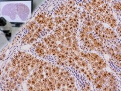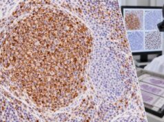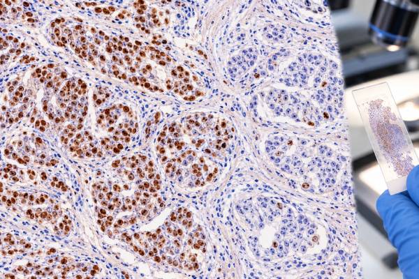
The progesterone receptor (PR) immunohistochemistry (IHC) test measures PR protein in the nuclei of breast cancer cells. It is usually performed with estrogen receptor (ER) and HER2 testing after a breast cancer diagnosis. Under current ASCO/CAP guidance, invasive breast cancer is considered PR positive when at least 1% of tumor-cell nuclei show staining and PR negative when fewer than 1% stain, assuming the assay and controls are adequate. The report may also describe staining intensity and the percentage of positive cells. PR is most useful as a prognostic marker in ER-positive breast cancer and as part of the overall hormone-receptor profile; it should not be used alone to decide whether endocrine therapy will work. A very unusual result, such as ER-negative/PR-positive invasive cancer, may prompt review or repeat testing because true cases are uncommon. Preanalytic factors such as cold ischemia and formalin fixation can affect receptor staining, so the final interpretation depends on both the score and the quality of the specimen.
- PR positive in breast cancer: at least 1% of invasive tumor-cell nuclei stain for progesterone receptor on a valid assay.
- PR negative: fewer than 1% of tumor-cell nuclei stain, but controls and specimen quality must be adequate before calling a result negative.
- What the score means: the percentage shows how many tumor nuclei express PR; intensity may also be reported, but the clinically used positive/negative cutoff is based on nuclear staining.
- Why PR matters: PR adds prognostic information, especially in ER-positive disease, and helps describe the tumor’s hormone-receptor biology.
- What PR cannot do alone: it does not determine breast cancer stage and should not replace ER, HER2, pathology, or other treatment-relevant tests.
Table of Contents
- What the PR IHC Test Measures
- What a Positive PR Score Means
- PR-Negative, Low, and Unusual Results
- How PR Fits With ER and HER2 Status
- How PR Testing Is Performed and Reported
- Limitations and Diagnostic Pitfalls
- What Happens After a PR Result
What the PR IHC Test Measures
Progesterone receptor is a nuclear hormone receptor regulated in part by estrogen signaling. In breast tissue, PR expression provides information about the biology of the tumor and the activity of the hormone-receptor pathway. Pathologists detect PR with an antibody on a tissue section from a core biopsy, surgical specimen, or metastatic lesion.
The test evaluates nuclear staining in invasive tumor cells. Brown staining in the cytoplasm is not the intended scoring signal. The pathologist estimates the percentage of tumor-cell nuclei that stain and may also record average intensity as weak, moderate, or strong. Some laboratories use an H-score or Allred score in addition to the percentage, but the current positive-versus-negative definition for routine breast cancer reporting is based on the fraction of stained tumor nuclei.
PR testing is normally interpreted alongside the estrogen receptor IHC test. ER is the primary predictive hormone-receptor marker for endocrine therapy, while PR contributes important biologic and prognostic context. A tumor can be ER and PR positive, ER positive and PR negative, or, much less commonly, show another combination.
PR is not unique to breast tissue. Normal endometrium and many gynecologic tumors can express progesterone receptor, and selected tumors from other organs may also stain. Therefore, PR positivity in a metastasis does not prove breast origin. If the primary site is uncertain, markers such as GATA3, mammaglobin-related markers, PAX8, and morphology may be considered depending on the differential diagnosis.
The test also does not measure circulating progesterone. Blood hormone levels and tissue PR expression answer different questions. A person can have normal serum progesterone while a breast tumor is PR negative, or vice versa.
What a Positive PR Score Means
For invasive breast cancer, current ASCO/CAP guidance defines PR positive as nuclear staining in 1% to 100% of tumor cells on a technically valid IHC assay. A result below 1% is considered negative. This threshold is important because readers sometimes assume that only strongly or extensively stained tumors count as positive.
A report might say “PR positive, 80% strong nuclear staining,” “PR positive, 10% moderate staining,” or simply provide a percentage and intensity. All of these can meet the positive definition if at least 1% of invasive tumor nuclei are convincingly stained and appropriate controls work.
Unlike ER, the formal ER-low-positive reporting category for tumors with 1% to 10% staining does not create a separate PR-low category in the same way. PR can certainly be present in a low percentage of cells, but the special guideline language about limited endocrine benefit applies specifically to ER-low-positive tumors. This distinction matters when patients compare the two receptor scores.
A higher PR percentage often reflects a more intact estrogen-regulated hormone pathway in ER-positive cancer and is generally associated with more favorable tumor biology than loss of PR, but the score is not interpreted as a simple linear prediction of outcome. Tumor grade, stage, ER level, HER2 status, genomic assays when indicated, proliferation, patient factors, and treatment all contribute to prognosis.
PR positivity also does not guarantee response to endocrine therapy. Endocrine treatment decisions rely principally on ER status and the overall clinical situation. PR helps refine the picture but should not be used as the sole predictive marker.
In ductal carcinoma in situ, hormone-receptor testing may be performed for a different purpose than in invasive cancer, and local practice can differ. The pathology report should identify which component was tested and scored.
PR-Negative, Low, and Unusual Results
A PR-negative result means fewer than 1% of invasive tumor-cell nuclei stain, assuming the specimen is adequate and controls are valid. In an ER-positive breast cancer, PR loss can indicate different tumor biology from an ER-positive/PR-positive cancer and may be associated with higher grade or other less favorable features. It does not, however, make an ER-positive cancer automatically resistant to endocrine therapy.
A low but positive PR percentage should be reported accurately rather than rounded into a vague label. A tumor with 3% nuclear PR staining is positive by the guideline threshold, but the low level can be clinically relevant context. The treating team interprets it with ER, HER2, grade, stage, and other features.
The ER-negative/PR-positive phenotype deserves special attention. True examples appear to be uncommon. Because ER normally helps regulate PR, an isolated PR-positive result can raise concern about analytic or interpretive error, tumor heterogeneity, or specimen differences. Pathology review, checking controls, comparing another block, or repeating receptor testing may be appropriate when the result is unexpected.
Discordance between a core biopsy and surgical specimen can also occur. Most cases are concordant, but heterogeneity, sampling, fixation, or borderline percentages can produce different results. A 2023 study and literature review reported high overall PR concordance between biopsy and excision, while still recognizing that clinically relevant discrepancies occur.
Metastatic disease may differ from the original breast tumor. Hormone-receptor expression can be lost or gained during tumor evolution and treatment. When feasible and clinically appropriate, guidelines often support reassessing predictive biomarkers in a metastatic specimen because current tumor biology may affect management.
A negative PR result should therefore be read as a property of the tested specimen, not as a permanent whole-body label. The pathology team considers whether the sample is representative and whether repeating the test would change a treatment decision.
How PR Fits With ER and HER2 Status
Breast cancer biomarker reporting commonly combines ER, PR, and HER2 because the three markers describe clinically important tumor biology. Ki-67 may also be reported in selected settings, and genomic assays may provide additional information for certain early-stage hormone-receptor-positive cancers.
A simplified way to think about the receptor combinations is:
| Typical profile | General interpretation | Important caution |
|---|---|---|
| ER positive / PR positive | Hormone-receptor-positive biology | Treatment still depends on stage, HER2, patient factors, and other risk features |
| ER positive / PR negative | Still hormone-receptor positive because ER is positive | PR loss can carry prognostic information but does not cancel ER-directed endocrine treatment considerations |
| ER negative / PR positive | Uncommon pattern | Review or repeat testing may be considered to confirm the result |
| ER negative / PR negative | No hormone-receptor expression by IHC | HER2 and other tumor features remain essential |
HER2 answers a different question. The HER2 IHC test assesses HER2 protein overexpression and can trigger in situ hybridization in equivocal breast cancer cases. HER2 status has direct treatment implications that are separate from PR expression.
PR should also not be confused with proliferation. A tumor may be strongly PR positive and still have a relatively high proliferation index. Conversely, a PR-negative cancer can have a lower Ki-67 in some cases. The Ki-67 IHC test estimates proliferative activity rather than hormone-receptor expression.
These markers are combined because cancer biology is multidimensional. No one result should be expected to summarize the entire tumor.
How PR Testing Is Performed and Reported
Accurate PR testing begins before the stain is applied. The tissue should be handled promptly and fixed in neutral buffered formalin according to validated breast biomarker protocols. Excessive delay before fixation, inadequate fixation, or other processing problems can reduce antigen preservation and create a false-low or false-negative result.
After processing, the laboratory applies a validated PR antibody and detection system. Positive and negative controls demonstrate that the assay performed as expected. The pathologist identifies the invasive tumor, excludes in situ or normal epithelium from the invasive score when appropriate, and estimates the percentage of positive tumor nuclei.
A complete report commonly includes:
- the percentage of invasive tumor cells with nuclear PR staining;
- staining intensity or an accepted composite score if the laboratory reports it;
- a positive or negative interpretation using the validated cutoff;
- comments about unusual results, limited tissue, controls, or technical concerns when needed.
Some normal breast epithelial cells can express hormone receptors and may provide internal context, but controls must be interpreted according to the laboratory protocol. If the slide lacks convincing internal or external control performance, the pathologist may repeat the assay rather than issue a definitive negative result.
The percentage is an estimate made across representative invasive tumor. Heterogeneity can make scoring harder when strongly positive and negative areas coexist. Digital image analysis may assist in some laboratories, but it also requires validation and pathologist oversight.
When a specimen contains very little invasive tumor, the report may state that the result is limited or uninterpretable. Preserving enough viable tumor for ER, PR, HER2, and possible molecular testing is an important part of biopsy handling.
Limitations and Diagnostic Pitfalls
Preanalytic handling is one of the most important limitations. Hormone receptors are sensitive to tissue processing. A delayed or poorly fixed specimen can look falsely negative even when the tumor biologically expresses PR. This is why pathology laboratories follow standardized fixation and quality-control procedures.
Sampling is another limitation. Breast cancers can be heterogeneous, and a small core biopsy represents only part of the lesion. Neoadjuvant treatment can alter receptor expression, and a recurrence years later may not match the original tumor exactly. When a new specimen is available and the result could change therapy, reassessment may be clinically useful.
Interpretation near the 1% cutoff requires care. Scattered weak nuclei can be difficult to distinguish from artifact, and counting very small tumor populations creates uncertainty. The pathologist should verify that the stained cells are invasive carcinoma and that the assay controls are appropriate.
PR is not breast-specific. Endometrial and other gynecologic tissues commonly express it, so a PR-positive metastasis needs additional lineage markers. The PAX8 IHC test can help support Müllerian origin in the appropriate differential, while breast-associated markers and clinical history may support breast origin.
Another pitfall is assuming that a higher PR percentage directly predicts a larger endocrine-treatment benefit. PR contributes prognostic information, but ER status and the overall disease setting remain more important for endocrine treatment decisions. There is no validated rule that an 80% PR result means eight times the benefit of a 10% result.
Finally, receptor status should not be used to infer stage. A small localized PR-negative tumor and a metastatic PR-positive tumor can exist. Stage is based on anatomy and spread, not receptor percentage.
What Happens After a PR Result
Once ER, PR, and HER2 results are complete, the oncology team combines them with tumor size, lymph-node status, grade, histologic type, patient age, menopausal status, comorbidities, and other relevant biomarkers. In early-stage ER-positive/HER2-negative disease, a genomic recurrence assay may be considered in selected patients to refine chemotherapy decisions.
A PR-positive result generally confirms that the tumor expresses part of the hormone-receptor pathway. If ER is also positive, endocrine therapy is commonly considered according to stage and clinical circumstances. The exact drug and duration depend on factors such as menopausal status, recurrence risk, prior therapy, and current guidelines.
If PR is negative but ER is positive, the cancer is still considered hormone-receptor positive on the basis of ER. PR negativity may influence prognostic assessment but does not by itself remove endocrine therapy from consideration.
If the pattern is ER negative and PR positive, the pathology team may review the slide, controls, and specimen handling or repeat testing when appropriate. Confirmation is useful because this phenotype is uncommon and could affect how the tumor is categorized.
Patients reviewing their report can ask: What percentage of tumor nuclei were PR positive? What was the ER result? What is the HER2 result? Were the receptor results performed on the original tumor or a metastasis? Do these markers change the recommended systemic treatment? Is additional genomic testing useful in this stage of disease?
The PR score is most informative as part of a complete breast cancer profile. It provides meaningful biologic and prognostic information, but its clinical value comes from integration with ER, HER2, morphology, stage, and the patient’s overall treatment context.
PR can also provide context when tumor biology changes over time. A primary breast cancer and a later metastasis may not have identical receptor profiles because tumors evolve under natural selection and treatment pressure. A change from PR positive to PR negative does not automatically mean either test was wrong. The difference may reflect genuine biologic evolution, sampling of a different clone, or preanalytic variation. When the new result could affect management, the pathology and oncology teams consider the quality of both specimens and the total receptor pattern.
Another practical issue is intratumoral heterogeneity. Some cancers contain strongly PR-positive regions next to weak or negative regions. The pathologist estimates the percentage across representative invasive tumor rather than selecting only the darkest area. If the biopsy is tiny, the report may note that the score could differ in a larger specimen. This is especially relevant near the 1% cutoff, where a small number of stained nuclei can change the formal positive/negative category.
Patients may also encounter composite scoring systems such as the Allred score or H-score in older reports, studies, or specific laboratories. These systems combine proportion and intensity in different ways. They should not be converted casually into the modern percentage-based positive/negative definition because the scales are not interchangeable. The safest approach is to use the interpretation provided by the testing laboratory and, when available, the reported percentage of invasive tumor nuclei.
References
- Estrogen and Progesterone Receptor Testing in Breast Cancer: ASCO/CAP Guideline Update 2020 (Guideline)
- Hormone receptors in breast cancer: An update on the uncommon subtypes 2023 (Review)
- The Role of Progesterone Receptors in Breast Cancer 2022 (Review)
- Assessment of Predictive Biomarkers in Breast Cancer: Challenges and Updates 2022 (Review)
- Concordance of immunohistochemistry for predictive and prognostic factors in breast cancer between biopsy and surgical excision: a single-centre experience and review of the literature 2023
Disclaimer
PR IHC is a pathology biomarker test that must be interpreted with ER, HER2, specimen quality, tumor morphology, and clinical findings. A PR percentage alone cannot determine stage, prognosis, or the best treatment for an individual patient. Treatment decisions should be made with the oncology team using the complete pathology report.