
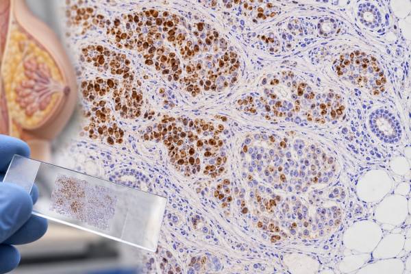
Estrogen receptor (ER) immunohistochemistry is the standard tissue test used to determine whether a breast cancer expresses estrogen receptors and may benefit from endocrine therapy. The stain is performed on biopsy or surgical tissue and is interpreted by counting the percentage of invasive tumor-cell nuclei that show ER staining. Under current ASCO/CAP guidance, invasive breast cancers with 1% to 100% ER-positive tumor nuclei are considered ER-positive, while tumors with less than 1% staining are ER-negative. Cancers with 1% to 10% staining are reported as ER Low Positive because their biology and response to hormone therapy may differ from strongly ER-positive tumors. ER results should be interpreted with the progesterone receptor (PR), HER2 status, tumor grade, histologic type, and clinical setting. The stain is not a blood test and has no serum “normal range”; the important values are the percentage of positive tumor nuclei, staining intensity, and laboratory quality controls.
- ER-positive breast cancer means at least 1% of invasive tumor nuclei stain for estrogen receptor using a validated IHC assay.
- ER Low Positive means 1% to 10% nuclear staining, a group with less certain endocrine sensitivity than cancers with higher ER expression.
- ER-negative means less than 1% tumor nuclear staining, assuming the specimen and controls are adequate.
- ER is a predictive biomarker, mainly used to estimate potential benefit from endocrine treatments such as tamoxifen or aromatase inhibitors.
- No special patient preparation is needed for ER IHC because the test is performed on tissue already obtained by biopsy or surgery.
Table of Contents
- What ER IHC Measures
- How ER Is Scored and Reported
- What an ER-Positive Result Means
- ER Low Positive Breast Cancer
- ER-Negative and Discordant Results
- Testing Quality, Controls, and Pitfalls
- How ER Results Guide Treatment
What ER IHC Measures
ER IHC detects estrogen receptor protein in the nuclei of breast cancer cells. Estrogen receptor is a transcription factor activated by estrogen. When the receptor is present and biologically active, estrogen signaling can help drive tumor growth. That makes ER both a classification marker and a treatment-predictive biomarker.
The test is performed on formalin-fixed, paraffin-embedded tissue from a core biopsy, excision, mastectomy, or metastatic-site biopsy. A laboratory applies a validated antibody to the tissue section. If ER protein is present, tumor-cell nuclei develop a visible stain. The pathologist evaluates only the invasive tumor component when assigning the ER status of invasive breast cancer.
Normal breast epithelial cells and some non-tumor cells can show nuclear ER staining. Those cells can help confirm that staining is technically successful, but they are not counted as malignant cells. The pathologist must distinguish invasive carcinoma from benign glands, ductal carcinoma in situ, inflammatory cells, and stroma before calculating the tumor percentage.
ER IHC is different from measuring estrogen in the blood. There is no ER concentration in pg/mL or a “healthy range.” Instead, the report describes the percentage of invasive tumor nuclei that stain, often together with intensity. A result might read “ER positive, 90%, strong” or “ER low positive, 5%, weak to moderate.”
ER is usually assessed alongside progesterone receptor IHC and HER2 testing. These biomarkers divide breast cancers into clinically important groups and help determine systemic treatment options. A HER2 IHC result answers a different question—HER2 protein expression rather than hormone dependence—so the tests complement rather than replace one another.
How ER Is Scored and Reported
Current ASCO/CAP recommendations classify invasive breast cancer as ER-positive when 1% or more of tumor-cell nuclei show specific staining. Tumors with less than 1% are ER-negative. The 1% threshold is based on evidence that very low expression may still identify some tumors with potential endocrine responsiveness, although benefit is less certain at the lowest levels.
| ER staining | Reporting category | General meaning |
|---|---|---|
| Less than 1% of tumor nuclei | ER-negative | Endocrine benefit is not expected from ER expression |
| 1% to 10% | ER Low Positive | ER is present, but endocrine sensitivity may be limited or heterogeneous |
| More than 10% to 100% | ER-positive | Usually supports hormone-responsive biology; strength of benefit depends on the whole clinical picture |
Many pathology reports also include staining intensity—weak, moderate, or strong. Intensity is informative but does not replace the percentage. A tumor with 90% weak-to-moderate nuclear staining is still ER-positive, while a tumor with 0% staining is negative.
Some laboratories report an Allred score, which combines the proportion of positive cells with average staining intensity. The total score ranges from 0 to 8. Allred scoring is still encountered in practice and research, but the clinically central ASCO/CAP classification is based on the percentage of positive tumor nuclei and the low-positive category.
The report may also mention whether internal controls are adequate. If a specimen has very low or absent ER staining and no normal breast elements are available, laboratory controls and pre-analytic conditions become especially important. When a low or negative result looks unexpected for the tumor type, the pathologist may repeat the stain on another block or recommend additional review.
What an ER-Positive Result Means
An ER-positive result means that at least 1% of invasive tumor cells show nuclear estrogen receptor protein. In most cases with clearly positive expression—especially moderate or strong staining in a large percentage of cells—ER positivity supports a tumor that may respond to endocrine therapy.
Endocrine treatments reduce estrogen signaling in different ways. Tamoxifen blocks estrogen receptor activity in breast tissue. Aromatase inhibitors reduce estrogen production in postmenopausal patients and in premenopausal patients when combined with ovarian suppression. Other endocrine agents, including selective estrogen receptor degraders, may be used in advanced disease depending on prior therapy and tumor characteristics.
The percentage matters because ER expression exists on a continuum. A cancer with 95% strong nuclear staining is biologically different from many tumors with only 2% weak staining, even though both meet the technical definition of ER-positive. This is one reason pathology reports should include the actual percentage rather than simply “positive.”
ER status also contributes to breast cancer subtype discussions. ER-positive/HER2-negative cancers are commonly treated as hormone receptor–positive disease, while ER-positive/HER2-positive cancers can require both endocrine and HER2-directed strategies. ER-negative, PR-negative, HER2-negative cancers fall into the clinical triple-negative category if all three markers meet negative criteria.
ER positivity does not tell you the cancer stage, grade, or recurrence risk by itself. A small node-negative ER-positive cancer and a metastatic ER-positive cancer can have identical staining percentages. Stage comes from tumor size and spread, while prognosis and treatment intensity depend on a combination of anatomy, histology, biomarkers, genomic assays in selected cases, age, menopausal status, and health factors.
ER Low Positive Breast Cancer
ER Low Positive is the specific reporting category for 1% to 10% of invasive tumor nuclei staining for ER. This category deserves separate attention because these cancers often resemble ER-negative tumors more than typical strongly ER-positive cancers at the molecular and clinical level.
Studies show that ER-low cancers are uncommon, often representing only a few percent of breast cancers. They are more likely than strongly ER-positive cancers to be high grade, PR-negative, and biologically similar to basal-like or triple-negative disease. However, the group is heterogeneous. Some tumors retain meaningful estrogen signaling, while others may have only limited ER expression that does not translate into strong endocrine dependence.
ASCO/CAP recommends reporting these tumors as ER Low Positive and including a comment about the limited evidence for endocrine therapy benefit. This is not the same as calling the tumor ER-negative. The 1% threshold remains the formal cutoff for positivity, but clinicians interpret a 1% to 10% result with greater nuance.
Low-level results also deserve technical attention. When staining is close to the threshold, pre-analytic factors and interobserver differences can change classification. A repeat stain on another block may be reasonable if the result is unexpected, controls are questionable, or the difference between 0% and 2% would change treatment strategy. The decision is individualized rather than automatic.
Patients should not infer from an ER-low result that endocrine therapy is useless. Evidence suggests possible benefit in at least some patients, but the magnitude is less certain than in strongly ER-positive disease. Oncology teams often weigh endocrine therapy’s relatively favorable risk profile against the tumor’s overall biology, stage, chemotherapy sensitivity, HER2 status, and current treatment guidelines.
ER-Negative and Discordant Results
ER-negative invasive breast cancer has less than 1% specific tumor nuclear staining. In this setting, treatment decisions generally do not rely on ER-directed endocrine therapy. The tumor is instead managed according to HER2 status, stage, histologic subtype, genomic features, and other biomarkers.
A negative result must still be technically credible. Inadequate fixation, prolonged cold ischemia, decalcification, very small samples, or heavy treatment effect can reduce antigen detection. Bone metastases are a particular challenge because strong acid decalcification can damage hormone-receptor antigens. When possible, laboratories use validated decalcification methods and interpret results cautiously.
Discordance can occur between a core biopsy and surgical specimen, between primary and metastatic tumors, or between different metastatic sites. Tumor evolution, intratumoral heterogeneity, treatment pressure, and sampling differences can all contribute. If a new metastatic lesion is safely accessible, retesting ER, PR, and HER2 may provide information relevant to current therapy.
Discordance also occurs when morphology and biomarkers do not fit. For example, a low-grade classic lobular carcinoma is usually ER-positive. A completely ER-negative result in such a tumor may prompt verification of tumor identity, controls, fixation, and repeat staining. Conversely, some uncommon breast cancer subtypes are genuinely ER-negative.
ER IHC is not used to prove breast origin in a metastatic tumor because many non-breast tissues and cancers can express estrogen receptor. For tumor-origin questions, pathologists use morphology and a broader panel that may include GATA3, TRPS1, mammaglobin, SOX10, PAX8, and other markers selected for the differential. The GATA3 IHC test is commonly used when breast or urothelial origin is being considered.
Testing Quality, Controls, and Pitfalls
Reliable ER testing begins before the stain is placed on the slide. Tissue should be handled promptly and fixed in a validated manner, usually with neutral buffered formalin. Excessive delay before fixation or inappropriate fixation can cause false-negative or weaker results.
Laboratories also validate antibody clones, staining platforms, control tissues, and interpretation procedures. External proficiency testing helps ensure that results are reproducible across laboratories. These quality systems matter because an ER result can directly affect years of systemic therapy.
Common interpretive pitfalls include:
- counting benign breast epithelium or ductal carcinoma in situ instead of invasive tumor;
- mistaking cytoplasmic staining for true ER positivity, which requires nuclear staining;
- overlooking a small ER-positive tumor population in a heterogeneous specimen;
- overcalling weak nonspecific background as true nuclear staining;
- interpreting a negative specimen without adequate controls;
- assuming that intensity alone determines positivity;
- treating 1% to 10% staining as equivalent to 90% to 100% staining biologically.
Neoadjuvant treatment can alter biomarker expression, and small residual tumors may be difficult to assess. When there is enough viable tumor, repeat testing may be considered if the result could change adjuvant therapy.
Another important distinction is between ER protein IHC and ESR1 mutation testing. ESR1 is the gene encoding estrogen receptor alpha. ESR1 mutations can emerge in advanced ER-positive breast cancer after aromatase-inhibitor exposure and may influence endocrine treatment choices. They are detected by molecular testing, not by routine ER immunostaining.
How ER Results Guide Treatment
ER IHC primarily answers whether the tumor has enough estrogen receptor expression to consider endocrine therapy. It does not prescribe one drug or determine treatment duration by itself.
For early-stage ER-positive breast cancer, treatment may include surgery, radiation when indicated, endocrine therapy, and sometimes chemotherapy. The choice of endocrine agent depends on menopausal status, recurrence risk, tolerance, and other factors. Some patients with hormone receptor–positive/HER2-negative early cancer also have a genomic recurrence assay to help decide whether chemotherapy adds meaningful benefit.
In metastatic ER-positive/HER2-negative breast cancer, endocrine therapy is often combined with targeted agents such as CDK4/6 inhibitors, depending on prior therapy and current standards. Molecular testing may identify ESR1, PIK3CA, AKT1, PTEN, germline BRCA1/2, or other alterations that affect later-line options. None of these molecular results changes the basic meaning of an ER IHC percentage, but together they produce a more complete treatment profile.
For ER-low cancers, treatment planning is more individualized. Clinicians may consider the tumor’s resemblance to triple-negative biology, chemotherapy sensitivity, stage, and evidence for possible endocrine benefit. The pathology report’s exact percentage and comment are therefore more useful than a simple positive/negative label.
If you are reviewing your own report, useful questions include: What percentage of tumor cells stained? Was the intensity weak, moderate, or strong? Is the tumor classified as ER Low Positive? What are the PR and HER2 results? Were the controls adequate? Does the oncology team recommend endocrine therapy, and what benefit is expected in this specific clinical setting?
The final treatment decision belongs to the whole clinical picture. ER is one of the most important breast cancer biomarkers, but it works in combination with stage, pathology, HER2, patient factors, and—when appropriate—molecular tests.
The source of the tested tissue can also affect interpretation. Primary breast cancers and later metastases are usually related, but receptor expression can change because of tumor evolution, treatment pressure, sampling, or technical factors. When a recurrence is biopsied, repeating ER along with PR and HER2 can provide information that is more relevant to the current disease than a result from many years earlier. A discordant result should be reviewed in context rather than automatically assuming one specimen was wrong.
ER results also need to be separated from menopausal status. A tumor can be ER-positive in a premenopausal or postmenopausal patient; the IHC result describes the cancer cells, not the patient’s circulating estrogen level. Menopausal status matters because it can influence which endocrine treatment is used and whether ovarian suppression is considered. It does not change the basic pathology definition of ER positivity.
For very small percentages near the cutoff, preanalytic quality and tumor-cell counting become particularly important. If only a few invasive tumor nuclei stain, the pathologist may review controls, compare another block, or comment on limited tissue. That extra care matters because a shift from below 1% to above 1% changes the formal ER category, while the expected endocrine benefit for ER-low disease remains less certain than for strongly ER-positive cancers.
The ER percentage also should not be used as a stand-alone measure of recurrence risk. A breast cancer with 95% ER-positive nuclei may still have high-risk features, while a lower percentage does not by itself define stage. Tumor size, lymph-node status, grade, HER2, genomic assays when indicated, patient factors, and response to therapy all contribute to risk assessment and treatment planning.
References
- Estrogen and Progesterone Receptor Testing in Breast Cancer: ASCO/CAP Guideline Update 2020 (Guideline)
- Hormone Receptor and HER2 Testing in Breast Cancer: Latest Questions Answered 2025 (Review)
- Estrogen receptor-low positive breast cancer: Historical perspective and recent advancements 2025 (Review)
- The “lows”: Update on ER-low and HER2-low breast cancer 2024 (Review)
- Estrogen-Receptor-Low-Positive Breast Cancer: Pathological and Clinical Perspectives 2023 (Review)
- Clinical Behavior, Management, and Treatment Response of Estrogen Receptor Low (1-10%) Breast Cancer 2023
Disclaimer
ER IHC is a predictive pathology test and must be interpreted with the complete breast cancer diagnosis, PR and HER2 results, specimen quality, stage, and clinical factors. An ER percentage alone cannot determine prognosis or the best treatment for an individual patient. Discuss your pathology report and endocrine-therapy options with your oncology team.





