
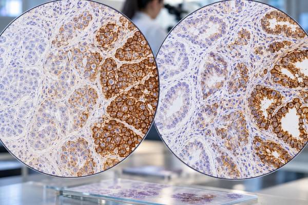
HER2 immunohistochemistry (IHC) measures HER2 protein expression on tumor-cell membranes and helps determine whether a cancer may qualify for HER2-directed therapy. It is used most routinely in breast cancer and gastric or gastroesophageal junction adenocarcinoma, but the scoring rules are not identical across tumor types. In breast cancer, HER2 IHC is scored 0, 1+, 2+, or 3+. A 3+ result is HER2-positive, while a 2+ result is equivocal and usually requires in situ hybridization (ISH) to assess ERBB2 gene amplification. Scores of 0 or 1+ are traditionally HER2-negative, although low levels of expression can now matter for some antibody-drug conjugate treatments in advanced disease. Gastric cancer uses a modified scoring system that allows incomplete basolateral or lateral membrane staining and gives special attention to tumor heterogeneity. HER2 results must therefore be interpreted using the correct organ-specific criteria and clinical setting.
- HER2 IHC 3+ is generally considered HER2-positive in breast and gastroesophageal adenocarcinoma when organ-specific criteria are met.
- HER2 IHC 2+ is an equivocal result, so ERBB2 gene testing by ISH is usually needed before final HER2 classification.
- HER2 IHC 0 and 1+ are not amplified/overexpressed categories, but distinguishing 0 from 1+ can matter for some metastatic breast cancer therapies.
- Breast and gastric cancers use different HER2 scoring rules, especially for membrane completeness and biopsy interpretation.
- No patient preparation is needed for HER2 IHC because testing is performed on tumor tissue from biopsy or surgery.
Table of Contents
- What HER2 IHC Measures
- HER2 Scoring in Breast Cancer
- HER2-Low and HER2-Ultralow
- HER2 Scoring in Gastric Cancer
- IHC Versus ISH and Gene Amplification
- Heterogeneity and Testing Pitfalls
- How HER2 Results Affect Treatment
What HER2 IHC Measures
HER2 is a receptor tyrosine kinase encoded by the ERBB2 gene. When the gene is amplified or the protein is overexpressed, signaling through HER2 can promote tumor-cell growth and survival. HER2-targeted medicines exploit that biology, which is why accurate testing directly influences treatment.
IHC measures protein expression, not the gene itself. A laboratory applies an antibody to a formalin-fixed tumor section, and the pathologist evaluates membrane staining in viable invasive tumor cells. The amount, intensity, and pattern of staining determine the IHC score.
ISH answers a related but different question. It evaluates the number of ERBB2 gene copies, often in relation to a chromosome 17 control probe. In breast cancer, IHC and ISH are integrated when the IHC result is equivocal or when specific unusual ISH patterns need correlation with protein expression.
HER2 is not a blood “level” with a normal range. The result is a tissue score. In breast cancer, the same specimen usually also receives estrogen receptor and progesterone receptor testing. The estrogen receptor IHC test assesses hormone dependence, while HER2 evaluates a growth-factor receptor pathway.
The test may be performed on a core biopsy, surgical resection, or metastatic biopsy. If a specimen is small, heterogeneous, poorly fixed, or heavily treated, another block or later specimen may be tested if the result does not fit the clinical picture.
HER2 Scoring in Breast Cancer
Breast cancer HER2 IHC uses a four-tier score: 0, 1+, 2+, or 3+. The interpretation focuses on membrane staining in invasive carcinoma.
| IHC score | Typical staining pattern | Usual interpretation |
|---|---|---|
| 0 | No staining, or incomplete faint/barely perceptible staining in 10% or fewer tumor cells | HER2-negative by traditional overexpression criteria |
| 1+ | Incomplete faint/barely perceptible membrane staining in more than 10% of tumor cells | HER2-negative; may fall into a HER2-low treatment category in metastatic disease |
| 2+ | Weak to moderate complete membrane staining in more than 10% of tumor cells | Equivocal; reflex ISH is generally required |
| 3+ | Strong complete membrane staining in more than 10% of tumor cells | HER2-positive |
A 3+ result reflects clear protein overexpression and normally supports eligibility for HER2-directed treatment when clinically indicated. A 2+ result is not itself positive; it triggers gene-amplification assessment. A 0 or 1+ result is not considered HER2-positive under conventional ASCO/CAP classification.
The wording “more than 10%” matters. Strong complete membrane staining in only a tiny focus does not automatically make the whole tumor 3+. Pathologists assess the size and distribution of the positive population and may select another block when heterogeneity is suspected.
Breast cancer reports should preserve the actual IHC score rather than reporting only “positive” or “negative,” because lower-expression categories can have treatment implications. HER2 testing is also interpreted alongside hormone receptors, histologic type, grade, and prior treatment.
HER2-Low and HER2-Ultralow
The terms HER2-low and HER2-ultralow became clinically important with antibody-drug conjugates that can work in tumors without classic HER2 overexpression or ERBB2 amplification.
HER2-low breast cancer generally refers to IHC 1+ or IHC 2+ with a negative ISH result. These cancers are still HER2-negative under the traditional positive-versus-negative classification. The term describes a treatment-relevant expression range rather than a new biologic subtype with one consistent molecular identity.
HER2-ultralow has been used for cancers scored as IHC 0 but showing faint, incomplete membrane staining in a small proportion of cells. This creates a practical challenge because older scoring systems focused on separating positive from negative disease, not on distinguishing tiny amounts of expression near the bottom of the scale.
For pathologists, the distinction between no staining and very faint incomplete staining requires careful slide review, adequate controls, and sometimes repeat testing on a different specimen. Interobserver agreement is lower around the 0-versus-1+ boundary than it is for obvious 3+ cases.
Patients should not interpret “HER2-low” as “slightly HER2-positive.” A HER2-low tumor does not meet standard overexpression/amplification criteria. Rather, certain newer drugs can deliver a cytotoxic payload to cells expressing lower levels of HER2. Eligibility depends on the specific drug, disease setting, prior therapy, regulatory indication, and current oncology guidance.
HER2 Scoring in Gastric Cancer
Gastric and gastroesophageal junction adenocarcinomas require a modified HER2 approach because their staining patterns differ from breast cancer. Gastric tumors often show incomplete basolateral or lateral membrane staining and more pronounced intratumoral heterogeneity.
For surgical resections, 3+ generally requires strong complete, basolateral, or lateral membrane reactivity in a meaningful proportion of tumor cells. For biopsies, a cohesive cluster of strongly staining tumor cells can be sufficient even when the positive focus represents a small fraction of the sampled tumor. This biopsy rule reflects the patchy expression common in gastric cancer.
IHC 2+ gastric cancers usually undergo ISH to determine whether ERBB2 is amplified. IHC 0 and 1+ are generally considered negative for classic HER2 overexpression. As in breast cancer, treatment development is expanding interest in lower-expression categories, but organ-specific trial criteria and current approvals matter.
A key practical point is that breast scoring should not be copied directly onto gastric tissue. Incomplete membrane staining that would not qualify as strong positivity in breast cancer can be diagnostically meaningful in gastric cancer when it has the characteristic lateral or basolateral pattern.
Heterogeneity also means specimen selection matters. Multiple tumor-containing biopsies improve the chance of detecting a positive clone. If a gastric tumor is negative on a limited sample but clinical suspicion remains high, additional tissue or a metastatic site may sometimes reveal different HER2 expression.
A broader gastric cancer biomarker workup may also include PD-L1, mismatch-repair proteins, and claudin 18.2, depending on disease stage and treatment setting. HER2 is one component of that predictive profile rather than the only clinically relevant marker.
IHC Versus ISH and Gene Amplification
IHC and ISH are complementary. IHC asks, “How much HER2 protein is present on the tumor-cell membrane?” ISH asks, “Is the ERBB2 gene amplified?”
In breast cancer, an unequivocal IHC 3+ result generally establishes HER2 positivity without a separate ISH test. An IHC 2+ result requires ISH because protein expression alone is indeterminate. ISH results are interpreted using gene copy number and, in dual-probe assays, the HER2-to-CEP17 ratio according to current guideline categories.
A common misconception is that every 2+ cancer is “borderline positive.” In reality, many 2+ tumors are not amplified and are classified as HER2-negative for conventional HER2-targeted therapy. The 2+ score simply identifies a group that needs gene-level clarification.
Discordant cases require additional attention. Examples include unusually strong staining without expected amplification, equivocal morphology, or gene results that fall into uncommon categories. The pathologist may recount a targeted area, review the IHC and ISH slides together, test another block, or obtain expert consultation.
Molecular next-generation sequencing can detect ERBB2 amplification in some tumors, but it does not automatically replace validated IHC/ISH algorithms. HER2 therapeutic evidence is often tied to the assay and scoring method used in clinical trials.
Heterogeneity and Testing Pitfalls
HER2 is vulnerable to both biologic and technical variability. Pre-analytic tissue handling can change staining quality. Delayed fixation, inadequate fixation, over-fixation, crushing, necrosis, or decalcification can weaken membrane staining and increase interpretive uncertainty.
Biologic heterogeneity means different areas of the same tumor can have different HER2 levels. This is particularly important in gastric cancer but also occurs in breast cancer. A small biopsy may sample only a HER2-negative or HER2-positive region.
Other pitfalls include:
- scoring ductal carcinoma in situ instead of invasive breast carcinoma;
- interpreting cytoplasmic staining as membrane positivity;
- ignoring incomplete membrane patterns that are meaningful in gastric cancer;
- overcalling weak granular background as true 1+ staining;
- using breast criteria for gastric cancer or vice versa;
- assuming a historical HER2 result always represents a later recurrence;
- overlooking treatment-related changes in HER2 expression.
Discordance between primary and metastatic sites can occur. If metastatic disease is newly diagnosed and tissue is available, retesting can be useful because current treatment choices depend on the present tumor phenotype. The oncology team decides whether a new biopsy is safe and clinically worthwhile.
Quality assurance is central because one category change can alter therapy. Laboratories use validated antibodies, standardized fixation requirements, external proficiency testing, and trained pathologist interpretation. Digital image analysis may improve consistency in some settings, but pathologist oversight remains important, especially in heterogeneous or low-expression cases.
How HER2 Results Affect Treatment
The treatment meaning of HER2 depends on cancer type, stage, and the exact result. In HER2-positive breast cancer, therapies may include trastuzumab and other HER2-directed antibodies, tyrosine kinase inhibitors, and antibody-drug conjugates depending on early-stage or metastatic setting and prior therapy.
In advanced gastroesophageal adenocarcinoma, HER2-positive status can support HER2-targeted treatment combined with systemic therapy. Current regimens may also depend on PD-L1 and other biomarkers. Because gastric HER2 expression can be heterogeneous and can change after therapy, reassessment at progression may sometimes be considered.
For metastatic breast cancer that is HER2-low or HER2-ultralow, selected antibody-drug conjugates have expanded the relevance of low-level protein expression. This makes accurate distinction among IHC 0, 1+, 2+/ISH-negative, and 3+ increasingly important.
A HER2 result does not determine prognosis by itself. HER2-positive cancers historically had more aggressive behavior, but effective targeted therapy has substantially changed outcomes. Prognosis now depends on stage, treatment response, hormone receptor status, comorbidities, and many other factors.
If your report mentions HER2, useful questions include: What is the exact IHC score? If it is 2+, was ISH performed? Is ERBB2 amplified? Which scoring system was used for this organ? Was the sample a biopsy or resection? Is there evidence of heterogeneity? Does my current treatment depend on classic HER2 positivity, HER2-low expression, or another category?
The most clinically useful interpretation is the final integrated line in the pathology report—such as “HER2-positive,” “HER2-negative,” or “IHC 2+/ISH non-amplified”—combined with the oncology team’s current treatment criteria.
The exact report wording matters because “HER2-positive,” “HER2-low,” and “HER2-ultralow” are not interchangeable categories. In breast cancer, HER2-positive disease remains defined by protein overexpression or ERBB2 amplification under validated criteria. HER2-low describes tumors that are not HER2-positive but show IHC 1+ or IHC 2+ without amplification. HER2-ultralow has been used clinically for tumors scored IHC 0 with faint, incomplete membrane staining in a small proportion of tumor cells. These lower-expression labels were developed to identify patients who may benefit from antibody-drug conjugates; they do not redefine the traditional biology of HER2-amplified cancer.
Because treatment options can depend on subtle distinctions at the low end of staining, laboratories pay close attention to fixation, controls, microscope interpretation, and agreement between pathologists. Distinguishing true faint membrane staining from background can be difficult, especially in small biopsies or previously treated tumors. A report may be reviewed or testing repeated on a more recent specimen when the difference could affect eligibility for therapy.
Breast and gastroesophageal tumors also differ in heterogeneity. Gastric and gastroesophageal adenocarcinomas often show patchy HER2 expression, so a biopsy may contain positive and negative tumor fragments. Organ-specific criteria account for this by allowing scoring based on clusters of tumor cells in biopsy material and by accepting basolateral or lateral membrane staining patterns that would not satisfy breast-cancer rules. Adequate sampling and careful block selection are therefore central to gastric HER2 testing.
A change in HER2 status over time can be biologically real. Metastases may differ from the primary tumor, and treatment can select different tumor-cell populations. When feasible, contemporary metastatic tissue can be informative if the result will change treatment. A new result should be interpreted with the prior report, specimen site, intervening therapies, and assay quality rather than assuming that one time point must represent all disease sites.
Finally, HER2 IHC is not a measure of how much cancer is present in the body. A 3+ score describes membrane protein expression, not tumor burden, stage, or response. Likewise, a change from 2+ to 1+ is not by itself a validated measure of shrinking disease. Imaging, clinical examination, and other disease-specific measures are used to assess response, while HER2 testing answers whether the tumor has a targetable expression/amplification phenotype.
Testing algorithms also depend on what happens after an equivocal result. In breast cancer, an IHC 2+ result is not treated as a final positive call; an in situ hybridization assay evaluates ERBB2 gene amplification under guideline criteria. In gastroesophageal adenocarcinoma, IHC is commonly used first and 2+ tumors are likewise reflexed to ISH. This two-step approach helps avoid giving HER2-targeted therapy based on borderline protein staining alone.
The pathology laboratory’s validated assay and reporting language therefore matter. Different antibody clones and automated platforms can perform well when properly validated, but a score should always be interpreted using the criteria for that tumor type. If a report does not state whether a 2+ result had follow-up ISH, that is a useful point to clarify before assuming the final HER2 status.
If treatment is being planned from an older or outside HER2 report, the oncology team may request slide review or repeat testing when specimen quality, scoring criteria, or disease evolution could matter. This is particularly relevant when a narrow expression category changes access to a specific therapy.
References
- Human Epidermal Growth Factor Receptor 2 Testing in Breast Cancer: ASCO-College of American Pathologists Guideline Update 2023 (Guideline)
- Hormone Receptor and HER2 Testing in Breast Cancer: Latest Questions Answered 2025 (Review)
- Best practices for achieving consensus in HER2-low expression in breast cancer: current perspectives from practising pathologists 2024 (Review)
- HER2 evaluation for clinical decision making in human solid tumours: pearls and pitfalls 2024 (Review)
- Best-Practice Biomarker Testing of Oesophago-Gastric Cancer in the UK: Expert Consensus Recommendations Developed Using a Modified Delphi 2024 (Position Statement)
- Spatial and Temporal Tumor Heterogeneity in Gastric Cancer: Discordance of Predictive Biomarkers 2025 (Review)
Disclaimer
HER2 IHC is a predictive pathology test whose scoring depends on tumor type, specimen quality, and validated laboratory methods. An isolated IHC score cannot determine treatment without the complete pathology report, ISH when required, disease stage, and current treatment criteria. Discuss HER2 results with your pathologist and oncology team.





