
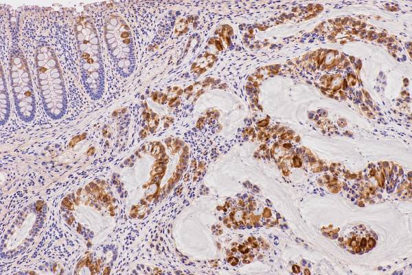
MUC2 immunohistochemistry (IHC) detects mucin 2, the major gel-forming mucin produced by intestinal goblet cells. In tumor pathology, MUC2 is used mainly as a marker of intestinal or goblet-cell-type mucinous differentiation. It is commonly expressed in colorectal and appendiceal mucinous neoplasms and can help identify the intestinal pathway in pancreatic intraductal papillary mucinous neoplasms (IPMNs) and related colloid carcinoma. However, a positive MUC2 stain does not prove that a tumor arose in the colon, does not prove malignancy, and does not by itself define a mucinous carcinoma. MUC2 can be present in normal intestinal epithelium, benign or precursor lesions, and mucinous tumors from several organs. Conversely, many conventional colorectal adenocarcinomas show reduced MUC2 compared with normal bowel. Pathologists therefore interpret the stain with morphology and a panel that may include CDX2, SATB2, CK7, CK20, MUC1, MUC5AC, PAX8, and other site-specific markers.
- MUC2 is an intestinal goblet-cell mucin: positive cytoplasmic or intraluminal staining supports intestinal-type or mucinous differentiation.
- MUC2 is often strong in mucinous colorectal and appendiceal tumors, but many non-mucinous colorectal cancers show reduced expression.
- In pancreatic IPMN, MUC2 with CDX2 supports intestinal-type differentiation and is commonly associated with the colloid carcinoma pathway.
- MUC2 positivity is not organ-specific or cancer-specific: normal bowel and mucinous tumors from other organs may stain.
- No special patient preparation is needed: MUC2 IHC is performed on tissue already obtained by biopsy or surgery.
Table of Contents
- What MUC2 IHC Detects
- What Positive MUC2 Staining Means
- MUC2 in Colorectal and Appendiceal Tumors
- MUC2 in Pancreatic and Other GI Tumors
- How MUC2 Fits Into IHC Panels
- Limitations and Common Pitfalls
- How to Read a MUC2 Pathology Result
What MUC2 IHC Detects
MUC2 IHC detects a secreted gel-forming mucin that is normally abundant in intestinal goblet cells. MUC2 is a very large glycoprotein that helps form the mucus barrier over the intestinal surface. In healthy colon, goblet cells synthesize and secrete large amounts of MUC2 into the lumen.
Because MUC2 is closely tied to goblet-cell differentiation, pathologists use it as a phenotypic clue. A tumor that makes MUC2 may be reproducing an intestinal or mucinous secretory program even when it arises outside the colon. That is why MUC2 can be informative in pancreatic, ovarian, pulmonary, and other mucinous tumors as well as in gastrointestinal neoplasms.
The stain typically highlights mucin within the cytoplasm of tumor cells and may also mark extracellular or luminal mucin. The pathologist focuses on whether viable lesional cells show convincing expression and how widespread the staining is. A report may describe MUC2 as diffuse, patchy, focal, strong, weak, positive, or negative.
MUC2 IHC is not a blood test and has no serum reference range. It also does not measure the amount of extracellular mucin used to classify a carcinoma histologically. For example, colorectal mucinous adenocarcinoma is defined by the proportion of the tumor composed of extracellular mucin on routine microscopy, not by MUC2 positivity alone.
MUC2 should also be distinguished from other mucins. MUC1 is a membrane-associated mucin commonly upregulated in many invasive epithelial cancers. MUC5AC is a gastric-type secreted mucin. MUC2 points more strongly toward intestinal goblet-cell differentiation, although overlap among these markers is common.
What Positive MUC2 Staining Means
A positive MUC2 stain means that the cells being tested express mucin 2 and therefore show a secretory phenotype compatible with intestinal or goblet-cell differentiation. The result becomes clinically meaningful only after the pathologist identifies the tumor type and specimen site.
In a colon tumor, positive MUC2 may be expected, especially in a mucinous or MSI-associated phenotype. In a pancreatic cystic neoplasm, MUC2 positivity can help support an intestinal-type IPMN. In an ovarian mucinous tumor, however, MUC2 may suggest intestinal differentiation without proving that the lesion is metastatic from the gastrointestinal tract.
Positive staining does not establish:
- that a lesion is malignant;
- that the primary site is colon or appendix;
- that a tumor meets histologic criteria for mucinous carcinoma;
- that a colorectal tumor has microsatellite instability;
- the tumor stage or grade;
- an individual prognosis.
A negative result also needs context. Conventional non-mucinous colorectal adenocarcinoma can show decreased or absent MUC2 compared with normal colonic mucosa. Loss of goblet-cell differentiation during tumor progression can reduce expression. Therefore, a negative MUC2 stain does not exclude colorectal origin.
The antibody clone and scoring method also matter. Studies use different cutoffs for the percentage and intensity of positive cells, so a research label such as “MUC2-high” cannot be translated into a universal diagnostic threshold.
How the staining pattern is described
MUC2 is usually interpreted as a cytoplasmic secretory stain, often with accompanying luminal or extracellular mucin. In well-formed glands, staining may be concentrated toward the luminal side of the cell, while mucin-rich tumors can show broad cytoplasmic labeling. Signet-ring cells, when they truly produce MUC2, may show staining around the intracytoplasmic mucin vacuole. The pathologist also notes whether the reaction is diffuse across most of the tumor or confined to small areas.
The percentage of positive cells can be important in research, but routine reports often use descriptive terms rather than a single numeric score. A focal result may still support intestinal differentiation if the morphology and companion markers agree, while diffuse strong staining carries more weight when the competing diagnosis is expected to be MUC2 negative. The location of staining is equally important. Extracellular pools of mucin can remain positive after tumor cells have degenerated, so the most useful evidence comes from intact viable lesional cells.
A practical internal control is nearby normal intestinal mucosa when present. Goblet cells should show the expected reaction. If normal goblet cells fail to stain, an apparently negative tumor result may be technically unreliable and the laboratory may repeat the assay.
MUC2 in Colorectal and Appendiceal Tumors
MUC2 is strongly linked to the biology of colorectal and appendiceal mucin-producing tumors, but its expression is heterogeneous across these diseases. Normal colorectal goblet cells are an important internal positive control because they usually contain abundant MUC2.
Mucinous colorectal adenocarcinomas frequently show prominent MUC2 expression. These tumors are defined morphologically by more than 50% extracellular mucin, not by immunostaining. MUC2 helps explain the secretory phenotype but is not required to recognize extracellular mucin on routine sections.
By contrast, non-mucinous colorectal adenocarcinomas often have lower MUC2 expression than the adjacent normal mucosa. This apparent paradox is important: MUC2 is both the normal intestinal mucin and a marker that can be overexpressed in selected mucinous cancers while being reduced in other colorectal cancers. The direction of change therefore depends on the histologic and molecular subtype.
Recent analyses also emphasize that mucinous colorectal cancer is not a single biologic group. MUC2 and MUC5AC expression can be enriched in proximal, serrated-pathway, mucinous, and microsatellite-instability-associated tumors, but outcomes are influenced by many additional features such as stage, grade, MSI status, BRAF or RAS alterations, and the broader molecular phenotype.
Appendiceal mucinous neoplasms
Appendiceal mucinous neoplasms often express an intestinal immunophenotype that includes MUC2, CK20, CDX2, and SATB2. This pattern can help establish lower gastrointestinal differentiation when a mucinous tumor presents in the ovary or peritoneum.
The distinction between a primary ovarian mucinous neoplasm and metastatic appendiceal or colorectal disease is a classic diagnostic problem. MUC2 may support a gastrointestinal source, but it is not enough by itself because primary ovarian mucinous tumors can also show intestinal-type differentiation. Pathologists combine the stain with tumor size and laterality, gross appearance, clinical imaging, and markers such as SATB2, CDX2, CK7, CK20, and PAX8.
In pseudomyxoma peritonei, abundant extracellular mucin can contain very sparse epithelial cells. MUC2 staining may highlight the mucin-producing epithelial component, but diagnosis and grading still depend on the cellularity, architecture, cytology, and established appendiceal/peritoneal classification criteria.
MUC2 in Pancreatic and Other GI Tumors
In pancreatic pathology, MUC2 is especially useful for recognizing intestinal-type IPMN and the related colloid carcinoma pathway. This is one of the most clinically recognizable uses of the stain outside the colon.
Pancreatic IPMNs are mucin-producing epithelial neoplasms that arise within ducts. Their histologic subtypes differ in morphology and immunophenotype. Intestinal-type IPMN typically forms villous or papillary structures resembling intestinal epithelium and commonly expresses MUC2 and CDX2. When invasive carcinoma develops from this pathway, it is often colloid carcinoma, characterized by large pools of extracellular mucin containing relatively sparse neoplastic cells.
Gastric-type IPMN more commonly expresses MUC5AC and lacks strong MUC2. Pancreatobiliary-type lesions tend to favor MUC1 and MUC5AC. These are tendencies rather than absolute rules, so the pathologist uses morphology first and IHC as support.
MUC2 can also be expressed in gastric cancers showing intestinal differentiation and in some small-bowel or ampullary adenocarcinomas. In ampullary tumors, an intestinal profile may include CDX2, CK20, and MUC2, whereas pancreatobiliary differentiation more often includes CK7 and MUC1. Because mixed patterns occur, no single mucin should be used as an organ-of-origin test.
Outside the gastrointestinal tract, mucinous carcinomas of the breast, lung, and ovary can show ectopic MUC2 expression. This broader distribution is another reason that positive staining should be interpreted as a differentiation clue, not a site-specific label.
How MUC2 Fits Into IHC Panels
MUC2 is most useful when paired with markers that establish anatomic lineage and distinguish competing mucinous phenotypes. The panel should be chosen around the actual diagnostic question.
| Marker | Common association | How it complements MUC2 |
|---|---|---|
| MUC2 | Goblet-cell/intestinal mucin | Supports intestinal or mucinous secretory differentiation |
| CDX2 | Intestinal transcription factor | Nuclear marker supporting intestinal differentiation |
| SATB2 | Lower GI differentiation | Can increase specificity for colorectal/appendiceal origin in the right context |
| CK20 | Intestinal and urothelial phenotypes | Provides a cytokeratin pattern that can support GI origin |
| CK7 | Many pancreatobiliary, ovarian, lung, and upper GI tumors | Helps distinguish broader epithelial phenotypes |
| MUC1 | Membrane-associated epithelial mucin | Often favors invasive pancreatobiliary-type differentiation over an intestinal MUC2-dominant pattern |
| MUC5AC | Gastric/foveolar-type mucin | Helps identify gastric-type mucinous differentiation and is common in pancreatic mucinous precursors |
A useful panel is small and hypothesis-driven. If the question is whether an ovarian mucinous tumor is primary or metastatic from the lower GI tract, SATB2 and PAX8 may be more discriminating than simply adding several mucins. If the question is IPMN subtype, the combination of morphology, MUC2, MUC1, MUC5AC, and CDX2 may be more informative.
The strongest diagnosis comes from concordance. A tumor with intestinal morphology plus diffuse CDX2 and MUC2 and appropriate CK20/SATB2 expression is more convincing than MUC2 positivity alone. Discordant patterns should prompt reconsideration of the differential rather than forced interpretation.
Limitations and Common Pitfalls
The main MUC2 pitfall is assuming that an intestinal mucin identifies the organ of origin. MUC2 can be expressed in tumors from several sites, while some true colorectal cancers lose it.
Other limitations include:
- normal-tissue positivity: intestinal goblet cells stain strongly and can be mistaken for lesional cells in small or contaminated specimens;
- extracellular mucin staining: secreted mucin may be positive even when only a few viable tumor cells are present;
- heterogeneity: different areas of one tumor can have different mucin profiles;
- assay variation: antibody clones, antigen retrieval, percentage cutoffs, and scoring systems differ among studies and laboratories;
- treatment effects: treated tumors may lose recognizable glandular or mucinous differentiation;
- prognostic uncertainty: associations between MUC2 expression and outcome vary by tumor type, stage, molecular subtype, and study method.
MUC2 should also not be used as a surrogate for molecular testing. A mucinous colorectal carcinoma may be enriched for MSI-high disease, but mismatch-repair IHC or molecular MSI testing is needed to establish that status. Similarly, a pancreatic intestinal-type IPMN may carry characteristic molecular alterations, but MUC2 does not identify a specific mutation.
When tissue is limited, a pathologist may prioritize markers that answer the highest-value questions first. Using too many overlapping mucin stains can consume tissue needed for predictive molecular tests. This is particularly important in small biopsies from advanced cancer.
How to Read a MUC2 Pathology Result
Interpret MUC2 by asking what phenotype the pathologist was trying to confirm. A result such as “MUC2 positive” is meaningful only when paired with the final diagnosis and the rest of the IHC profile.
When reviewing the report, check:
- The specimen site and final diagnosis. MUC2 has different implications in colon, appendix, pancreas, stomach, ovary, and metastatic sites.
- Whether staining is in viable tumor cells. Positive extracellular mucin alone is less informative than convincing tumor-cell expression.
- The extent of staining. Diffuse versus focal expression may change how strongly the marker supports a phenotype.
- The companion markers. CDX2, SATB2, CK7, CK20, MUC1, MUC5AC, and PAX8 can narrow the differential.
- Whether the stain changes classification. In pancreatic IPMN, MUC2 may support an intestinal subtype; in many colorectal cancers, it is descriptive rather than classification-defining.
If MUC2 is negative in a suspected colorectal metastasis, the diagnosis should not be discarded automatically. The pathologist may rely more heavily on SATB2, CDX2, cytokeratin patterns, morphology, and clinical findings. If MUC2 is positive in an ovarian or pancreatic tumor, the result should trigger comparison with plausible primary and metastatic sources rather than an automatic colorectal label.
The practical takeaway is that MUC2 is a strong marker of goblet-cell and intestinal-type mucin production, especially useful in mucinous GI pathology and pancreatic intestinal-type IPMN. It is not a universal colorectal marker, not a malignancy test, and not a substitute for morphology or organ-specific IHC. Its value comes from how well the staining pattern fits the tumor’s architecture, site, and broader immunophenotype.
A practical point in mucin-rich tumors is to separate true tumor-cell staining from mucin that has accumulated outside the cells. MUC2 is a secreted gel-forming mucin, so pools of extracellular mucin may contain immunoreactive material. The most convincing evidence of an intestinal or goblet-cell phenotype is staining that can be localized to viable neoplastic cells, especially when those cells also show compatible morphology. In a sparsely cellular mucinous specimen, this distinction can prevent an acellular mucin pool from being overinterpreted as proof of tumor origin.
Sampling can also change the apparent result. Appendiceal, colorectal, gastric, and pancreatic mucinous neoplasms may contain more than one epithelial phenotype, and a small biopsy may capture only a focal MUC2-positive component. A negative result on limited tissue is therefore less persuasive when the morphology and other markers strongly support an intestinal tumor. Review of a resection or another block may show a broader pattern.
MUC2 should also remain separate from molecular biomarkers that affect prognosis or therapy. In colorectal cancer, for example, mucinous morphology or MUC2 expression may occur in tumors with particular molecular patterns, but the stain does not establish mismatch-repair deficiency, microsatellite instability, RAS or BRAF status, or another actionable alteration. Those questions require their own validated assays. The same principle applies to pancreatic and appendiceal tumors: MUC2 can help define differentiation, while molecular tests answer different biologic and treatment questions.
References
- The role of MUC2 in cancer – Biological significance and therapeutic implications: A review 2025 (Review)
- Clinicopathological and prognostic significance of mucin signatures in lower gastrointestinal cancer-a systematic review and meta-analysis 2026 (Systematic Review)
- Molecular and immunohistochemical markers in appendiceal mucinous neoplasms: A systematic review and comparative analysis with ovarian mucinous neoplasms and colorectal adenocarcinoma 2025 (Systematic Review)
- Intraductal neoplasms of the pancreas: Current concepts and controversies regarding definitions, evolving classifications, diagnostic challenges and clinical significance 2026 (Review)
- Intraductal Papillary Mucinous Carcinoma Versus Conventional Pancreatic Ductal Adenocarcinoma: A Comprehensive Review of Clinical-Pathological Features, Outcomes, and Molecular Insights 2021 (Review)
Disclaimer
MUC2 IHC must be interpreted by a qualified pathologist together with morphology, specimen site, technical controls, and other immunostains. A positive or negative MUC2 result alone cannot establish malignancy, prove a colorectal primary, determine stage, or predict an individual outcome. This article is for general education and does not replace individualized medical or pathology advice.





