
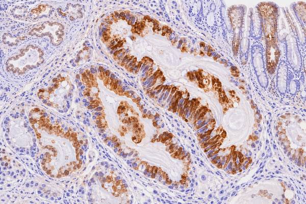
MUC5AC immunohistochemistry (IHC) detects mucin 5AC, a secreted gel-forming mucin normally produced by gastric foveolar cells and other mucus-secreting epithelia. In pathology, MUC5AC is useful as a marker of gastric-type and mucinous differentiation and is commonly expressed in pancreatic ductal adenocarcinoma, pancreatic mucinous precursor lesions, gastric adenocarcinoma, esophageal adenocarcinoma, and several other mucin-producing tumors. A positive stain can support a diagnostic pattern, but it does not prove that a tumor started in the stomach or pancreas, and it does not by itself prove malignancy. MUC5AC is also present in normal gastric mucosa and can appear in benign or premalignant lesions. Results vary with tumor subtype, antibody clone, glycosylation state, and whether staining is apical, cytoplasmic, or extracellular. Pathologists therefore interpret MUC5AC with morphology and a panel that may include MUC1, MUC2, MUC6, CK7, CK20, CDX2, SATB2, PAX8, and organ-specific markers.
- MUC5AC is a gastric-type secreted mucin: positive staining supports gastric/foveolar or mucinous differentiation rather than one specific cancer.
- Pancreatic ductal adenocarcinoma frequently expresses MUC5AC, while normal pancreatic parenchyma has little or no expression with many assays.
- Pancreatic IPMNs and other mucinous precursor lesions commonly express MUC5AC, so positivity does not distinguish benign or noninvasive lesions from invasive cancer.
- Gastric mucosa normally expresses MUC5AC: loss, retention, or redistribution in gastric cancer depends on histologic phenotype and tumor progression.
- No special patient preparation is required: MUC5AC IHC is performed on biopsy or surgical tissue already collected for pathology.
Table of Contents
- What MUC5AC IHC Detects
- What Positive MUC5AC Staining Means
- MUC5AC in Pancreatic Tumors
- MUC5AC in Gastric and Upper GI Tumors
- MUC5AC in Other Mucinous Tumors
- Staining Patterns, Assays, and Pitfalls
- How to Read a MUC5AC Pathology Result
What MUC5AC IHC Detects
MUC5AC IHC detects a secreted mucin associated with gastric foveolar and other mucus-producing epithelial differentiation. MUC5AC is one of several mucin proteins that help form protective mucus layers. In the normal stomach, it is strongly expressed by surface and foveolar epithelium, where it contributes to the mucus barrier over the gastric lining.
Tumors can retain this normal gastric program or activate it in organs where MUC5AC is not normally prominent. That makes the marker useful for recognizing a gastric-type phenotype rather than identifying a single anatomic origin. Pancreatic neoplasms are a major example: MUC5AC appears early in several pancreatic precursor lesions and is common in pancreatic ductal adenocarcinoma even though normal pancreatic ducts and acini generally show little expression with standard diagnostic assays.
The stain usually appears in the cytoplasm, along the apical/luminal surface, or within secreted extracellular mucin. Different antibodies may recognize immature versus mature or differently glycosylated forms of MUC5AC, so localization and intensity can vary by assay.
MUC5AC is not a blood tumor marker in routine practice. Research has explored circulating MUC5AC and MUC5AC-targeted imaging, particularly in pancreatic cancer, but tissue IHC is a separate test performed on fixed tumor material.
The marker also does not replace histologic classification. A “mucinous” tumor is defined according to organ-specific microscopic criteria. MUC5AC positivity can support the phenotype but cannot create a mucinous diagnosis when the required architecture or extracellular mucin is absent.
What Positive MUC5AC Staining Means
A positive MUC5AC stain means the cells or mucin being evaluated contain detectable MUC5AC protein. The result suggests gastric-type or mucus-secreting differentiation, but its diagnostic weight depends on where the tumor is located and what other markers show.
In a pancreatic mass, MUC5AC positivity is compatible with pancreatic ductal adenocarcinoma, IPMN, pancreatic intraepithelial neoplasia, mucinous cystic neoplasm, and other mucin-producing lesions. It therefore does not separate invasive cancer from a precursor lesion by itself. In a gastric biopsy, positivity may simply reflect retained gastric differentiation because normal foveolar epithelium is also positive.
A positive result does not independently establish:
- malignant versus benign behavior;
- stomach versus pancreas as the primary site;
- tumor stage or grade;
- a specific molecular alteration;
- response to chemotherapy;
- eligibility for a MUC5AC-targeted drug or imaging agent.
Negative staining also needs caution. Gastric cancers may lose gastric mucins as they become less differentiated, and pancreatic tumors can be heterogeneous. A small biopsy can miss a positive area. Technical factors, tissue fixation, and the antibody clone may also affect the result.
The most useful question is therefore not “Is MUC5AC positive?” but “Does the MUC5AC pattern fit the morphologic and immunophenotypic diagnosis being considered?”
MUC5AC in Pancreatic Tumors
MUC5AC is one of the most frequently expressed mucins in pancreatic ductal neoplasia and can be present from precursor stages through invasive pancreatic ductal adenocarcinoma. This broad expression makes it biologically interesting and diagnostically supportive, but not a stand-alone cancer discriminator.
Large tissue-series studies have found MUC5AC in a substantial proportion of pancreatic ductal adenocarcinomas. Some reviews report expression in roughly two-thirds to 70% of cases, although exact rates vary with antibody and scoring method. Importantly, normal pancreatic tissue generally lacks the strong tumor-type expression seen in ductal neoplasia.
Pancreatic intraepithelial neoplasia can begin expressing MUC5AC early. IPMNs also commonly express the marker. Gastric-type IPMN is characteristically MUC5AC positive, while intestinal-type IPMN often coexpresses MUC5AC but is better defined by MUC2 and CDX2. Pancreatobiliary-type lesions may express MUC1 and MUC5AC. This overlap shows why a mucin panel works better than a single stain.
Pancreatic ductal adenocarcinoma
In pancreatic ductal adenocarcinoma, MUC5AC may show apical, luminal, cytoplasmic, or extracellular staining. Researchers have investigated whether the amount or location predicts outcome or drug response, but results have not been consistent enough to create a universal clinical scoring rule.
A major reason is assay biology. Antibodies such as CLH2 and 45M1 can recognize different maturation or glycosylation states of MUC5AC. One study may measure a perinuclear immature form while another emphasizes mature apical or secreted mucin. Percentage cutoffs have also varied. Therefore, a finding such as “high MUC5AC predicts poor prognosis” cannot be applied safely without knowing which antibody, localization, cutoff, and patient group were studied.
MUC5AC is also being explored as a tumor target because pancreatic cancers can express forms that are much more abundant than in normal pancreas. These experimental uses include serum detection, molecular imaging, and therapeutic targeting. A routine diagnostic MUC5AC stain, however, is not automatically equivalent to a validated companion diagnostic for those approaches.
Pancreatic cystic and intraductal lesions
MUC5AC is frequently positive in IPMN and can also appear in mucinous cystic neoplasms. Because these lesions range from low-grade dysplasia to invasive carcinoma, MUC5AC positivity cannot determine grade. The pathologist evaluates epithelial atypia, architecture, invasion, cyst wall features, and other markers.
This is an important practical pitfall. If a small pancreatic biopsy or cyst specimen is MUC5AC positive, the finding may confirm mucinous epithelial differentiation but does not answer whether an invasive carcinoma is present. Imaging, cytology, histology, molecular findings, and the clinical setting may all be needed.
MUC5AC in Gastric and Upper GI Tumors
In the stomach, MUC5AC is a normal gastric mucin, so its diagnostic value comes from the pattern of retention, loss, or aberrant expression rather than simple positivity. Normal surface and foveolar cells provide a built-in reference for what a gastric phenotype looks like.
Gastric adenocarcinomas are heterogeneous. Some retain MUC5AC strongly, especially tumors with gastric or foveolar differentiation, while others lose expression as they acquire intestinal or poorly differentiated phenotypes. Studies have linked reduced MUC5AC with adverse clinicopathologic features in some cohorts, but published results vary.
Intestinal metaplasia illustrates the phenotypic shift. Complete intestinal metaplasia tends to express intestinal markers such as MUC2, while incomplete forms may retain a mixture of gastric and intestinal mucins, including MUC5AC. Gastric dysplasia and carcinoma can likewise show mixed profiles.
MUC5AC is also expressed in a substantial proportion of esophageal adenocarcinomas. Barrett-related neoplasia develops in a background of metaplastic glandular mucosa and can show gastric and intestinal mucin programs. MUC5AC can contribute to phenotyping, but it is not specific enough to distinguish gastric from esophageal origin in a tumor spanning the gastroesophageal junction.
In upper-GI pathology, MUC5AC is therefore best viewed as one component of a differentiation profile. The diagnosis still depends on anatomic location, morphology, endoscopic findings, and broader IHC when the primary site is uncertain.
MUC5AC in Other Mucinous Tumors
MUC5AC can be positive well beyond the stomach and pancreas, especially in tumors with mucinous or glandular differentiation. A broad tumor survey found expression across many entities, with notable positivity in esophageal adenocarcinoma, ovarian mucinous carcinoma, ampullary adenocarcinoma, gastric adenocarcinoma, lung adenocarcinoma subtypes, and colorectal lesions.
In colorectal pathology, normal colon does not use MUC5AC as its dominant mucin, but adenomas, serrated lesions, and carcinomas can acquire expression. MUC5AC is particularly associated with some serrated and mucinous pathways. Therefore, positivity in a metastatic adenocarcinoma does not exclude colorectal origin.
Ovarian mucinous tumors may also express MUC5AC, often reflecting gastric or intestinal-type mucinous differentiation. When the differential includes metastatic GI adenocarcinoma, the pathologist needs a broader panel including PAX8, SATB2, CDX2, CK7, and CK20 rather than relying on MUC5AC.
Some lung adenocarcinomas, especially mucinous forms, can express MUC5AC. This is another reason MUC5AC cannot serve as a pancreas-specific or stomach-specific marker in metastatic disease.
| Setting | Typical role of MUC5AC | Main caution |
|---|---|---|
| Pancreatic ductal neoplasia | Supports ductal/mucinous phenotype | Precursor lesions and invasive cancer can both be positive |
| Gastric tumors | Shows gastric/foveolar differentiation | Normal gastric epithelium is also positive |
| Ovarian mucinous tumors | Supports mucinous phenotype | Does not distinguish primary ovarian from metastatic GI tumor alone |
| Colorectal lesions | May mark serrated or mucinous differentiation | Not a standard colorectal lineage marker |
| Lung adenocarcinoma | May occur in mucinous/glandular tumors | Can mimic GI or pancreatobiliary mucin profiles |
Staining Patterns, Assays, and Pitfalls
MUC5AC interpretation depends heavily on where the stain appears and which antibody was used. A report that simply says “positive” may omit information that matters in research or difficult diagnostic cases.
Mature secreted MUC5AC is often apical or extracellular, while less mature forms can show more cytoplasmic or perinuclear staining. Antibodies differ in which forms they detect. Consequently, two studies using the same tumor type can report different positivity rates or opposite prognostic associations without necessarily contradicting each other biologically.
Common pitfalls include:
- normal gastric positivity: benign foveolar epithelium can be strongly positive;
- secreted mucin: extracellular mucin can stain even when viable tumor cells are sparse;
- tumor heterogeneity: small biopsies may underrepresent expression;
- clone-specific staining: mature and immature glycoforms are not interchangeable;
- no universal cutoff: routine pathology has no single percentage that defines clinically meaningful MUC5AC positivity across tumors;
- site overinterpretation: pancreatic, gastric, ovarian, lung, and colorectal tumors can overlap in expression;
- prognostic overreach: research associations are not equivalent to validated individual-risk tools.
Tissue handling can also affect results. Poor fixation, decalcification, necrosis, and scant tumor reduce confidence in a negative stain. Internal positive controls are helpful when normal MUC5AC-expressing epithelium is present.
A related issue is how much weight to give a focal stain. A tiny cluster of positive glands may be biologically real, especially in a heterogeneous tumor, but it is less persuasive for site assignment than broad staining that matches the expected morphology. Conversely, diffuse positivity is still not specific if several competing tumors commonly express MUC5AC. Pathologists therefore avoid turning intensity into a shortcut for origin. They also distinguish true intracellular or luminal staining from nonspecific background in necrotic debris, thick mucus, or damaged tissue. When a result is clinically important and the internal controls are weak, repeating the stain on another block can be more useful than interpreting a technically marginal slide.
When a MUC5AC result conflicts with morphology, the solution is usually to broaden the diagnostic view rather than repeat the same conclusion. The pathologist may review deeper sections, add organ-specific markers, compare prior material, or request molecular testing if the primary site remains uncertain.
How to Read a MUC5AC Pathology Result
Read MUC5AC as a phenotype marker: it tells you that a tumor is producing a gastric-type mucin, not where the tumor definitely began. The final diagnosis and the full IHC panel remain more important than the individual stain.
A practical approach is to ask:
- What is the specimen site and proposed diagnosis? Pancreatic, gastric, ovarian, lung, and metastatic tumors have different competing explanations.
- Is staining in tumor cells, extracellular mucin, or both? Tumor-cell localization is often more diagnostically informative.
- Is it apical, cytoplasmic, diffuse, or focal? The pattern may reflect differentiation and assay type.
- Which antibody clone was used? This matters most if the result is being compared with a research study or therapeutic assay.
- What do MUC1, MUC2, MUC6 and site-specific markers show? A coherent panel is more reliable than one mucin result.
- Does the result change management? Most routine MUC5AC stains support diagnosis rather than directly selecting therapy.
A pancreatic lesion that is MUC5AC positive may fit IPMN, PanIN, or ductal adenocarcinoma depending on the architecture and evidence of invasion. A gastric cancer that has lost MUC5AC may simply have shifted away from a foveolar phenotype. An ovarian mucinous tumor that is strongly positive still requires assessment for a primary ovarian versus metastatic GI source.
The essential takeaway is that MUC5AC is a widely useful marker of gastric-type and mucinous differentiation with particular relevance to pancreatic and gastric pathology. Its greatest strength is phenotyping; its greatest weakness is lack of disease and organ specificity. Correct interpretation combines staining location, antibody characteristics, tissue morphology, and a focused IHC panel.
In practice, that means the stain is most informative when it answers a narrow pathology question rather than when it is used as a broad general cancer screening tool.
Normal and metaplastic tissues provide important context for MUC5AC. Gastric foveolar epithelium is a familiar physiologic source of the protein and can serve as an internal positive control when present on the slide. Gastric-type metaplasia in other organs may also express MUC5AC. As a result, positivity is not synonymous with malignancy; it describes a mucin phenotype that can occur in both benign and neoplastic epithelium.
This is especially relevant in pancreatic pathology. Many pancreatic precursor lesions and mucinous neoplasms express MUC5AC, so the stain can support mucinous or gastric-type differentiation but cannot by itself distinguish a low-grade precursor from invasive carcinoma. The diagnosis of invasion depends on architecture, cytologic features, stromal response, and the relationship of abnormal glands to the surrounding tissue. MUC1, MUC2, MUC6, CDX2, and other markers may then help refine the epithelial subtype when that distinction is clinically useful.
Technical differences can influence results as well. Antibody clone, fixation, antigen retrieval, and scoring method may change the intensity or percentage reported. There is no single universal MUC5AC cutoff that applies to every tumor type and every clinical question. This is why prognostic studies that divide tumors into “high” and “low” expression groups may not translate directly to a routine pathology report. For an individual patient, the key issue is whether MUC5AC adds a coherent piece of evidence to the morphology and the broader organ-specific panel, not whether the stain exceeds a research threshold.
References
- MUC5AC Expression in Various Tumor Types and Nonneoplastic Tissue: A Tissue Microarray Study on 10 399 Tissue Samples 2021
- Understanding the Clinical Impact of MUC5AC Expression on Pancreatic Ductal Adenocarcinoma 2021 (Review)
- Predictive Value of MUC5AC Signature in Pancreatic Ductal Adenocarcinoma: A Hypothesis Based on Preclinical Evidence 2023 (Review)
- The Role of Mucin Expression in the Diagnosis of Oesophago-Gastric Cancer: A Systematic Literature Review 2023 (Systematic Review)
- Prognostic and clinicopathological significance of mucin family members expression in gastric cancer: a meta-analysis 2024 (Systematic Review)
- The Role of Mucins in Cancer and Cancer Progression: A Comprehensive Review 2025 (Review)
Disclaimer
MUC5AC IHC must be interpreted by a qualified pathologist together with morphology, specimen site, assay method, and other immunostains. A positive or negative MUC5AC result alone cannot diagnose cancer, establish the primary organ, determine stage, or predict an individual treatment response. This article is for general education and does not replace individualized medical or pathology advice.





