
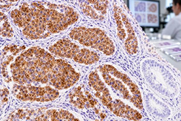
Chromogranin immunohistochemistry (IHC) detects proteins found in neurosecretory granules and is one of the classic tissue markers used to confirm neuroendocrine differentiation. Pathologists most often interpret chromogranin A staining together with morphology and other markers such as synaptophysin and INSM1. Strong granular cytoplasmic staining is common in well-differentiated neuroendocrine tumors, while poorly differentiated neuroendocrine carcinomas may show weaker, focal, or even absent chromogranin despite being genuinely neuroendocrine. A positive result does not identify the primary organ by itself, and chromogranin expression can occur in several normal neuroendocrine cell populations and in tumors from different sites. The result is therefore most useful as part of a panel that answers three separate questions: Is the tumor neuroendocrine? Is it well or poorly differentiated? And, if metastatic, where is it most likely to have started?
- Chromogranin-positive tumor cells support neuroendocrine differentiation, especially when staining is granular and matches the tumor cells.
- Strong diffuse staining is common in many well-differentiated neuroendocrine tumors, but staining intensity is not a universal grade or prognosis score.
- High-grade neuroendocrine carcinomas can be weak or negative for chromogranin, so synaptophysin and INSM1 are often used alongside it.
- Chromogranin IHC is a tissue stain and should not be confused with a blood chromogranin A level.
- A positive stain does not reveal the tumor’s primary site; organ-specific markers, imaging, and clinical findings may still be needed.
Table of Contents
- What chromogranin IHC detects
- Staining pattern and normal expression
- Chromogranin in neuroendocrine tumors
- Chromogranin and tumor differentiation
- Marker panels and primary site
- Pitfalls and limitations
- How to read the pathology report
- Practical chromogranin interpretation scenarios
What chromogranin IHC detects
Chromogranins are acidic proteins stored in dense-core secretory granules of neuroendocrine cells. Chromogranin A is the member most commonly targeted in routine IHC. Because many neuroendocrine cells package peptide hormones or amines in these granules, chromogranin can serve as a visible marker of neurosecretory differentiation.
IHC is performed on a tissue section from a biopsy, resection, or cell block. The antibody binds to chromogranin protein inside cells, producing a colored reaction that the pathologist evaluates under the microscope. The result is generally described as positive or negative, often with an assessment of intensity and distribution. There is no universal numeric cutoff that applies to every neuroendocrine tumor.
Chromogranin IHC is different from a serum chromogranin A test. Blood chromogranin A has been used in selected neuroendocrine tumor settings, but circulating levels can be affected by kidney function, proton-pump inhibitors, gastric conditions, and other factors. Tissue IHC asks whether the tumor cells show neurosecretory granules; it does not measure a circulating concentration.
Pathologists rarely use chromogranin alone. Synaptophysin IHC detects a synaptic-vesicle protein and is generally very sensitive for neuroendocrine differentiation. INSM1 is a nuclear marker that can add sensitivity, particularly in high-grade neuroendocrine carcinoma. A coherent panel is more reliable than any one stain.
Staining pattern and normal expression
Classic chromogranin staining is granular cytoplasmic because the target protein is stored in secretory granules. In a well-differentiated neuroendocrine tumor, the staining may be strong and diffuse through most tumor cells. Focal granular staining can still be meaningful, especially in a tumor with convincing neuroendocrine morphology.
Normal neuroendocrine cells also express chromogranin. Examples include enteroendocrine cells in the gastrointestinal tract, pancreatic islet cells, adrenal medullary cells, and neuroendocrine cells in the respiratory tract. These cells can serve as internal controls depending on the specimen.
The pathologist must confirm that the abnormal cells—not only scattered normal neuroendocrine cells—are staining. This is particularly relevant in mucosal biopsies, where normal endocrine cells may sit between non-neuroendocrine tumor glands. A few positive normal cells should not be misread as evidence that the carcinoma itself is neuroendocrine.
Staining quality depends on fixation, antibody clone, antigen retrieval, tissue viability, and technical controls. Necrotic or severely crushed tissue can be difficult to assess. A negative result is more credible when appropriate internal or external controls stain as expected.
Chromogranin in neuroendocrine tumors
Well-differentiated neuroendocrine tumors arise in many organs, especially the gastrointestinal tract, pancreas, and lung. They typically show organoid nests, trabeculae, rosettes, or gland-like structures and cells with finely stippled chromatin. Chromogranin and synaptophysin often confirm the neuroendocrine phenotype in these tumors.
The current WHO framework emphasizes that “neuroendocrine” describes a family of neoplasms rather than one disease. A well-differentiated neuroendocrine tumor is biologically different from a poorly differentiated small cell or large cell neuroendocrine carcinoma. Chromogranin can be positive in both, but the pattern often differs.
In gastroenteropancreatic well-differentiated neuroendocrine tumors, chromogranin is frequently strong and diffuse, although expression varies by tumor type and site. Some tumors produce specific hormones, but hormone IHC is generally used only when clinically or diagnostically relevant. Neuroendocrine phenotype is established with general markers rather than by requiring a hormone stain.
Pulmonary carcinoid tumors also usually express chromogranin and synaptophysin. Small cell lung carcinoma often expresses neuroendocrine markers but may show reduced chromogranin compared with well-differentiated tumors. This is one reason INSM1 IHC has become a valuable addition to modern panels.
Chromogranin and tumor differentiation
Differentiation describes how closely tumor cells resemble their normal cell counterpart. Well-differentiated neuroendocrine tumors retain organoid architecture and many neurosecretory features, so chromogranin staining is often robust. Poorly differentiated neuroendocrine carcinomas are more primitive and may lose some dense-core granules, resulting in weaker or patchier staining.
That relationship is useful but should not be turned into a rigid rule. A strong chromogranin stain does not automatically mean low grade, and a weak stain does not by itself mean high grade. Grading is based on site-specific criteria that can include mitotic rate, necrosis, and the Ki-67 IHC proliferation index. Morphology remains central.
| Setting | Common chromogranin pattern | Interpretive point |
|---|---|---|
| Well-differentiated NET | Often strong, diffuse, granular cytoplasmic | Supports mature neuroendocrine differentiation |
| Small cell neuroendocrine carcinoma | Can be focal or weak | Negative/weak staining does not exclude the diagnosis |
| Large cell neuroendocrine carcinoma | Variable | Use morphology plus synaptophysin/INSM1 and other markers |
| Non-neuroendocrine carcinoma with scattered positive cells | Focal isolated staining | May not justify a neuroendocrine diagnosis |
A tumor can also contain mixed components. Some carcinomas have distinct neuroendocrine and non-neuroendocrine areas. In those cases, the percentage and distribution of each component may be diagnostically important, but classification follows organ-specific criteria rather than a universal chromogranin percentage.
Marker panels and primary site
General neuroendocrine markers answer whether a tumor shows neuroendocrine differentiation. They do not reliably identify where a metastatic neuroendocrine tumor started. Once lineage is established, site-of-origin markers can be added selectively.
CDX2 may support intestinal origin in some neuroendocrine tumors, while TTF-1 can support pulmonary origin in an appropriate setting. Islet-1, PAX8-family reactivity depending on antibody clone, SATB2, serotonin, calcitonin, and other markers can contribute in selected differentials. The choice depends on tumor morphology, metastatic pattern, and imaging.
The pathologist also needs to exclude mimics. Paraganglioma can express neuroendocrine markers but differs from epithelial neuroendocrine tumors because chief cells are usually keratin negative and sustentacular cells can be highlighted by S100 or SOX10. Merkel cell carcinoma is a cutaneous neuroendocrine carcinoma with a characteristic keratin profile. Some non-neuroendocrine tumors can show focal synaptophysin or chromogranin, so the full pattern matters.
CD56 is sometimes included as a general neuroendocrine marker and can be highly sensitive, but its specificity is lower. The CD56 IHC test is also used in NK-cell and other hematologic contexts. A panel of chromogranin, synaptophysin, and/or INSM1 generally provides more reliable evidence than CD56 alone.
Pitfalls and limitations
The most important limitation is false reassurance from a negative stain. Poorly differentiated neuroendocrine carcinomas may be chromogranin negative or only focally positive. If morphology strongly suggests small cell carcinoma, the pathologist should not abandon the diagnosis until more sensitive neuroendocrine markers and epithelial markers have been assessed.
The opposite problem is overcalling neuroendocrine differentiation based on rare positive cells. Conventional adenocarcinomas in several organs may contain scattered neuroendocrine cells or focal neuroendocrine marker expression. A diagnosis of neuroendocrine carcinoma requires the right morphology and a convincing phenotype, not merely one tiny chromogranin-positive focus.
Preanalytic factors can alter staining. Delayed fixation, prolonged fixation, decalcification, necrosis, cautery, and crush artifact may reduce antigen preservation. Small biopsies can also miss heterogeneous expression. Internal controls and comparison with other neuroendocrine stains help determine whether an unexpected negative result is believable.
Another limitation is that chromogranin does not determine treatment target status. It is not a predictive biomarker in the same way as an actionable mutation or receptor score. Treatment choices depend on tumor site, differentiation, grade, stage, somatostatin receptor status when relevant, molecular features, and patient factors.
How to read the pathology report
When a report lists chromogranin, first identify what the pathologist concluded. “Tumor cells are positive for chromogranin and synaptophysin” supports neuroendocrine differentiation. “Chromogranin highlights only scattered cells” may be interpreted as focal differentiation or may be considered insufficient for a neuroendocrine classification, depending on morphology.
If the stain is negative, look for synaptophysin and INSM1 results. A high-grade tumor can still be neuroendocrine when those markers are positive and morphology is characteristic. If all neuroendocrine markers are negative, the pathologist may broaden the differential and consider non-neuroendocrine mimics.
For a confirmed neuroendocrine neoplasm, the clinically important report elements often include anatomic site, well versus poorly differentiated morphology, grade, mitotic rate, necrosis, Ki-67 index where applicable, tumor size, margins, lymphovascular invasion, nodal status, and stage. Chromogranin is primarily part of establishing phenotype rather than replacing those prognostic variables.
Patients with a metastatic neuroendocrine tumor of uncertain origin can ask whether site-specific IHC was performed and how strongly it supports a particular primary site. Imaging and endoscopic evaluation may still be required because IHC probabilities are not absolute.
Finally, terminology matters. “Chromogranin-positive carcinoma” is not synonymous with “well-differentiated neuroendocrine tumor.” The distinction between NET and NEC rests on morphology and classification criteria. A pathology report that integrates those elements is more informative than the stain result alone.
Practical chromogranin interpretation scenarios
A pancreatic mass with nested cells and finely stippled chromatin is a typical setting in which chromogranin can be highly informative. If the tumor is strongly and diffusely positive for chromogranin and synaptophysin, the immunophenotype supports a well-differentiated neuroendocrine tumor. The pathologist then shifts from proving neuroendocrine lineage to classifying grade and documenting features such as Ki-67, mitotic activity, size, invasion, and stage. Additional hormone stains are not automatically required; they are selected when a functional syndrome or a specific differential diagnosis makes them useful.
A small lung biopsy suspected of small cell carcinoma demonstrates the opposite staining pattern. The tumor may be strongly positive for synaptophysin and INSM1 but only focally positive—or even negative—for chromogranin. That does not invalidate a neuroendocrine diagnosis because poorly differentiated carcinomas may have fewer mature dense-core granules. Morphology, keratin expression, and the broader neuroendocrine panel carry more weight than requiring all classic markers to be positive.
A liver biopsy containing a metastatic well-differentiated neuroendocrine tumor creates a third question: origin. Chromogranin confirms differentiation but cannot tell whether the primary is ileal, pancreatic, pulmonary, or another site. The pathologist may add CDX2 or SATB2 for intestinal support, TTF-1 in selected pulmonary differentials, or other site-associated markers. Imaging and endoscopy remain essential because site-of-origin IHC is probabilistic rather than absolute.
Scattered chromogranin-positive cells within an otherwise conventional adenocarcinoma illustrate a common pitfall. Many epithelial tumors can contain isolated neuroendocrine cells. If the tumor lacks neuroendocrine morphology and the staining is limited to a tiny minority of cells, pathologists may describe focal neuroendocrine differentiation without reclassifying the entire tumor as a neuroendocrine carcinoma. Organ-specific classification rules determine when a mixed tumor category is appropriate.
Specimen quality influences the result. Neuroendocrine carcinomas often undergo necrosis, and small biopsies may be heavily crushed. Chromogranin can be particularly vulnerable to reduced staining in poorly preserved tissue. A more robust positive marker such as synaptophysin or a nuclear INSM1 stain may remain interpretable when chromogranin is weak. Pathologists therefore choose complementary markers rather than simply repeating the same biological signal.
The difference between tissue chromogranin IHC and serum chromogranin A is also clinically important. A blood level can be affected by non-tumor factors and, even when elevated in a patient with a known neuroendocrine tumor, it does not tell the pathologist what a biopsy looks like. Tissue IHC confirms protein expression in specific cells. Serum testing, when used, is a separate monitoring question and should not be substituted for histologic diagnosis.
Finally, chromogranin intensity should not be used as a shortcut for prognosis. Strong granular staining may reflect preserved neurosecretory differentiation, but prognosis is driven by the diagnosed entity, grade, stage, proliferative activity, molecular features, and treatment response. A pathology report that says “chromogranin strongly positive” still needs the formal tumor type and grade before the result can be translated into clinical meaning.
A related issue is the distinction between well-differentiated grade 3 neuroendocrine tumor and poorly differentiated neuroendocrine carcinoma in sites where both categories exist. Both can have a high Ki-67 index, so chromogranin positivity alone does not resolve the problem. Well-differentiated tumors usually retain the architecture and cytology of lower-grade NETs and may preserve stronger chromogranin, while NECs show overtly high-grade cytology and may have abnormal p53 or loss of RB expression in some organs. The distinction matters because the tumors differ biologically and may be treated differently.
When a pathology report is borderline or the biopsy is very small, the most useful next action is often review of the complete panel rather than focusing on one negative marker. Asking whether synaptophysin and INSM1 support the diagnosis, whether Ki-67 was assessed in a representative hotspot, and whether the morphology is well or poorly differentiated usually gives a clearer picture. If the biopsy and imaging are discordant, a repeat or larger sample may occasionally be needed to establish both lineage and grade confidently.
The amount of chromogranin can also change with tumor biology. Well-differentiated neuroendocrine tumors often retain abundant dense-core secretory granules and therefore show strong granular cytoplasmic staining. Poorly differentiated neuroendocrine carcinomas may have much less granular differentiation, so chromogranin can be weak, focal, or absent even when other neuroendocrine markers are convincing. This is why a negative stain does not end the workup when the morphology strongly suggests neuroendocrine carcinoma.
In metastatic disease, chromogranin confirms phenotype more reliably than site of origin. A positive liver metastasis, for example, may still require CDX2, SATB2, TTF-1, PAX8, or other site-directed markers chosen from the clinical differential. Imaging and endoscopic findings may be equally important. The goal is to separate two questions: “Is this tumor neuroendocrine?” and “Where did it arise?” Chromogranin mainly helps answer the first.
Chromogranin IHC should also be separated from serum chromogranin A testing. IHC is performed on fixed tissue to show neuroendocrine granule-associated protein within tumor cells; a blood chromogranin A level is a different laboratory test with different limitations and can be affected by non-tumor factors. A positive tissue stain does not predict a particular blood level, and an elevated blood level does not replace histologic diagnosis. This distinction matters because patients may see the same marker name in both a pathology report and a laboratory result and assume they measure the same thing. For tumor classification, the tissue pattern is interpreted with morphology and other IHC markers, while blood biomarkers—when used at all—are considered in a separate clinical monitoring context.
Another useful clue is internal heterogeneity. A well-differentiated neuroendocrine tumor may contain areas with stronger granular chromogranin staining and other areas with less expression, especially after treatment or in higher-grade components. Pathologists focus on viable representative tumor and do not usually interpret small intensity differences as separate diagnoses unless morphology and proliferation findings also change. When a metastasis looks more aggressive than the known primary, repeating the neuroendocrine panel and proliferation assessment on the new specimen can help determine whether the biology has evolved.
References
- Overview of the 2022 WHO Classification of Neuroendocrine Neoplasms 2022 (Review)
- Comparison of INSM1 immunostaining with established neuroendocrine markers synaptophysin and chromogranin A in over 14,000 neuroendocrine and non-neuroendocrine tumors 2024
- Small Biopsy and Cytology of Pulmonary Neuroendocrine Neoplasms: Brief Overview of Classification, Immunohistochemistry, Molecular Profiles, and World Health Organization Updates 2022 (Review)
- Update from the 5th Edition of the World Health Organization Classification of Head and Neck Tumors: Overview of the 2022 WHO Classification of Head and Neck Neuroendocrine Neoplasms. 2022 (Review)
- [Current WHO classification (2022) of neuroendocrine neoplasms] 2024 (Review)
- DOG1 expression in neuroendocrine neoplasms: Potential applications and diagnostic pitfalls 2023
Disclaimer
Chromogranin IHC is a diagnostic tissue stain and cannot classify or grade a neuroendocrine neoplasm by itself. The result should be interpreted with morphology, other immunostains, proliferation measures, imaging, and clinical findings by the treating pathology and clinical teams.





