
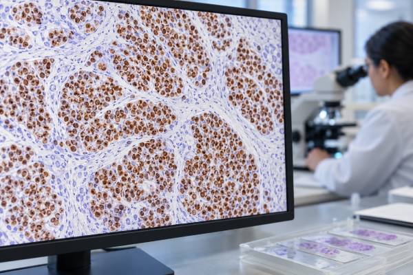
INSM1 immunohistochemistry (IHC) is a tissue stain that helps pathologists identify neuroendocrine differentiation in tumors. INSM1 is a transcription factor involved in neuroendocrine development, so a positive result appears in the nuclei of many neuroendocrine neoplasms. It is especially useful when a tumor is poorly differentiated, the biopsy is small, or older neuroendocrine markers give weak or conflicting results. INSM1 can support diagnoses such as small cell neuroendocrine carcinoma, large cell neuroendocrine carcinoma, and well-differentiated neuroendocrine tumor, but it is not completely specific. Some non-neuroendocrine tumors can express INSM1, and occasional neuroendocrine tumors are negative. Pathologists therefore interpret the stain with microscopic morphology and companion markers such as synaptophysin and chromogranin A. The result helps answer whether a tumor shows neuroendocrine lineage; it does not by itself determine the tumor’s grade, primary site, stage, prognosis, or best treatment.
- INSM1-positive staining is nuclear and usually supports neuroendocrine differentiation in a compatible tumor.
- INSM1 is often more sensitive than traditional markers in poorly differentiated neuroendocrine carcinoma, but no single stain is perfect.
- A positive result does not identify the primary organ by itself, because neuroendocrine neoplasms can arise at many sites.
- A negative INSM1 result does not completely exclude a neuroendocrine tumor, especially when morphology and other markers support the diagnosis.
- There is no blood “normal range” for INSM1 IHC; it is interpreted on tumor tissue, usually as the presence, distribution, and intensity of nuclear staining.
Table of Contents
- What INSM1 IHC Measures
- When INSM1 Is Used
- How INSM1 Staining Is Interpreted
- INSM1 With Other Neuroendocrine Markers
- INSM1 in Different Neuroendocrine Tumors
- Limitations and Diagnostic Pitfalls
- Questions After an INSM1 Result
What INSM1 IHC Measures
INSM1 stands for insulinoma-associated protein 1. It is a zinc-finger transcription factor that plays a role in the development and differentiation of neuroendocrine cells. Because it functions in the nucleus, true INSM1 immunoreactivity is interpreted as nuclear staining. That nuclear localization is one practical advantage of the marker: it can be easier to distinguish from nonspecific cytoplasmic background in small or crushed specimens.
The test is performed on tissue from a biopsy, cytology cell block, or surgical specimen. A laboratory applies an antibody against INSM1 to a thin section and develops the reaction for microscopic assessment. The pathologist evaluates whether the tumor nuclei stain and, when useful, describes the proportion and strength of staining.
INSM1 IHC is not a blood test. There is no serum reference interval, no fasting requirement, and no therapeutic or toxic range. The result depends on tumor biology and tissue quality. Fixation, crush artifact, necrosis, scant tumor, prior therapy, and technical platform can affect what is seen.
The marker is used mainly to establish neuroendocrine differentiation. Neuroendocrine cells share features of nerve and endocrine cells and can produce peptide hormones or biogenic amines, although many neuroendocrine tumors do not cause a hormone syndrome. Their tumors range from relatively indolent well-differentiated neuroendocrine tumors to aggressive poorly differentiated neuroendocrine carcinomas.
A positive INSM1 result therefore answers a lineage question rather than a staging question. It can strengthen the case that an undifferentiated tumor belongs to the neuroendocrine family, but it does not tell whether disease has spread, whether a tumor is surgically removable, or what drug will work. Those decisions require the complete diagnosis and clinical workup.
When INSM1 Is Used
INSM1 is useful when routine histology suggests neuroendocrine differentiation or when a poorly differentiated tumor needs lineage classification. Small cell morphology is a common trigger: tightly packed cells, scant cytoplasm, nuclear molding, brisk mitotic activity, and necrosis can suggest small cell neuroendocrine carcinoma, but similar appearances occur in lymphoma, some sarcomas, and other poorly differentiated cancers.
Pathologists may use INSM1 in:
- suspected small cell carcinoma of the lung or an extrapulmonary site;
- suspected large cell neuroendocrine carcinoma;
- well-differentiated neuroendocrine tumors of the gastrointestinal tract, pancreas, lung, or other organs;
- metastatic tumors in which neuroendocrine lineage is possible;
- small biopsies or cell blocks where conventional markers are equivocal;
- unusual tumors that show partial neuroendocrine morphology or immunophenotype.
The stain can be particularly helpful when synaptophysin or chromogranin A is weak. Poorly differentiated neuroendocrine carcinomas sometimes lose the dense secretory granules that make chromogranin strongly positive. INSM1 reflects a nuclear differentiation program, so it may remain detectable in some tumors with limited chromogranin expression.
INSM1 can also reduce ambiguity created by broad markers. Synaptophysin is sensitive but can show focal expression in some non-neuroendocrine cancers. A coherent pattern of INSM1 nuclear staining, neuroendocrine morphology, and companion-marker positivity provides stronger evidence than one isolated result.
Tumor location still matters. A positive INSM1 stain in a lung mass supports neuroendocrine differentiation but does not by itself prove the tumor began in the lung. A metastasis from a gastrointestinal, pancreatic, or other neuroendocrine primary can share the same lineage markers. Organ-of-origin markers, imaging, endoscopy, and clinical history may be needed to determine the primary site.
For this reason, INSM1 often sits inside a broader tumor IHC panel rather than functioning as a solitary diagnostic test.
How INSM1 Staining Is Interpreted
The expected INSM1 pattern is nuclear. Pathologists look for convincing staining inside the nuclei of tumor cells and compare it with internal and external controls. Cytoplasmic color without nuclear labeling is not the characteristic positive pattern.
There is no universal percentage cutoff that defines every neuroendocrine neoplasm as INSM1-positive. Studies and laboratories may use different thresholds depending on tumor type, antibody clone, platform, and research purpose. In routine diagnosis, pathologists usually interpret staining qualitatively in combination with morphology and other IHC results.
| Finding | Typical interpretation | Important limitation |
|---|---|---|
| Strong, diffuse nuclear staining | Strong support for neuroendocrine differentiation in a compatible tumor | Does not identify one specific neuroendocrine tumor or primary site |
| Patchy or focal nuclear staining | May support neuroendocrine differentiation | Needs greater caution because non-neuroendocrine tumors can show limited expression |
| No nuclear staining | Reduces support from INSM1 | Does not fully exclude a neuroendocrine neoplasm |
| Only cytoplasmic/background staining | Not the expected diagnostic pattern | Should not be counted as true INSM1 positivity |
The percentage of positive cells can add context, but a larger number is not automatically a worse prognosis. INSM1 is primarily a diagnostic lineage marker. Tumor grade and behavior are determined by the classification system for the organ and tumor type, often using mitotic rate, necrosis, morphology, and proliferation measures such as Ki-67.
Internal controls and specimen quality matter. A tiny biopsy with extensive crush artifact may distort nuclear detail. If the immunophenotype is incomplete, the pathologist may repeat the stain on another block or interpret it with more weight on well-preserved areas. A negative result in damaged tissue should not be treated as stronger evidence than it deserves.
Reports may use wording such as “positive,” “diffusely positive,” or “supports neuroendocrine differentiation.” That wording reflects evidence for lineage. It is the final integrated diagnosis, not the INSM1 phrase by itself, that should guide clinical decisions.
INSM1 With Other Neuroendocrine Markers
INSM1 complements rather than universally replaces established neuroendocrine markers. The most common companions are synaptophysin and chromogranin A. Each marker detects a different aspect of neuroendocrine differentiation, so the pattern across them can be more reliable than one stain alone.
Synaptophysin IHC detects a protein associated with neurosecretory vesicles and is often highly sensitive. It commonly shows diffuse cytoplasmic staining in neuroendocrine tumors, including many poorly differentiated carcinomas. However, focal synaptophysin can occur in some non-neuroendocrine malignancies, so morphology remains important.
Chromogranin A is associated with dense-core secretory granules. Well-differentiated neuroendocrine tumors often express it strongly, while poorly differentiated neuroendocrine carcinoma may show weaker or more focal expression. That is one setting where INSM1 can add diagnostic confidence.
| Marker | Expected staining | Typical role |
|---|---|---|
| INSM1 | Nuclear | Neuroendocrine lineage marker with strong performance across many neuroendocrine neoplasms |
| Synaptophysin | Cytoplasmic/membranous granular | Sensitive conventional neuroendocrine marker |
| Chromogranin A | Cytoplasmic granular | Supports neurosecretory granule differentiation, often strong in well-differentiated tumors |
| Ki-67 | Nuclear | Measures proliferation for grading in selected neuroendocrine tumor systems rather than lineage |
The panel may also include cytokeratins to establish epithelial differentiation in a carcinoma, TTF-1 in selected pulmonary differentials, CDX2 or other site-associated markers, and markers used to exclude lymphoma or sarcoma. In a pancreatic lesion, hormones such as insulin or glucagon may be tested when a functioning tumor is clinically suspected, but hormone expression and neuroendocrine lineage are separate questions.
Blood tests also answer different questions. A serum chromogranin A test may be affected by medications and other conditions and is not interchangeable with tissue IHC. Tissue INSM1 helps classify the cells present in a specimen; it is not a monitoring level that rises and falls in a standardized way with tumor burden.
INSM1 in Different Neuroendocrine Tumors
INSM1 is expressed in many well-differentiated and poorly differentiated neuroendocrine neoplasms, but the clinical meaning depends on tumor type. A well-differentiated neuroendocrine tumor and a small cell neuroendocrine carcinoma can both be INSM1-positive while having very different behavior, grading rules, and treatment.
In well-differentiated neuroendocrine tumors of the gastrointestinal tract and pancreas, INSM1 commonly supports the neuroendocrine phenotype along with synaptophysin and chromogranin. Grading then relies on organ-specific criteria, often incorporating mitotic count and Ki-67 proliferation index. The INSM1 intensity itself is not the grade.
In small cell carcinoma, INSM1 can be highly useful because the cells may be crushed and the classic granular markers can be variable. The diagnosis still rests on characteristic morphology and an appropriate immunophenotype. Small cell carcinoma can arise in the lung and at extrapulmonary sites, so a positive INSM1 result establishes neuroendocrine differentiation more readily than it establishes anatomic origin.
Large cell neuroendocrine carcinoma is another aggressive, poorly differentiated category in which INSM1 can support lineage. Morphology distinguishes it from small cell carcinoma, and site-specific classification criteria matter. A pathologist may need to separate it from a poorly differentiated non-neuroendocrine carcinoma that happens to show one neuroendocrine marker.
INSM1 is also useful in neuroendocrine neoplasms from less common sites, but performance is not identical across every entity. Merkel cell carcinoma, medullary thyroid carcinoma, pheochromocytoma/paraganglioma, and other tumors can participate in neuroendocrine marker differentials, although the optimal panel differs by site and morphology.
Because expression is broad across the neuroendocrine family, INSM1 usually does not solve a carcinoma-of-unknown-primary problem on its own. Once neuroendocrine differentiation is established, the next questions are whether the tumor is well or poorly differentiated, what grade it has, where it most likely began, and which molecular or predictive biomarkers are needed for treatment.
Limitations and Diagnostic Pitfalls
INSM1 is a powerful marker, but its growing use can create overconfidence. The most important limitation is incomplete specificity. Some tumors outside the conventional neuroendocrine category can express INSM1, especially focally. A pathologist should be cautious when only a small subset of cells stains and the morphology does not otherwise look neuroendocrine.
Important pitfalls include:
- Treating any nuclear positivity as diagnostic. The strength and distribution of staining should fit the tumor and the rest of the panel.
- Ignoring morphology. A marker cannot compensate for a fundamentally incompatible microscopic pattern.
- Using INSM1 to assign primary site. Neuroendocrine tumors from different organs can share the same INSM1-positive phenotype.
- Equating positivity with high grade. Both well-differentiated and poorly differentiated tumors may be positive.
- Ruling out neuroendocrine disease after one negative stain. Sensitivity is high in many settings but not 100%.
- Confusing lineage with secretion. INSM1 positivity does not mean a patient has a hormone-producing syndrome.
- Overinterpreting damaged tissue. Crush artifact and necrosis can complicate nuclear assessment.
Another challenge is “neuroendocrine marker expression” in otherwise non-neuroendocrine cancers. Some carcinomas can show focal synaptophysin or INSM1 without meeting criteria for a neuroendocrine neoplasm. Classification depends on the degree of morphologic neuroendocrine differentiation, marker pattern, organ-specific definitions, and sometimes molecular features.
Tumor heterogeneity can also matter. A mixed neoplasm may contain a neuroendocrine component and a non-neuroendocrine component. Staining only one small area can miss or overrepresent a component. The pathologist may compare multiple blocks and correlate IHC with distinct morphologic regions.
Finally, diagnostic terminology evolves. Classification systems increasingly integrate morphology, proliferation, molecular biology, and site-specific criteria. INSM1 is an important tool within that framework, not a replacement for it. Difficult cases may benefit from subspecialty pathology review, especially when classification will substantially change chemotherapy, surgery, or eligibility for targeted approaches.
Questions After an INSM1 Result
When a report says a tumor is INSM1-positive, the next step is to understand what diagnosis the stain supports. Helpful questions include:
- Was INSM1 staining diffuse or focal, and was it clearly nuclear?
- Did the tumor also express synaptophysin, chromogranin A, or other neuroendocrine markers?
- Is the final diagnosis a well-differentiated neuroendocrine tumor or a poorly differentiated neuroendocrine carcinoma?
- If it is a neuroendocrine tumor, what is the grade and how was the grade determined?
- Does the IHC panel suggest a likely primary organ, or is the primary site still uncertain?
- Is Ki-67 reported, and is it relevant to grading for this particular tumor type and site?
- Are additional imaging, molecular testing, or hormone-related studies needed?
A patient may also see several different test names in the same report. INSM1, synaptophysin, and chromogranin are primarily lineage markers. Ki-67 measures proliferation. Organ-associated markers help with tumor origin. Molecular tests can identify genomic changes with diagnostic or treatment implications. These results should not be expected to answer the same question.
If the biopsy is very small, a report may appropriately use cautious language such as “supports neuroendocrine carcinoma” and recommend additional sampling or correlation. Small specimens can establish a clinically useful diagnosis while still leaving uncertainty about exact subtype or origin.
For treatment discussions, focus on the integrated pathology diagnosis, grade, stage, primary site, and any predictive biomarkers rather than the INSM1 percentage alone. INSM1 is valuable because it helps the pathology team identify neuroendocrine differentiation accurately; it is not usually a stand-alone prognostic score or routine treatment target.
INSM1 is also useful because its nuclear pattern can preserve interpretive clarity when cytoplasm is scant. This is common in small cell carcinoma, where crushed cells may have only a thin rim of cytoplasm and cytoplasmic markers can be difficult to assess. Even so, severe crush artifact can distort nuclei too, so a strong stain cannot rescue a specimen that lacks recognizable viable tumor. The pathologist may need deeper sections, another block, or repeat sampling.
A positive INSM1 result should not be confused with a positive functional-hormone test. Many neuroendocrine neoplasms are nonfunctioning and cause symptoms from mass effect or spread rather than hormone excess. Conversely, a patient’s biochemical syndrome may require blood or urine hormone testing that is not answered by INSM1. Tissue phenotype, hormone secretion, grade, and stage are separate dimensions of the disease and may each require different tests.
References
- Comparison of INSM1 immunostaining with established neuroendocrine markers synaptophysin and chromogranin A in over 14,000 neuroendocrine and non-neuroendocrine tumors 2024
- INSM1 expression in neuroendocrine tumors in a tertiary care hospital 2024
- Evaluation of immunohistochemical expression of novel neuroendocrine marker INSM1 and histological tumor growth pattern in well-differentiated neuroendocrine tumors of the appendix: 15-year single tertiary center experience 2024
- INSM1, a Novel Biomarker for Detection of Neuroendocrine Neoplasms: Cytopathologists’ View 2022 (Review)
- Immunohistochemistry in the diagnosis and classification of neuroendocrine neoplasms: what can brown do for you? 2020 (Review)
Disclaimer
INSM1 IHC is a diagnostic pathology test and should be interpreted with morphology, other immunostains, and clinical findings. A positive or negative result alone cannot determine a neuroendocrine tumor’s grade, primary site, stage, prognosis, or treatment. Discuss the integrated pathology report with your treating team.





