
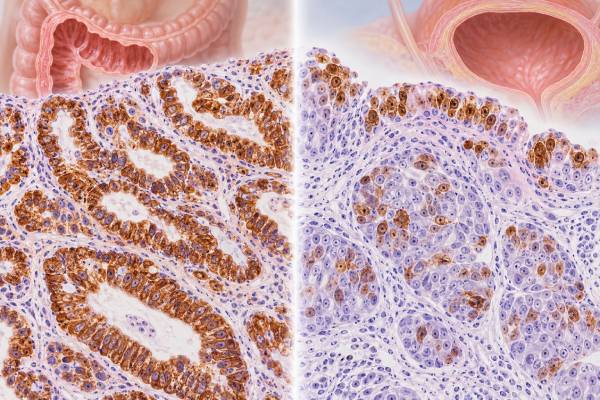
Cytokeratin 20 (CK20) immunohistochemistry (IHC) detects keratin 20, an intermediate-filament protein normally expressed by selected epithelial cells, especially in the lower gastrointestinal tract and urothelium. Pathologists use CK20 most often as part of a panel to help determine the origin of a carcinoma, particularly when distinguishing colorectal, urothelial, gastric, pancreaticobiliary, ovarian mucinous, Merkel cell, and other tumors. The result is not interpreted alone. A classic colorectal adenocarcinoma often shows diffuse CK20 positivity together with CDX2 and SATB2 and is frequently CK7 negative, while urothelial carcinoma can be CK20 positive but usually requires markers such as GATA3, p63/p40, or uroplakin for stronger site support. Some tumors show unexpected patterns, and a metastatic cancer can lose or gain staining. CK20 is therefore best used as a probability-shifting marker: it narrows the differential diagnosis but does not by itself identify the primary organ.
- CK20-positive staining often supports colorectal, urothelial, Merkel cell, or other selected epithelial differentiation, depending on morphology and the rest of the panel.
- A CK7-negative/CK20-positive pattern strongly raises colorectal origin but is not absolutely specific.
- Urothelial carcinoma may be CK20 positive, especially in more differentiated areas, but CK20 alone cannot confirm bladder origin.
- Normal colorectal epithelium and superficial urothelial cells can be CK20 positive, so normal tissue provides important context and controls.
- There is no universal CK20 percentage cutoff for tumor origin; distribution, intensity, morphology, and companion markers matter.
Table of Contents
- What CK20 IHC detects
- Normal expression and staining pattern
- CK20 in colorectal and GI cancers
- CK20 in bladder and urothelial cancer
- Using CK7 and CK20 together
- Other CK20-positive tumors and pitfalls
- How to read the report and next steps
- Practical CK20 scenarios and diagnostic decisions
What CK20 IHC detects
CK20 is a type I intermediate-filament protein encoded by KRT20. Cytokeratins form part of the structural framework of epithelial cells, and different epithelial tissues express characteristic combinations. Pathologists use those patterns as clues to lineage and site of origin.
IHC applies an antibody to a formalin-fixed tissue section. A true tumor-cell reaction is cytoplasmic, often with membranous accentuation because keratin filaments extend through the cytoplasm. The pathologist records whether the malignant cells stain and whether the distribution is diffuse, patchy, or focal.
CK20 is not a mutation test and does not measure a blood tumor marker. It is a tissue-expression test. A positive result means the tumor cells contain detectable keratin 20 protein. That can be highly useful, but several unrelated epithelial tumors share this phenotype.
The most powerful use of CK20 is combinatorial. When a metastatic adenocarcinoma has no obvious primary site, the CK7/CK20 profile can guide the next set of organ-specific markers. Newer evidence also shows why CK7 and CK20 should not be overused: site-specific markers often provide more direct information, and many tumors do not follow textbook patterns perfectly.
Normal expression and staining pattern
Normal colonic and rectal epithelium commonly expresses CK20, often with strong cytoplasmic staining. Gastric and small-intestinal epithelium can show variable expression depending on cell type and region. Urothelium usually shows CK20 in superficial umbrella cells, with limited or absent staining in deeper layers under normal conditions.
That normal urothelial pattern can be diagnostically useful. In reactive or non-neoplastic urothelium, CK20 restricted to the superficial layer is expected. More diffuse full-thickness expression can occur in urothelial dysplasia or carcinoma in situ, although diagnosis must also rely on morphology and other markers because inflammation and technical factors can complicate interpretation.
Merkel cells in skin are CK20 positive and typically show a characteristic perinuclear dot-like pattern. Merkel cell carcinoma often preserves this dot-like CK20 pattern, which becomes useful when distinguishing it from metastatic small cell carcinoma of lung, especially when combined with TTF-1 and other markers.
Normal internal controls matter. If adjacent expected epithelium stains appropriately, a negative tumor result is more trustworthy. If the internal controls fail, technical issues such as fixation or antigen retrieval may need to be considered.
CK20 in colorectal and GI cancers
Colorectal adenocarcinoma is the classic CK20-positive carcinoma. Many conventional colorectal cancers show diffuse CK20 expression, often together with strong nuclear CDX2 and SATB2. The traditional profile is CK7 negative and CK20 positive, but exceptions occur, particularly in poorly differentiated, mucinous, medullary, microsatellite-unstable, and BRAF-mutated tumors.
For this reason, current practice increasingly combines CK20 with more site-specific markers. CDX2 IHC supports intestinal differentiation, while SATB2 IHC adds specificity for lower gastrointestinal origin. A metastatic adenocarcinoma that is CK20+/CDX2+/SATB2+ and CK7 negative strongly favors colorectal or appendiceal origin in the right clinical setting.
CK20 can also be positive in gastric, pancreatic, and biliary adenocarcinomas, but the patterns are more variable. Gastric cancers may be CK7+/CK20+, CK7+/CK20−, or other combinations. Pancreaticobiliary adenocarcinomas are more often CK7 positive and may have variable CK20. Therefore, CK20 positivity does not automatically mean colon.
Mucinous tumors create another important overlap. Primary ovarian mucinous tumors can express CK20, often together with CK7, while metastases from colorectal or appendiceal primaries can involve the ovary and mimic a primary ovarian tumor. Pathologists integrate tumor laterality, size, growth pattern, CK7/CK20, SATB2, CDX2, PAX8, and clinical imaging to resolve that differential.
CK20 in bladder and urothelial cancer
Urothelial carcinoma frequently expresses CK20, but expression varies by grade, differentiation, and tumor area. In noninvasive urothelial lesions, the distribution of CK20 can be informative because normal urothelium typically restricts expression to umbrella cells, whereas dysplastic or carcinoma-in-situ epithelium may show abnormal extension into deeper layers.
In invasive urothelial carcinoma, CK20 can be diffuse, focal, or absent. Therefore, a negative result does not rule out urothelial origin. More specific urothelial markers include GATA3, uroplakin II or III, and p63/p40 in an appropriate panel. The GATA3 IHC test is sensitive for many urothelial carcinomas but is also expressed in breast and other tumors, so no single marker is definitive.
The CK7/CK20 combination can help because urothelial carcinoma is often CK7 positive and may be CK20 positive, while conventional colorectal adenocarcinoma is often CK7 negative and CK20 positive. When a pelvic or retroperitoneal metastasis is CK20 positive, CK7 and more specific markers can meaningfully narrow the likely origin.
Histologic variants complicate the picture. Micropapillary, plasmacytoid, squamous, glandular, and other variants of urothelial carcinoma may alter the expected immunophenotype. A pathologist gives the morphology and clinical location more weight than a single unexpected stain.
Using CK7 and CK20 together
CK7 and CK20 were historically used as a four-pattern matrix for carcinoma of unknown primary. The approach remains useful as a starting point, but it should be interpreted as a set of probabilities rather than a lookup table.
| Pattern | Common examples | Important caution |
|---|---|---|
| CK7− / CK20+ | Colorectal adenocarcinoma, Merkel cell carcinoma | Use morphology and CDX2/SATB2 or neuroendocrine markers |
| CK7+ / CK20+ | Urothelial carcinoma, some gastric/pancreatic and mucinous ovarian tumors | Broad overlap; needs site-specific markers |
| CK7+ / CK20− | Many lung, breast, Müllerian, thyroid, and pancreaticobiliary carcinomas | Very common pattern and not specific |
| CK7− / CK20− | Prostate, hepatocellular, renal, some squamous and other tumors | Also broad; morphology-directed markers are essential |
Large tissue-microarray studies confirm that these patterns are useful but imperfect. CK7 positivity is widespread among carcinomas, and CK20 expression clusters in gastrointestinal, urothelial, and Merkel cell tumors but is not confined to them. More recent studies have also questioned routine reflex use of CK7/CK20 when a more specific marker can answer the clinical question directly.
The companion CK7 IHC test is therefore most valuable when the expected CK7 status separates the leading diagnoses. If all major candidates are typically CK7 positive, adding CK7 contributes little.
Other CK20-positive tumors and pitfalls
Merkel cell carcinoma is an important non-GI CK20-positive tumor. It usually shows punctate perinuclear staining rather than the broad diffuse cytoplasmic pattern typical of many adenocarcinomas. Neuroendocrine markers such as synaptophysin and INSM1 support the diagnosis, while TTF-1 is generally negative, helping separate Merkel cell carcinoma from pulmonary small cell carcinoma.
Some ovarian mucinous, gastric, pancreaticobiliary, and upper-GI tumors can be positive. Rare lung and breast carcinomas can show unexpected CK20. Tumor heterogeneity means a metastasis can also show a different intensity or distribution from the primary.
A frequent pitfall is treating “focal positive” as equivalent to “diffuse classic positive.” Extent matters. A few weak cells may have much less diagnostic weight than uniform strong staining in nearly all tumor cells. The pathology report may therefore qualify the result rather than simply list it as positive.
Poorly differentiated colorectal cancers can lose CK20 and CDX2, particularly in certain molecular subtypes. This means a CK20-negative liver metastasis is not enough to exclude a colorectal primary if morphology and clinical history remain compelling. SATB2, molecular profile, prior pathology, and imaging may still support lower-GI origin.
Technical factors also matter. Necrosis, fixation, scant tissue, and antigen loss can weaken staining. Internal controls help determine whether the assay worked.
How to read the report and next steps
When CK20 appears in a pathology report, look for three details: whether the malignant cells are positive, whether the staining is focal or diffuse, and which other markers were used. A line such as “CK20 diffuse positive, CK7 negative, CDX2 and SATB2 positive” carries much stronger colorectal support than “CK20 focal positive” alone.
If bladder origin is suspected, ask whether GATA3 and uroplakin were assessed and whether the morphology is compatible with urothelial carcinoma. If colorectal origin is suspected, CDX2 and SATB2 are often more informative than repeating CK20. If a small blue-cell tumor is being evaluated, a dot-like CK20 pattern may prompt consideration of Merkel cell carcinoma with a neuroendocrine panel.
For metastatic carcinoma, IHC narrows the likely primary site but may not establish it with absolute certainty. Imaging, endoscopy, prior cancer history, molecular testing, and comparison with previous pathology can be decisive. The pathologist may phrase the conclusion as “consistent with,” “favors,” or “supports” a site rather than claiming proof.
CK20 status generally does not directly determine therapy. Treatment is driven by the final cancer diagnosis, stage, molecular biomarkers, and patient factors. In colorectal cancer, for example, RAS/BRAF, mismatch-repair or MSI status, HER2 in selected cases, and other molecular findings are much more relevant to systemic treatment decisions than CK20 expression.
The most useful interpretation is therefore contextual: CK20 tells the pathologist something about epithelial differentiation and probable origin, but its value comes from how well that finding matches the rest of the case.
Practical CK20 scenarios and diagnostic decisions
CK20 is most informative when its distribution is interpreted within a defined differential diagnosis. Because normal gastrointestinal epithelium, urothelium, Merkel cells, and several tumor types can express it, the practical question is rarely “Is CK20 positive?” The better question is whether the CK20 pattern fits the morphology, CK7 status, and organ-specific markers expected for the competing diagnoses.
Metastatic adenocarcinoma in the liver. A CK7-negative, CK20-positive tumor immediately raises lower-GI origin, especially when CDX2 and SATB2 are also strong and diffuse. This combination can be highly persuasive for colorectal or appendiceal origin. CK20 alone is much weaker evidence because urothelial, gastric, pancreatic, ovarian mucinous, and other carcinomas can express it. If CK7 is also positive or SATB2 is absent, the pathologist usually broadens the differential rather than forcing a colorectal label.
A bladder biopsy with flat atypical urothelium. In normal bladder mucosa, CK20 expression is usually concentrated in superficial umbrella cells. Urothelial carcinoma in situ often shows abnormal extension of CK20 into deeper layers, sometimes with diffuse full-thickness staining. That pattern can support carcinoma in situ when morphology is suspicious, but reactive atypia and technical variation require correlation with other markers and the microscopic appearance. CK20 should not be interpreted as a binary cancer test in this setting; the location of positive cells is as important as the fact that staining is present.
A metastatic tumor where urothelial carcinoma is possible. CK20 can support urothelial differentiation, but many urothelial carcinomas are CK7 positive and CK20 expression is variable. GATA3, p63 or p40, uroplakin, thrombomodulin, and morphology may contribute more site-specific evidence. A metastasis that is CK7 positive and CK20 positive can fit urothelial carcinoma, yet the same broad pattern also occurs in pancreaticobiliary, gastric, and some ovarian tumors. Clinical history and imaging therefore remain part of the diagnosis.
A small blue-cell tumor in skin or lymph node. Merkel cell carcinoma often shows a distinctive paranuclear dot-like CK20 pattern. That finding is especially helpful when paired with neuroendocrine markers and absence of TTF-1, helping separate Merkel cell carcinoma from metastatic pulmonary small-cell carcinoma. Not every Merkel cell carcinoma is CK20 positive, however, and unusual small-cell carcinomas can create overlap. The diagnosis still rests on morphology and a complementary panel rather than the dot pattern alone.
A mucinous ovarian tumor. CK20 expression can occur in both metastatic lower-GI tumors and primary ovarian mucinous neoplasms. Diffuse CK20 with CDX2 and SATB2 and little or no CK7 favors lower-GI origin, while diffuse CK7 with less extensive CK20 supports a primary ovarian mucinous process in the right morphologic setting. PAX8 can add support for Müllerian origin when positive. Tumor size, laterality, surface deposits, and invasive pattern remain critical because immunostains are not perfectly specific.
Known colorectal cancer with unexpected CK20 loss. Some colorectal carcinomas, particularly poorly differentiated or molecularly distinctive tumors, can be CK20 negative. A negative metastasis should not automatically be declared unrelated to a known colorectal primary. Comparison with the original tumor, CDX2 and SATB2, mismatch-repair status, and morphology may provide stronger evidence of relatedness. This is a common example of why expected marker patterns are probabilities rather than absolute rules.
The familiar CK7/CK20 four-pattern scheme is best used as a routing tool. A CK7−/CK20+ phenotype points toward one group of tumors, while CK7+/CK20−, CK7+/CK20+, and CK7−/CK20− patterns direct attention elsewhere. The CK7 IHC pattern therefore adds context, but modern diagnosis usually moves quickly from the keratin pair to more specific lineage markers. A site-specific positive marker generally carries more diagnostic weight than a broad keratin pattern that many cancers share.
CK20 also should not be confused with a predictive biomarker. Its presence does not select a colorectal, bladder, or other cancer therapy, and changes in staining intensity are not used as a routine treatment-response measure. Once tissue origin and tumor type are established, treatment decisions depend on stage, histologic subtype, molecular alterations, receptor status, and disease-specific biomarkers. For a patient reading the report, the useful questions are what diagnosis the full panel supports, whether CK20 was diffuse or focal, which markers were used to confirm the favored site, and whether the tumor still needs molecular testing for treatment planning.
Sampling can also change the apparent pattern. A tiny core may capture a CK20-negative component of a heterogeneous tumor, while a resection or another block shows clear positivity. When a result conflicts with strong clinical or morphologic evidence, repeating the stain on better-preserved tissue or comparing the original tumor can be more useful than expanding immediately to a very large panel.
CK20 staining is usually interpreted as cytoplasmic keratin labeling, and pathologists pay attention to which cells stain and how much of the tumor is involved. A few positive cells at a mucosal surface can mean something very different from diffuse labeling throughout an invasive carcinoma. In bladder specimens, for example, normal umbrella-cell staining provides an internal pattern to compare with deeper abnormal expression. In metastatic adenocarcinoma, diffuse CK20 carries more weight in a lower-GI profile than isolated weak cells. The stain’s distribution therefore helps distinguish a coherent phenotype from incidental or heterogeneous expression. Reports that describe CK20 as focal, patchy, or diffuse are giving information that is more useful than the word “positive” alone.
References
- Cytokeratin 7 and cytokeratin 20 expression in cancer: A tissue microarray study on 15,424 cancers 2022
- Revisiting the use of CK7 and CK20 immunohistochemical stains in pathological diagnoses 2025
- Carcinoma of Unknown Primary Origin: Application of Immunohistochemistry With Emphasis to Different Cytokeratin 7 and 20 Staining Patterns. 2022
- Real-world histopathological approach to malignancy of undefined primary origin (MUO) to diagnose cancers of unknown primary (CUPs) 2023
- Use of SATB2 and CDX2 Immunohistochemistry to Characterize and Diagnose Colorectal Cancer 2024
- Diagnostic and Prognostic Roles of CDX2 Immunohistochemical Expression in Colorectal Cancers 2022
Disclaimer
CK20 IHC is a diagnostic pathology stain and cannot identify a cancer’s primary site by itself. Individual results should be interpreted with morphology, CK7 and site-specific markers, clinical history, imaging, and molecular findings by the pathology and treating teams.





