
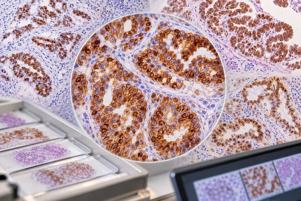
Cytokeratin 7 (CK7) immunohistochemistry (IHC) detects keratin 7, a structural protein expressed by many glandular and transitional epithelia. Pathologists use CK7 primarily to help classify carcinomas and estimate their likely site of origin, especially when a cancer is metastatic or poorly differentiated. CK7 is commonly positive in many lung adenocarcinomas, breast carcinomas, Müllerian tumors, thyroid carcinomas, pancreaticobiliary carcinomas, and urothelial carcinomas, while conventional colorectal and prostate adenocarcinomas are usually negative. Because CK7 expression is so widespread, a positive result is rarely diagnostic by itself. Its main value comes from the pattern created with CK20 and more specific markers such as TTF-1, Napsin A, GATA3, PAX8, ER, CDX2, SATB2, or uroplakin. A report that says “CK7 positive” should therefore be read as one clue in a larger tumor-origin panel, not as a stand-alone cancer label.
- CK7-positive staining is common in many carcinomas, including lung, breast, Müllerian, pancreaticobiliary, thyroid, and urothelial tumors.
- CK7 positivity alone does not identify a primary site because many unrelated cancers share the same pattern.
- A CK7-negative/CK20-positive profile strongly raises colorectal origin, while CK7-positive patterns usually require more specific markers.
- CK7 staining is typically cytoplasmic in tumor cells and may be diffuse or focal; there is no universal positive percentage cutoff for site-of-origin workups.
- Unexpected CK7 results can occur because tumors are heterogeneous, poorly differentiated, or biologically atypical.
Table of Contents
- What CK7 IHC detects
- Normal expression and staining pattern
- Cancers that are often CK7 positive
- Cancers that are often CK7 negative
- CK7 and CK20 patterns for tumor origin
- Pitfalls and limitations
- How to read the report and next steps
- Practical CK7 scenarios and diagnostic decisions
What CK7 IHC detects
CK7 is a type II intermediate-filament protein encoded by KRT7. Cytokeratins help maintain epithelial cell structure. Because different epithelial organs express characteristic keratin combinations, pathologists can use CK7 and related markers to infer differentiation and likely tumor origin.
The test is performed on a tissue section. The antibody produces cytoplasmic staining in cells that express CK7, sometimes with membranous accentuation. A pathologist confirms that the staining occurs in the malignant cells and describes the extent as diffuse, patchy, focal, or negative.
CK7 IHC does not measure a blood level, identify a gene mutation, or directly predict treatment response. There is no universal “normal range.” It is a qualitative or semiquantitative tissue marker whose meaning depends on the differential diagnosis.
The marker is especially useful when paired with CK20. However, the CK7/CK20 matrix is only an initial framework. Modern IHC panels increasingly use organ-specific transcription factors and lineage markers because those tests can provide stronger evidence than keratin profiles alone. The goal is to start broad and then narrow efficiently.
Normal expression and staining pattern
Normal CK7 expression is found in many glandular and ductal epithelia. Examples include breast ductal epithelium, lung and bronchial epithelium, biliary and pancreatic ducts, endometrial and other Müllerian epithelia, thyroid epithelium, and urothelium. In contrast, mature colorectal surface and glandular epithelium is generally CK7 negative or only rarely positive.
Because normal tissues may stain, the pathologist must distinguish tumor cells from surrounding structures. A liver metastasis, for example, may be CK7 positive while adjacent bile ducts also stain strongly. The bile ducts confirm that the stain worked but are not part of the metastasis.
True CK7 staining is cytoplasmic and can be diffuse in many adenocarcinomas. Some tumors show only focal expression. Extent matters because a diffuse pattern that fits a known phenotype carries more weight than a few isolated weak cells.
Fixation, necrosis, decalcification, and limited viable tissue can affect staining. Internal controls are useful when present. If expected normal epithelium is negative, an apparently negative tumor may need cautious interpretation.
Cancers that are often CK7 positive
Many common carcinomas are CK7 positive, which makes the marker sensitive but not specific. Lung adenocarcinoma is typically CK7 positive, but the more informative markers for pulmonary origin are TTF-1 and Napsin A. A metastatic adenocarcinoma that is CK7+/TTF-1+/Napsin A+ strongly supports lung origin in the right clinical setting. The TTF-1 IHC test is also positive in thyroid, so the broader panel and morphology remain important.
Breast carcinoma is often CK7 positive. GATA3, ER, PR, mammaglobin, or other markers may be used depending on the differential. GATA3 is also frequently positive in urothelial carcinoma, so breast versus urothelial origin cannot be settled by CK7 and GATA3 alone.
High-grade serous ovarian and other Müllerian carcinomas are generally CK7 positive. PAX8 is a key marker supporting Müllerian origin, while WT1 is commonly positive in high-grade serous carcinoma. The PAX8 IHC test also marks many renal and thyroid tumors, so site-specific interpretation is still required.
Pancreatic and biliary adenocarcinomas are commonly CK7 positive, with variable CK20. Gastric adenocarcinoma is heterogeneous and can show several CK7/CK20 combinations. Urothelial carcinoma is also often CK7 positive and may coexpress CK20.
Because this list is broad, CK7 positivity should usually trigger a more specific second step rather than a diagnosis. The most useful question is not “Which cancers can be CK7 positive?” but “Among the realistic candidates in this patient, which marker best separates them?”
Cancers that are often CK7 negative
Conventional colorectal adenocarcinoma is typically CK7 negative and CK20 positive. That combination, particularly with diffuse CDX2 and SATB2, strongly supports lower gastrointestinal origin. The CDX2 IHC test and SATB2 generally carry more site-specific information than CK7 negativity alone.
Prostate adenocarcinoma is also usually CK7 negative. PSA, NKX3.1, and other prostate-lineage markers are much more specific. Hepatocellular carcinoma is usually CK7 negative or only focally positive and is assessed with markers such as arginase-1 and HepPar-1. Many renal cell carcinomas have variable CK7 depending on subtype: clear cell renal cell carcinoma is often negative or focal, while papillary renal cell carcinoma is commonly positive.
Squamous cell carcinomas show variable CK7 and are generally classified with morphology and markers such as p40 rather than a CK7 profile. Merkel cell carcinoma is usually CK7 negative and CK20 positive with a dot-like pattern, which can help distinguish it from pulmonary small cell carcinoma.
The word “usually” matters. Poorly differentiated tumors can lose expected markers, and some colorectal cancers—especially unusual histologic or molecular subsets—can express CK7. Therefore, a CK7-positive result does not absolutely exclude colon, and a CK7-negative result does not absolutely exclude lung, breast, or Müllerian origin.
CK7 and CK20 patterns for tumor origin
The coordinate CK7/CK20 profile remains a useful orientation tool in carcinoma of unknown primary, but it is not a deterministic algorithm. A large modern tissue-microarray study showed substantial overlap across tumor types, and recent practice research suggests that site-specific markers often contribute more directly to the final diagnosis.
| Pattern | Examples that may fit | Best way to refine |
|---|---|---|
| CK7+ / CK20− | Lung, breast, Müllerian, thyroid, many pancreaticobiliary tumors | TTF-1/Napsin A, GATA3/ER, PAX8/WT1, organ-specific markers |
| CK7+ / CK20+ | Urothelial, some gastric/pancreaticobiliary, mucinous ovarian tumors | GATA3/uroplakin, PAX8, CDX2/SATB2, morphology |
| CK7− / CK20+ | Colorectal adenocarcinoma, Merkel cell carcinoma | CDX2/SATB2 versus neuroendocrine markers and TTF-1 |
| CK7− / CK20− | Prostate, hepatocellular, renal clear cell, many squamous tumors | Use direct lineage/site markers |
The CK20 IHC test is particularly useful when positive because CK20 expression is more concentrated in certain tumor families. CK7 positivity, by contrast, is extremely common, so it often has more value when it is unexpectedly absent or when it separates two realistic candidates.
Clinical context changes the pretest probability. A CK7+/CK20− liver metastasis in a patient with a lung mass and a CK7+/TTF-1+ profile is interpreted differently from the same keratin pattern in a patient with an ovarian mass and PAX8 positivity. The stains work as a network, not as isolated binary results.
Pitfalls and limitations
The biggest pitfall is over-specificity. CK7 is expressed by so many carcinomas that “CK7 positive” alone often narrows the differential only slightly. A report should therefore be interpreted with the more specific markers that follow.
Tumor heterogeneity is another limitation. Some cancers contain CK7-positive and CK7-negative regions. A tiny needle biopsy may sample only one component, and a metastasis can show a somewhat altered phenotype compared with the primary tumor. Focal staining should be weighted less heavily than a classic diffuse pattern when other evidence conflicts.
Technical factors can cause false-negative staining. Poor fixation, prolonged decalcification, extensive necrosis, and scant viable tumor may reduce antigen detection. Internal controls can help determine whether the stain performed adequately.
There is also a risk of mistaking normal CK7-positive structures for tumor. Bile ducts in liver, renal collecting ducts, glandular epithelium, and other normal components may stain strongly. The pathologist maps the immunostain back to the H&E slide to ensure the malignant cells are the ones being scored.
CK7 status is generally not a direct prognostic or predictive biomarker. A positive stain does not mean a tumor is more advanced, and a negative stain does not mean it is less aggressive. The clinical importance comes from correct tumor classification, which then determines the relevant molecular and treatment biomarkers.
How to read the report and next steps
When reading a CK7 result, focus on the complete sentence or panel. “CK7 positive” is a weak conclusion by itself. “CK7 positive, TTF-1 and Napsin A positive, PAX8 and GATA3 negative” strongly supports a different origin than “CK7 positive, PAX8 and WT1 positive.”
Terms such as diffuse, focal, patchy, and rare positive cells indicate how much weight the pathologist gives the result. Diffuse strong staining that matches an expected phenotype is generally more useful than weak focal staining.
If the primary site is unknown, the next step may include additional IHC, review of imaging, endoscopy, comparison with a previous tumor, or molecular profiling. IHC is often able to establish a favored origin, but sometimes the most accurate conclusion remains a carcinoma with a probable rather than proven primary site.
Patients can ask which diagnosis the whole panel supports, which competing diagnoses were excluded, and whether the origin is considered definitive or favored. If treatment depends on a tissue-of-origin assumption, it may be reasonable to ask whether the pathology has been reviewed by a specialist or whether molecular testing could add useful information.
The practical value of CK7 is therefore not that it names a cancer. It helps organize the diagnostic problem so that more specific evidence can be applied efficiently and tissue can be preserved for clinically important biomarkers.
Practical CK7 scenarios and diagnostic decisions
CK7 is a broad epithelial marker, so its strongest practical role is narrowing possibilities rather than naming a single cancer. Pathologists get the most value from CK7 when they combine its positive or negative pattern with morphology, CK20, and a small number of organ-directed markers. Several common scenarios illustrate how that reasoning works.
A lung mass or metastatic adenocarcinoma where lung origin is suspected. Most pulmonary adenocarcinomas are CK7 positive, but CK7 is far too common in other adenocarcinomas to establish lung origin. TTF-1 and Napsin A provide more specific support. A CK7-positive, TTF-1-positive, Napsin A-positive tumor in a compatible clinical setting strongly supports lung adenocarcinoma, whereas CK7 positivity with both lung markers negative leaves a much wider differential. Mucinous lung adenocarcinomas are an important exception because they may lose typical lung markers and can express gastrointestinal markers.
A pelvic or peritoneal adenocarcinoma. Müllerian serous carcinomas are commonly CK7 positive. PAX8 supports Müllerian origin, and WT1 often supports tubo-ovarian high-grade serous differentiation. CK7 alone cannot distinguish these tumors from breast, lung, pancreaticobiliary, or other CK7-positive cancers. If mesothelioma is in the differential, a balanced epithelial-versus-mesothelial panel is required because broad keratin expression overlaps and some mesothelial markers are not absolutely specific.
A breast or urothelial cancer differential. Both can be CK7 positive, so CK7 provides little separation by itself. GATA3 can support either lineage and must also be interpreted in context. Additional breast markers, uroplakins, p40/p63, ER, morphology, and the patient’s clinical history may be needed. This illustrates a general rule: once CK7 places a tumor into a broad phenotype, the next stain should usually test a specific competing hypothesis rather than repeat another broad epithelial marker.
Pancreaticobiliary or upper-GI adenocarcinoma. These tumors often express CK7, while CK20 and CDX2 may be variable. A CK7-positive, CK20-variable profile can therefore fit pancreatic, biliary, gastric, and several non-GI primaries. Site assignment depends on anatomic findings and a tailored panel, not on the CK7/CK20 combination alone. Strong intestinal markers may shift the probability toward GI differentiation, but focal CDX2 in a CK7-positive tumor does not by itself establish colorectal origin.
A suspected colorectal metastasis that is CK7 positive. Although the classic colorectal phenotype is CK7 negative and CK20 positive, exceptions occur. Some right-sided, poorly differentiated, mucinous, or otherwise unusual colorectal carcinomas can express CK7. Strong SATB2 and CDX2, prior-pathology comparison, and morphology can outweigh an atypical CK7 result. The CDX2 IHC pattern is especially useful when interpreted with SATB2 rather than as a solitary colon marker.
Cancer of unknown primary in a small biopsy. Tissue conservation is critical. A pathologist may first establish carcinoma with keratins, then use CK7 and CK20 to choose a direction, followed by a limited set of site-specific stains. Ordering a very large panel at once can exhaust tissue needed for molecular testing. Efficient testing is therefore a sequence of questions: what lineage is present, which sites are plausible, which one or two markers best separate them, and what predictive tests must be preserved for later?
Negative CK7 can be as informative as positivity when the morphology and comparison group are appropriate. For example, CK7 negativity supports the classic lower-GI phenotype when CK20, CDX2, and SATB2 are positive, but it cannot prove colon origin. Similarly, a CK7-negative tumor may fit prostate, hepatocellular, renal, or other lineages depending on morphology and the rest of the panel. The meaning comes from the combination, not from an isolated minus sign.
A pathology report may use phrases such as “immunophenotype favors” or “compatible with” because IHC works probabilistically. For patients, the most useful questions are which primary sites were seriously considered, which organ-specific markers support the favored site, whether the staining was diffuse or focal, and whether prior tumor tissue was compared. CK7 is often the first useful branch point in that process, but the final diagnosis should rest on a coherent set of independent findings.
The intensity of CK7 is usually less important than the distribution and the identity of the stained cells. Diffuse strong cytoplasmic labeling across the tumor carries different weight from a few positive glands or entrapped benign ducts. Internal controls and careful localization help avoid interpreting normal epithelial structures as evidence that the neoplasm itself is CK7 positive.
CK7 results can also differ between a primary tumor and a metastasis because cancers are heterogeneous and may change phenotype as they progress. A small core from one metastatic focus may not reproduce every stain seen in the original resection. When a new CK7 result conflicts with a well-documented prior cancer, pathologists often compare the specimens directly and ask whether the morphology, other lineage markers, and molecular findings still support a common origin. Repeating CK7 alone is rarely the whole solution. The purpose is to decide whether the discordance falls within expected biologic variation or is large enough to raise concern for a second primary tumor. This comparison is especially useful in patients with a history of more than one malignancy.
The laboratory’s validated antibody clone, fixation conditions, and scoring practice can also influence how weak or focal staining is reported. For that reason, subtle differences in CK7 intensity between institutions should not be overinterpreted. Diagnostic weight comes from a reproducible pattern in the tumor cells and agreement with the rest of the case.
References
- Cytokeratin 7 and cytokeratin 20 expression in cancer: A tissue microarray study on 15,424 cancers 2022
- Revisiting the use of CK7 and CK20 immunohistochemical stains in pathological diagnoses 2025
- Carcinoma of Unknown Primary Origin: Application of Immunohistochemistry With Emphasis to Different Cytokeratin 7 and 20 Staining Patterns. 2022
- Real-world histopathological approach to malignancy of undefined primary origin (MUO) to diagnose cancers of unknown primary (CUPs) 2023
- Use of SATB2 and CDX2 Immunohistochemistry to Characterize and Diagnose Colorectal Cancer 2024
- Diagnosing carcinoma origin in ultrasound-guided needle biopsies: a context-driven approach anchored in site-specific immunohistochemical markers 2026
Disclaimer
CK7 IHC is a diagnostic tissue stain and cannot determine a tumor’s primary site by itself. Results should be interpreted with morphology, CK20 and organ-specific markers, prior pathology, imaging, and other clinical or molecular information.





